Aggressive Sarcoma | 50% NF1-Associated | Worst Sarcoma Prognosis
- MPNST has worst prognosis of all soft tissue sarcomas - 5-year survival 40-60%
- 50% occur in NF1 patients from plexiform neurofibroma transformation (lifetime risk 8-13% - Evans; plexiform neurofibroma is the at-risk precursor)
- FDG-PET has 89% sensitivity and 95% specificity for MPNST in NF1 (Ferner); SUVmax over 3.5 is the conventional high-suspicion cut-off, not a decision rule on its own
- Wide surgical excision with 2 cm margins en bloc with nerve is only curative treatment
- Adjuvant radiotherapy improves local control 60-70% to 80-85% but NOT overall survival
- “NF1-associated MPNST is widely taught as having a worse prognosis (21% vs 42% 5-year survival, Evans) - but note that comparison was NOT significant (p=0.09), and NF1 did not survive multivariate analysis in the Mayo series. Size, grade, margin and local recurrence are the established predictors
- “S100 positive only 50-70% and FOCAL (not diffuse like schwannoma) - negative does not exclude
- “PRC2 (SUZ12/EED) loss in 70-90%, so H3K27me3 immunohistochemistry SUPPORTS the diagnosis - but it is a weak negative: 31% of MPNSTs RETAIN it, only about 60% of NF1-related tumours lose it, and ALL epithelioid MPNSTs retained it in Prieto-Granada's series
- “Rapid growth, severe pain 70%, neurological deficit - distinguish from stable neurofibroma
- “Chemotherapy response is SIMILAR to other soft-tissue sarcomas (Kroep EORTC: response 21% vs 22%), not intrinsically worse - the old 'chemo-resistant' label is refuted
Overview and Epidemiology
Malignant peripheral nerve sheath tumour (MPNST) is an aggressive soft-tissue sarcoma arising from the cells of the peripheral nerve sheath. It makes up 5-10% of soft-tissue sarcomas and carries one of the worst prognoses among them, with 5-year survival of 40-60% overall. About 50% arise in neurofibromatosis type 1 (NF1), typically by malignant transformation of a plexiform neurofibroma.
How common. The annual incidence in the general population is about 0.001% (1 per 100,000). In NF1 it is 0.16% overall and 2-5% in adults with a plexiform neurofibroma, and the lifetime risk in NF1 is 8-13% (Evans, population study), concentrated in those with a plexiform neurofibroma, the at-risk precursor lesion. There is a slight male predominance (1.2:1).
Who. NF1 patients present young, at a median of 26-30 years; sporadic disease presents at a median of 40-50 years. In Evans's population series the gap was 26 against 62 years.
Where. The site distribution:
- Proximal extremities: 40-50% (thigh most common, and upper arm)
- Trunk: 25-30% (paraspinal, retroperitoneum)
- Head and neck: 15-20%
- Distal extremities: 5-10%
The brachial and lumbosacral plexuses are commonly involved in NF1.
Three aetiologies. Half of MPNST is NF1-associated, most of the rest sporadic, and a tenth follows radiotherapy. The origin shapes the age of the patient, the genetics and the reported survival.
- Frequency
- 50%
- Median Age
- 26-30 years
- Pathogenesis
- Plexiform neurofibroma transformation, germline NF1 plus somatic TP53
- 5-Year Survival
- 21%
- Frequency
- 40%
- Median Age
- 40-50 years
- Pathogenesis
- Somatic NF1 inactivation, TP53, PRC2 mutations
- 5-Year Survival
- 42%
- Frequency
- 10%
- Median Age
- Variable
- Pathogenesis
- Prior radiation over 40 Gy, latency 10-20 years
- 5-Year Survival
- 20-30%
Is NF1-associated MPNST prognostically worse? The honest answer is that it is widely taught, plausible, and not established by the evidence on this page. Evans reported five-year survival of 21% in NF1 against 42% in sporadic disease, but at p=0.09, in 21 versus 37 patients. Ducatman (1986) did find neurofibromatosis independently adverse at p less than 0.05. Stucky's 175-patient Mayo series found NF1 associated with disease-specific survival on univariate analysis only; it did not appear among the independent multivariate predictors, where size, grade, local recurrence and truncal location did.
What is not in doubt is that NF1 patients present younger and often with larger, deeper, plexiform-derived tumours that are harder to resect, so any survival difference may be mediated by stage and resectability rather than by NF1 biology itself. Counsel on the size, grade and margin in front of you rather than on the NF1 label.
Pathophysiology and Molecular Pathogenesis
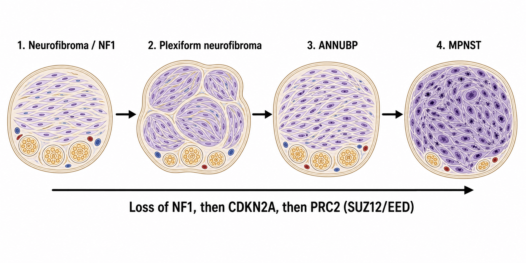
The NF1 pathway. Transformation is a sequence of genetic hits. A germline NF1 mutation plus a somatic second hit (loss of heterozygosity) produces the benign neurofibroma: slow-growing, soft, painless, stable for years, of low cellularity with wavy spindle cells. Additional genetic changes produce the atypical neurofibroma, hypercellular with mild atypia, still benign but at higher risk of progression and difficult to diagnose histologically; its consensus name, ANNUBP, has its own section below. TP53 inactivation (75% of MPNST) plus PRC2 complex mutations (SUZ12 or EED loss in 70-90%), with CDKN2A deletion in 50-60%, complete the transformation to MPNST.
What each hit does. Each loss contributes differently:
- NF1 loss: neurofibromin deficiency, so Ras-MAPK is hyperactivated and proliferation runs uncontrolled
- TP53 loss: cell-cycle checkpoints and apoptosis are lost, so mutations accumulate
- PRC2 loss (SUZ12/EED): epigenetic dysregulation, with loss of the H3K27 trimethylation mark
- CDKN2A deletion: loss of the p16 tumour suppressor and cell-cycle dysregulation
Sporadic MPNST. Arises with no prior neurofibroma or NF1 diagnosis, through somatic biallelic NF1 inactivation (not inherited), with TP53 mutations common, PRC2 mutations (SUZ12, EED) and CDKN2A deletions.
Radiation-induced MPNST. Median latency is 10-20 years after radiotherapy, at doses typically over 40 Gy, with the field including a peripheral nerve; examples are treatment for breast cancer, lymphoma and childhood malignancies. These tumours are often higher grade with a worse prognosis.
Which neurofibroma transforms. In NF1 the risk depends on the type and site of the neurofibroma; internal plexiform lesions carry the highest malignant potential and cutaneous neurofibromas rarely transform.
- Risk Level
- High
- Transformation Rate
- 8-13% lifetime (Evans)
- Management
- Annual surveillance, PET-CT if concerning
- Risk Level
- High
- Transformation Rate
- Higher than superficial
- Management
- MRI surveillance, low threshold for PET-CT
- Risk Level
- Moderate
- Transformation Rate
- Difficult to monitor
- Management
- Regular imaging, PET-CT surveillance
- Risk Level
- Low
- Transformation Rate
- Rare transformation
- Management
- Clinical surveillance only
Histopathology and Classification
Macroscopic. The tumour is usually over 5 cm at diagnosis (mean 8-10 cm), with poorly defined, infiltrative margins, unlike the encapsulated schwannoma. The cut surface is tan-grey, fleshy and heterogeneous; necrosis is common in high-grade tumours (50-70%) and haemorrhage is frequent. The nerve of origin may be visible in a smaller tumour and is obscured in a large mass.
High-grade MPNST (90% of cases). Dense fascicles of spindle cells in a whorled pattern, a "marbled" alternation of densely cellular and myxoid zones, perivascular accentuation (cells clustered around vessels) and infiltrative margins into the surrounding tissue. The cells have hyperchromatic, wavy, buckled nuclei like those of benign Schwann cells, with moderate to severe pleomorphism and a high nuclear-to-cytoplasmic ratio. Mitoses typically exceed 10 per 10 HPF with atypical forms common, and geographic necrosis is present in 50-70% and predicts aggressive behaviour.
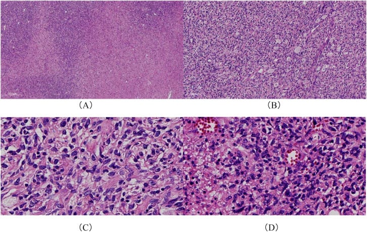
Low-grade MPNST (10% of cases). A rare variant that is difficult to separate from atypical neurofibroma: fascicles of spindle cells with mild to moderate nuclear atypia, a low mitotic count (under 5 per 10 HPF), minimal necrosis, hypercellularity relative to neurofibroma and, the key feature, an infiltrative growth pattern. Molecular markers (H3K27me3 loss) help, the tumour remains aggressive despite its grade, and the diagnosis requires expert sarcoma pathology review.
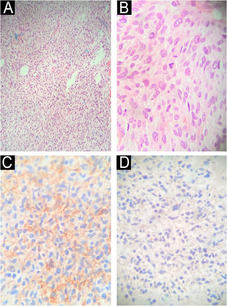
Immunohistochemistry. The neural markers are present but patchy, which is the recurring trap: S100 is positive in only 50-70% and focally, so a negative stain does not exclude MPNST, whereas a schwannoma stains strongly and diffusely.
- Positivity Rate
- 50-70%
- Pattern
- FOCAL and PATCHY (not diffuse)
- Clinical Significance
- Positive supports diagnosis but negative does NOT exclude
- Positivity Rate
- 50-60%
- Pattern
- Focal
- Clinical Significance
- Neural lineage marker
- Positivity Rate
- 50-70%
- Pattern
- Global loss
- Clinical Significance
- Indicates PRC2 mutation, supports MPNST diagnosis
- Positivity Rate
- Variable
- Pattern
- Greater than 10%, often 30-50%
- Clinical Significance
- High proliferation index confirms high-grade
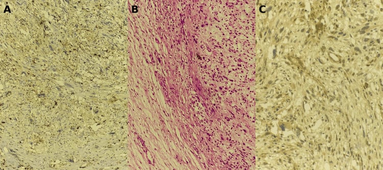
Reading H3K27me3. Loss of the mark reflects PRC2 inactivation and supports the diagnosis, and it is retained in neurofibroma and the main mimics, so complete loss in a spindle-cell lesion is meaningful. It is a weak negative: 31% of MPNSTs retained it in Prieto-Granada's series, only about 60% of NF1-related tumours lose it, and all the epithelioid MPNSTs in that series retained it. Retained staining therefore does not exclude MPNST, and least of all in an NF1 patient or an epithelioid tumour.
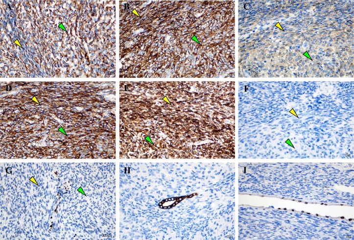
Negative markers. These exclude the look-alikes:
- Desmin negative (excludes rhabdomyosarcoma)
- Cytokeratin negative (excludes synovial sarcoma and epithelioid sarcoma)
- CD34 typically negative
- EMA negative (unlike perineurioma)
Variants. MPNST with rhabdomyoblastic differentiation, the malignant Triton tumour, is 5-10% of MPNST, contains a malignant skeletal-muscle component that is desmin, myogenin and MyoD1 positive in the rhabdomyoblastic areas, and has a worse prognosis than conventional MPNST. Epithelioid MPNST has epithelioid rather than spindle cytology, is more common in superficial locations, is often S100 positive and must be distinguished from melanoma and epithelioid sarcoma.
Differential diagnosis. MPNST is a spindle-cell sarcoma that overlaps morphologically with several other neoplasms, and immunohistochemistry and clinical context are what separate them.
- Key Distinguishing Feature
- Low mitotic rate, no necrosis, retains uniform architecture
- Immunohistochemistry
- S100 diffuse, H3K27me3 RETAINED
- Behaviour
- Benign / pre-malignant (atypical)
- Key Distinguishing Feature
- Encapsulated, Antoni A/B, Verocay bodies, hyalinised vessels
- Immunohistochemistry
- S100 STRONG and DIFFUSE, SOX10 diffuse
- Behaviour
- Benign
- Key Distinguishing Feature
- Uniform spindle cells, SS18-SSX fusion
- Immunohistochemistry
- Cytokeratin / EMA focal positive, TLE1 positive, S100 focal
- Behaviour
- Malignant
- Key Distinguishing Feature
- Herringbone pattern, no nerve origin
- Immunohistochemistry
- S100 negative, CD34 (DFSP), H3K27me3 retained
- Behaviour
- Malignant (variable grade)
- Key Distinguishing Feature
- Blunt-ended (cigar) nuclei, fascicles at right angles
- Immunohistochemistry
- SMA / desmin / h-caldesmon positive, S100 negative
- Behaviour
- Malignant
- Key Distinguishing Feature
- Junctional component, history of pigmented lesion
- Immunohistochemistry
- S100 diffuse, SOX10 diffuse, H3K27me3 RETAINED
- Behaviour
- Malignant
The discriminators to carry: diffuse strong S100 favours schwannoma or melanoma over MPNST; H3K27me3 loss favours MPNST and is retained in melanoma, neurofibroma and most mimics; epithelial markers (cytokeratin, EMA, TLE1) point to synovial sarcoma; and smooth-muscle markers point to leiomyosarcoma.
Grading. MPNST is graded by the FNCLCC system, summing differentiation, mitotic count and necrosis.
- Score 1
- Well-differentiated
- Score 2
- Moderately differentiated
- Score 3
- Poorly differentiated
- Score 1
- 0-9 per 10 HPF
- Score 2
- 10-19 per 10 HPF
- Score 3
- 20 or more per 10 HPF
- Score 1
- None
- Score 2
- Less than 50%
- Score 3
- 50% or greater
The total score gives the grade, and the distribution explains why most MPNST is called high grade at diagnosis:
- Grade 1 (low): 2-3 points, rare in MPNST (under 10%)
- Grade 2 (intermediate): 4-5 points (20%)
- Grade 3 (high): 6-8 points (70%)
Atypical Neurofibromatous Neoplasm of Uncertain Biologic Potential (ANNUBP)
The intermediate lesion on the road from neurofibroma to MPNST deserves a name and a definition, because it is the diagnostic grey zone that determines whether a lesion is watched, marginally excised, or treated as a sarcoma. The benign neurofibroma itself is covered in the dedicated neurofibroma topic; here the focus is the precursor.
The consensus term. A 2017 international consensus replaced the vague "atypical neurofibroma" with atypical neurofibromatous neoplasm of uncertain biologic potential (ANNUBP), the histological and biological bridge between a benign neurofibroma and MPNST. It is a recognised premalignant lesion, especially within large internal or plexiform neurofibromas in NF1.
Diagnostic criteria. ANNUBP is diagnosed when at least two of four features are present:
- Cytologic atypia
- Loss of the normal neurofibroma architecture
- Hypercellularity
- Low-level mitotic activity, present but below the MPNST threshold (fewer than 3 mitoses per 10 high-power fields), with no necrosis
The distinction from low-grade MPNST is essentially quantitative: a higher mitotic count (3 or more per 10 HPF) and/or necrosis push a lesion into MPNST. This is a genuinely difficult call that requires expert sarcoma pathology.
Molecular correlate. Progression from neurofibroma to ANNUBP is marked by biallelic CDKN2A (p16) inactivation; loss of p16 by immunohistochemistry supports the diagnosis and dovetails with the CDKN2A deletions seen in established MPNST. H3K27me3 is usually retained at the ANNUBP stage, its global loss being a later event that comes with PRC2 inactivation as the lesion becomes MPNST, so an atypical lesion that has lost H3K27me3 should raise concern for transformation.
Clinical relevance and management. ANNUBP often corresponds to a focally concerning area within a plexiform neurofibroma, for example a region of intermediate PET avidity (SUVmax 2.5-3.5) or new growth, and may be what a targeted biopsy of that area yields. Unlike MPNST, ANNUBP is managed by complete (marginal) excision with clear margins alone: it does not require the wide 2 cm margins, nerve sacrifice, radiotherapy or chemotherapy used for MPNST, but it does mandate negative margins and ongoing surveillance because of its malignant potential.
Investigations and Staging
MRI is the gold standard for local staging. The protocol is T1-weighted for anatomy, T2-weighted with fat suppression for tumour extent and oedema, and T1 post-gadolinium with fat suppression for the enhancement pattern, covering the entire compartment plus the joint above and below. It guides resection planning and biopsy location.
What MRI shows. On T1 the tumour is isointense to muscle with heterogeneous signal from necrosis and haemorrhage, irregular infiltrative margins unlike the well-defined benign tumours, and loss of the surrounding fat planes. On T2 the signal is heterogeneous, the uniform hyperintensity of a benign neurofibroma is lost, central necrosis gives fluid signal and aggressive tumours carry peritumoral oedema. After gadolinium the enhancement is heterogeneous, with non-enhancing necrotic areas, rim enhancement around them and enhancement of the involved nerve proximally and distally.
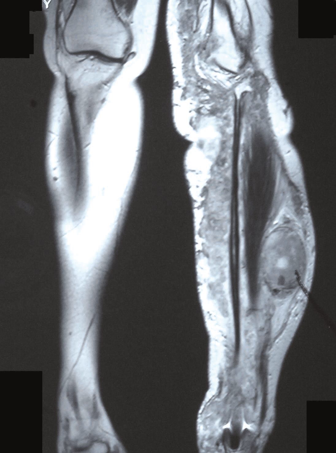
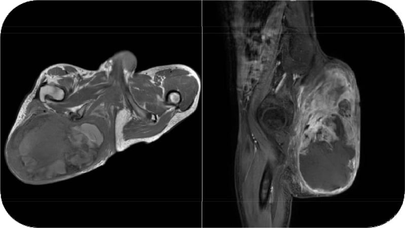
Benign neurofibroma or MPNST. The features that separate the two on MRI:
- Benign Neurofibroma
- Usually under 5 cm
- MPNST
- Greater than 5 cm (mean 8-10 cm)
- Benign Neurofibroma
- Uniform T2 hyperintense
- MPNST
- Heterogeneous signal
- Benign Neurofibroma
- Well-defined
- MPNST
- Irregular, infiltrative
- Benign Neurofibroma
- Absent
- MPNST
- Present 50-70%
- Benign Neurofibroma
- Present (central low, peripheral high T2)
- MPNST
- Lost
- Benign Neurofibroma
- Minimal
- MPNST
- Perilesional oedema common
FDG PET-CT. This is the critical tool for diagnosing malignant transformation in NF1, where the question is which of many plexiform neurofibromas has changed. Its indications:
- Known plexiform neurofibroma with concerning clinical features
- Surveillance of high-risk plexiform neurofibromas
- Preoperative staging
- Detection of metastases
Reading the SUV. Ferner showed that FDG-PET and PET-CT separate benign from malignant well (sensitivity 0.89, specificity 0.95) but also that SUVmax did not predict tumour grade, and the commonly quoted cut-offs (about 2.5 and 3.5) come from pooled and subsequent analyses rather than that primary report. Treat them as convention: a high SUV raises concern and a low SUV lowers it, but neither is a decision rule on its own, and a clinically worrying lesion is biopsied whatever the number.
- Interpretation
- Likely benign
- Sensitivity/Specificity
- NPV 100%
- Action
- Reassuring, continue surveillance
- Interpretation
- Intermediate risk
- Sensitivity/Specificity
- Indeterminate
- Action
- Close surveillance OR biopsy
- Interpretation
- High suspicion MPNST
- Sensitivity/Specificity
- Sens 89%, Spec 95%, PPV 77%
- Action
- URGENT biopsy and treatment planning
What PET adds and where it fails. It guides the biopsy to the highest-SUV area of a heterogeneous tumour, identifies occult metastases on whole-body staging, serves as a surveillance tool in high-risk NF1 patients and monitors treatment response. Inflammation and infection give false positives, low-grade MPNST may have a lower SUV and give false negatives, and a PPV of 77% means a 23% false-positive rate, so it is not a replacement for biopsy. Cost and radiation exposure limit its use for routine surveillance. An SUV over 3.5 should trigger urgent MDT discussion and biopsy.
Staging CT. The lung is the most common metastatic site (50% at presentation or during follow-up), the liver less common, bone rare, and retroperitoneal or mediastinal disease is looked for on the same study. The protocol is CT chest with intravenous contrast, and CT abdomen and pelvis for a trunk primary or concerning symptoms; a dedicated chest CT rather than a chest radiograph, because the lung metastasis risk is high. Staging is completed before biopsy so that treatment can be planned in full.
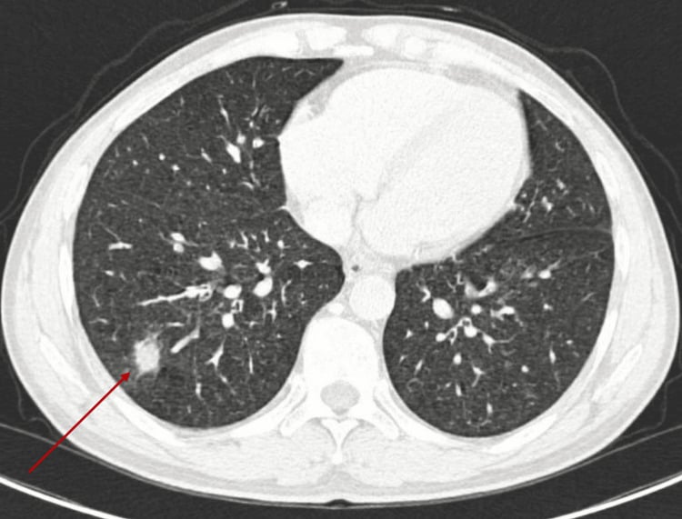
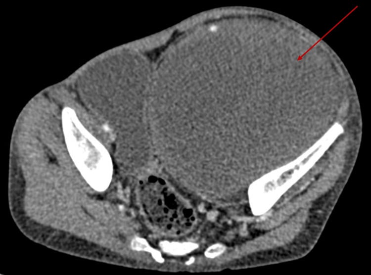
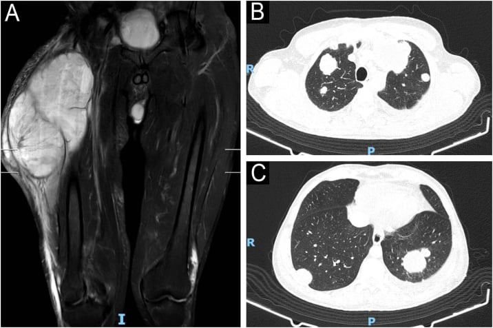
Biopsy. The principles are inviolable because the tract becomes part of the specimen.
- Image-guided core needle biopsy is preferred: 14-16 gauge, 4-6 cores
- The tract MUST lie in line with the planned surgical incision, to be excised en bloc
- Target solid areas and avoid necrosis; image guidance is essential
- NEVER perform an excisional biopsy for suspected MPNST, which violates oncological principles
- Send for H&E, immunohistochemistry (S100, SOX10, desmin, cytokeratin, Ki67, H3K27me3) and FISH or molecular studies if uncertain
Referral to a sarcoma centre before biopsy is ideal for optimal outcomes.
What to ask of the pathologist. The report must give the FNCLCC grade, which drives prognosis and adjuvant decisions, together with S100, Ki67 and H3K27me3 status, and it must exclude the other spindle-cell sarcomas, synovial sarcoma and fibrosarcoma in particular.
AJCC staging. Most MPNST presents as stage IIIB (large, deep, high grade) or stage IV (metastatic).
- T (Size/Depth)
- T1 (5 cm or less) superficial or deep
- Grade
- Grade 1 (low)
- 5-Year Survival
- 90%
- T (Size/Depth)
- T2-T4 (greater than 5 cm)
- Grade
- Grade 1 (low)
- 5-Year Survival
- 80%
- T (Size/Depth)
- T1 (5 cm or less)
- Grade
- Grade 2-3 (high)
- 5-Year Survival
- 70%
- T (Size/Depth)
- T2 (greater than 5 cm but 10 cm or less)
- Grade
- Grade 2-3 (high)
- 5-Year Survival
- 60%
- T (Size/Depth)
- T3-T4 (greater than 10 cm)
- Grade
- Grade 2-3 (high)
- 5-Year Survival
- 40-50%
- T (Size/Depth)
- Any T with N1 or M1
- Grade
- Any grade
- 5-Year Survival
- 10-15%
Clinical Assessment
History. The presentation is a rapidly enlarging mass over weeks to months, the key feature separating it from a benign lesion, with severe pain in 60-70% (neurogenic, in the distribution of the nerve) and a progressive neurological deficit. Symptoms have usually been present for less than a year. In NF1 the story is a neurofibroma that was stable for years and has suddenly grown.
Red flags in NF1. A neurofibroma that enlarges rapidly, changes from soft to firm, becomes newly and severely painful having been painless, or produces weakness or numbness is a red flag for transformation. Constitutional symptoms (weight loss, fatigue, in 10-20%) are rare in localised disease, suggest metastatic disease when present, and warrant full staging.
Examination. The mass is large, typically over 5 cm, firm and fixed to underlying structures; the overlying skin is normal or, in advanced disease, tethered or ulcerated, and large tumours may show venous engorgement. On palpation the firm to hard consistency against the soft neurofibroma is the critical distinguishing feature; the mass is non-mobile, fixed to deep tissues, poorly defined at its margins, tender in 60-70%, and without pulsation, which separates it from a vascular lesion.
Neurological assessment. A motor deficit in the distribution of the involved nerve is present in 30-40% and a sensory deficit in 40-50%, with muscle atrophy if chronic and reduced or absent deep tendon reflexes. Document the preoperative neurological status, because it is the baseline for informed consent. Brachial or lumbosacral plexus involvement gives weakness and sensory loss across multiple nerve distributions. Lymphadenopathy is rare, since MPNST rarely spreads to lymph nodes (under 5%).
Red flags requiring urgent investigation. Any of these calls for imaging now:
- Size greater than 5 cm
- Deep (subfascial) location
- Rapid growth over weeks to months
- Fixation to underlying structures
- Neurological deficit
Management Algorithm
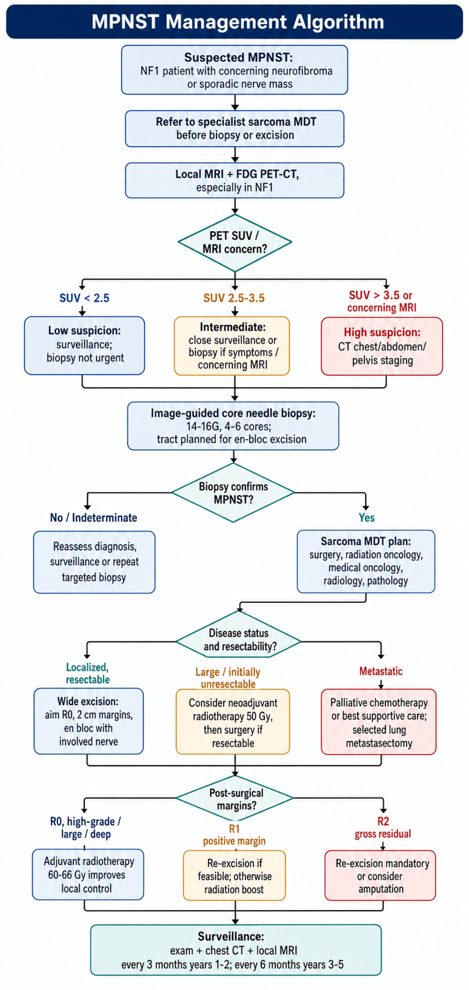
- 1Suspected MPNST
An NF1 patient with a neurofibroma that is growing, newly painful or causing a deficit, or any patient with a sporadic nerve-related mass. Do not biopsy first - image first, because the biopsy tract must sit inside the eventual resection field.
MRI for local staging AND FDG-PET/CT, the latter particularly in NF1 where multiple lesions must be triaged
- 2Imaging complete - triaging by PET
A high SUV raises concern and a low SUV lowers it, but neither is a decision rule on its own, and a clinically worrying lesion is biopsied whatever the number.
High SUV or concerning MRI features - proceed to CT chest, abdomen and pelvis for metastatic staging. Intermediate - close surveillance or biopsy if there is clinical concern. Low SUV with a reassuring clinical picture - surveillance
- 3Staging complete
Image-guided core needle biopsy, planned WITH the surgeon who will resect so the tract is excisable en bloc. Sample the enhancing or PET-avid component rather than a necrotic centre.
Histological diagnosis, with H3K27me3 immunohistochemistry as supportive evidence - remembering that retained staining does not exclude MPNST, least of all in NF1 or epithelioid tumours
- 4Biopsy confirms MPNST
Present every case at a sarcoma multidisciplinary meeting before committing to treatment. This is not a formality: margin planning, neoadjuvant sequencing and the amputation-versus-limb-salvage conversation all change with the discussion.
Surgical oncology, medical oncology, radiation oncology, radiology and pathology all represented, with NF1 specialist input where relevant
- 5MDT treatment plan
Localised and resectable - wide excision with the nerve taken en bloc, accepting the resulting deficit. Large or initially unresectable - consider neoadjuvant radiotherapy then reassess for surgery. Metastatic - palliative systemic therapy or best supportive care, noting that response rates are comparable to other soft-tissue sarcomas rather than worse.
Complete resection remains the only treatment associated with cure; plan the reconstruction and the functional consequence before the incision
- 6Post-surgical pathology
R0 with high grade - adjuvant radiotherapy improves local control. R1 - re-excise if feasible, otherwise adjuvant radiotherapy with a boost. R2 gross residual - re-excision is mandatory and amputation should be discussed. Adjuvant radiotherapy improves LOCAL CONTROL without a demonstrated overall survival benefit, and adjuvant chemotherapy is unproven - Kroep's data are from advanced disease only.
Local control optimised, with the survival conversation kept honest: size, grade, margin and local recurrence are what predict outcome
- 7Adjuvant therapy complete - surveillance
Intensive follow-up, because local recurrence is itself a strong adverse prognostic factor (HR 4.4 in the Mayo series) and detecting it early is the point of the schedule.
Examination with cross-sectional imaging every 3 months for years 1-2, then every 4-6 months to year 5, with chest imaging for pulmonary metastases
Adjuvant and Systemic Therapy
Radiotherapy. It is indicated for high-grade MPNST (most cases, 90%), positive or close margins (less than 1 mm), large tumours (over 5 cm) and deep (subfascial) location. It improves local control from 60-70% to 80-85% but does not improve overall survival, and it is recommended for most MPNST given the high local recurrence risk.
Before or after surgery. Preoperative radiotherapy is 50 Gy in 25 fractions: a smaller treatment volume and potentially better local control, against wound complications in 30-40% and a delay to surgery of 6-8 weeks after radiotherapy. Postoperative radiotherapy is 60-66 Gy in 30-33 fractions: immediate surgery and fewer wound complications (15-20%), against a larger treatment volume that includes the entire surgical bed. If a positive margin cannot be re-resected, a boost of a further 10-16 Gy is given to the positive margin area.
Chemotherapy. The settings and what they achieve:
- Indication
- Initially unresectable, downstaging
- Regimen
- Ifosfamide plus doxorubicin
- Response Rate
- 20-30%
- Survival Benefit
- No proven benefit
- Indication
- Controversial, young patients high-grade
- Regimen
- Ifosfamide plus doxorubicin
- Response Rate
- N/A
- Survival Benefit
- No proven benefit
- Indication
- Palliative for symptomatic metastases
- Regimen
- Ifosfamide plus doxorubicin OR gemcitabine plus docetaxel
- Response Rate
- 20-40%
- Survival Benefit
- Median 8-12 months
What the response rate means. Response rates of 20-30% were long read as evidence that MPNST is intrinsically chemoresistant. The EORTC pooled analysis (Kroep) refutes that: response was 21% in MPNST against 22% in other soft-tissue sarcomas, so the benefit is modest in the way it is modest for every soft-tissue sarcoma. Adjuvant chemotherapy remains unproven, and Kroep's data are from advanced disease only.
Novel therapies. MEK inhibitors (selumetinib, trametinib) target the Ras-MAPK pathway but have modest activity in established MPNST (response under 10%); EZH2 inhibitors (tazemetostat) target the PRC2 pathway with limited single-agent activity; combination strategies are under investigation. Checkpoint inhibitors (anti-PD1, anti-CTLA4) have limited data and low response rates (under 10%), and tumour mutational burden is typically low. No targeted therapy or immunotherapy is standard of care, and clinical trial enrolment is encouraged.
MEK inhibition treats the precursor, not the sarcoma. The real, practice-changing role of MEK inhibition is in the benign plexiform neurofibroma, and the distinction is a high-yield one. Selumetinib, an oral MEK1/2 inhibitor that blocks the Ras-MAPK pathway driven by NF1 loss, is approved for paediatric NF1 patients with symptomatic, inoperable plexiform neurofibromas. In the pivotal SPRINT phase II trial, 35 of 50 children (70%) had a confirmed partial response (a reduction of at least 20% in tumour volume), most responses were durable, and there were meaningful improvements in pain, function and quality of life (Gross et al., N Engl J Med 2020, PMID 32187457).
The logic is a two-compartment distinction. In the benign plexiform neurofibroma, MEK inhibition can shrink the tumour, relieve symptoms and reduce the at-risk substrate; in established MPNST the same drugs, and the other targeted and immune agents, have minimal activity, and complete surgical resection remains the only curative modality. The danger is mistaking a transforming or frankly malignant mass for a benign plexiform neurofibroma and managing it medically: a plexiform neurofibroma that grows, becomes painful, develops a focally intermediate-to-high SUV, or shows worrying MRI change must be biopsied to exclude ANNUBP or MPNST before it is attributed to benign disease and offered a MEK inhibitor. Detailed medical management of plexiform neurofibroma belongs to the neurofibromatosis topic; the point here is the boundary with sarcoma.
Surgical Technique
The MDT. The discussion that precedes planning must include:
- A sarcoma surgeon (orthopaedic oncology or surgical oncology)
- A medical oncologist
- A radiation oncologist
- A musculoskeletal radiologist
- A pathologist with sarcoma expertise
- An NF1 specialist, if applicable
Planning. The MRI is reviewed for tumour extent and neurovascular involvement, the relationship to major structures (vessels, bone, joints), and the feasibility of limb salvage against amputation. The functional cost of resecting the nerve is assessed and the reconstruction options discussed (nerve, soft tissue, bone if needed), with vascular surgery on standby if major vessel involvement is anticipated and an anaesthetic evaluation completed.
Consent. Realistic expectations are essential given the prognosis, and the discussion covers:
- The nature of MPNST and its poor prognosis (5-year survival 40-60%)
- Nerve sacrifice is mandatory, so a permanent neurological deficit is expected
- The functional consequence for the nerve concerned, for example foot drop with the sciatic nerve or hand weakness with the median or ulnar nerve
- The alternative of amputation, with its better local control and the same survival
- The need for adjuvant radiotherapy
- The risk of recurrence and of metastasis
Wide excision. The goal is an R0 resection, microscopically negative margins with a 2 cm margin in all directions. Under general anaesthesia, positioned for the tumour's location, with a tourniquet for an extremity (released before closure to assess perfusion) and vascular surgery available if a major vessel is involved, the operation runs in five steps:
- Incision. A longitudinal incision incorporating the biopsy site, with a wide ellipse around the tract so it is excised en bloc, and exposure at least 5 cm beyond the tumour margins proximally and distally.
- Dissection and margins. Develop the fascial planes 2 cm from the tumour edge, measured with a ruler, and resect the tumour en bloc with its cuff of normal tissue. The involved nerve is transected 2 cm proximal and distal to the tumour margins, involved muscle compartments are sacrificed, and major vessels are preserved if possible by dissecting them free with an adventitial margin.
- Vessel and bone. An encased vessel is resected and reconstructed with a vein graft or synthetic conduit; a vessel that is adherent but not invaded is dissected free with its adventitia, with frozen section of the adventitia if uncertain. Bone that is abutted is resected extraperiosteally; bone that is invaded is resected en bloc.
- Margin assessment. Orient the specimen with sutures or clips (superior, lateral, deep), send frozen section of any concerning margin, and re-resect if a margin is positive.
- Reconstruction and closure. Primary closure if tension-free, otherwise local flaps or free tissue transfer for a large defect. Nerve reconstruction is rarely performed, because this is a cancer operation and a nerve graft would lie within the radiation field; vascular and bone reconstruction follow if those structures were resected. Haemostasis, drains and a layered closure.
Limb salvage is achieved in over 95% of extremity MPNST, but with a significant functional deficit.
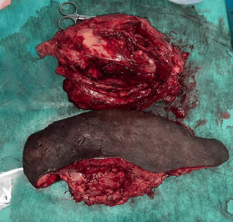
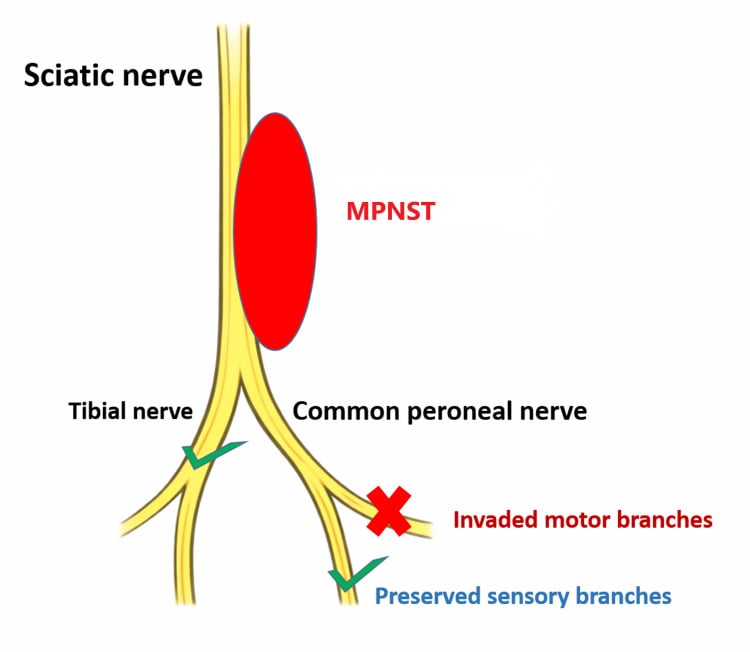
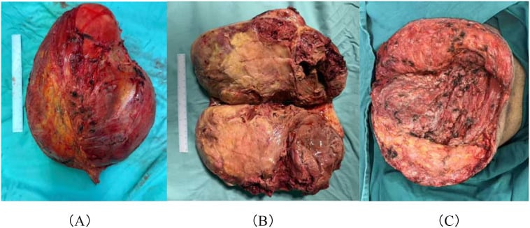
Amputation. It is indicated when the neurovascular bundle is completely encased with no possibility of limb salvage, when extensive soft-tissue involvement prevents a functional reconstruction, for recurrent MPNST after failed limb salvage, and by patient preference after informed discussion. The level is proximal to the tumour with an adequate margin, for example an above-knee amputation for a distal femoral MPNST, and the proximal margin is confirmed negative by frozen section of bone and soft tissue.
Why it is offered. Local control after amputation is superior to limb salvage (95% versus 80%), while overall survival is no different from limb salvage that achieves negative margins. Prosthetic rehabilitation is required and patient acceptance is often poor, so extensive counselling is needed: quality of life with a prosthesis may exceed that with a severely impaired limb, the decision is the patient's, and amputation is presented as an alternative option, not a failure.
Complications
Treatment-related. The neurological deficit is expected with nerve resection, which is why it sits in the consent discussion; the rest are managed as they arise.
- Incidence
- 20-30%
- Risk Factors
- Preoperative radiation (30-40%), large resection, poor nutrition
- Management
- VAC therapy, debridement, flap coverage
- Incidence
- 40-60%
- Risk Factors
- Expected with nerve resection (informed consent critical)
- Management
- Physiotherapy, tendon transfers, orthotics, occupational therapy
- Incidence
- 5-10%
- Risk Factors
- Vessel encasement, difficult dissection
- Management
- Vascular repair or reconstruction, anticoagulation
- Incidence
- Variable
- Risk Factors
- Postoperative radiation, high dose
- Management
- Physiotherapy, surgical release if severe
Local recurrence. It occurs in 30-50% at 5 years despite wide resection, the risk factors being positive margins, size over 10 cm, trunk location and high grade. The median time to recurrence is 12-24 months, with 80% within 2 years. Management is re-resection if feasible, radiotherapy if not previously given, amputation, or palliative care.
Distant metastasis. It occurs in 40-65% at 5 years, at a median of 18 months. The lung takes 80-90% of metastases; bone, liver and brain are rare sites. Management is palliative chemotherapy, metastasectomy in selected cases, or best supportive care.
Metastasectomy. In selected patients it improves survival, to 5-year survival of 30-40% against 10% with chemotherapy alone. The selection criteria:
- Limited lung metastases (1-3 nodules)
- Disease-free interval greater than 12 months
- Complete resection possible
- Good performance status
Postoperative Care and Surveillance
The first weeks. The inpatient phase is about the wound and the deficit; the outpatient phase is about the histology and what it changes.
Postoperative Management
Monitor the wound (flap viability, drain volume), give mechanical plus pharmacological DVT prophylaxis and multimodal analgesia that avoids opioid dependence, and examine the neurological deficit, documenting what was expected and anything that was not.
Remove drains once output is under 30 mL per 24 hours, watch the wound for infection, dehiscence and haematoma, and start early mobilisation with physiotherapy and occupational therapy for functional adaptation.
Wound check and suture removal, review of the final histology (margin status, grade), MDT discussion of adjuvant therapy and radiation oncology consultation if indicated.
If radiotherapy is indicated it starts 6-8 weeks after surgery once the wound has healed, delivered over 6-7 weeks, with physiotherapy continuing throughout. Completion of treatment marks the start of surveillance.
Surveillance. The schedule is front-loaded because most recurrence is early, and early detection may allow metastasectomy in selected cases.
- Clinical Exam
- Every 3 months
- Chest Imaging
- CT chest every 3 months
- MRI Primary Site
- Every 3-6 months
- Rationale
- 80% recurrence within 2 years
- Clinical Exam
- Every 4-6 months
- Chest Imaging
- CT chest every 4-6 months
- MRI Primary Site
- Every 6-12 months
- Rationale
- 90% recurrence within 5 years
- Clinical Exam
- Annually
- Chest Imaging
- CT chest annually
- MRI Primary Site
- As clinically indicated
- Rationale
- Late recurrence possible
Outcomes and Prognosis
Survival. MPNST has the worst prognosis among soft-tissue sarcomas. Five-year survival by aetiology is given in the table in the Overview: 21% NF1-associated, 42% sporadic and 20-30% radiation-induced, with the caveat there about how secure the NF1 comparison is.
Margin status. It is the single most important modifiable prognostic factor: 5-year survival is 65% after R0 (negative margins), 40% after R1 (microscopic positive) and 30% after R2 (gross residual).
Favourable factors. The features that predict a better outcome:
- Complete surgical resection (R0 margins)
- Small size (less than 5 cm)
- Superficial location (above the fascia)
- Low grade (rare, under 10% of MPNST)
- Distal extremity location
- Sporadic (non-NF1) origin, with the caveat in the Overview
Unfavourable tumour factors. Large size (over 10 cm), deep (subfascial) location, high grade (grade 3, 70% of MPNST), trunk or proximal extremity location, necrosis over 50% and a mitotic count over 20 per 10 HPF.
Unfavourable patient factors. NF1 association (taught as adverse, but not established, as set out in the Overview), radiation-induced disease, the malignant Triton variant and age over 50 years.
Unfavourable treatment factors. Inadequate initial surgery (marginal excision), delay in diagnosis and treatment, inability to deliver adjuvant radiotherapy and poor response to chemotherapy.
Guidelines, Registries & Global Practice
Global Epidemiology
MPNST is rare but disproportionately important because of its NF1 association and poor outcomes. The incidence in the general clinic population is approximately 0.001% versus 4.6% in neurofibromatosis (Ducatman et al., Cancer 1986, DOI), and the lifetime risk in NF1 is 8-13% (Evans et al., J Med Genet 2002, DOI). MPNST accounts for roughly 5-10% of all soft-tissue sarcomas. NF1-associated disease presents around two decades earlier (median 26 years) than sporadic disease (median 62 years), a pattern reproduced across European, North American and Asian series. Geographic incidence is broadly similar worldwide because NF1 prevalence (~1 in 3000 births) is consistent across populations; differences in reported outcomes largely reflect access to specialist sarcoma centres rather than true biological variation.
International Guideline Comparison
- Diagnostic Pathway
- MRI + core biopsy at sarcoma centre; consider FDG-PET in NF1
- Surgery
- Wide excision to negative margins; limb preservation when feasible
- Adjuvant Therapy
- Radiotherapy for high-grade / large / close margins; chemotherapy selective
- Evidence Level
- Category 2A (consensus)
- Diagnostic Pathway
- Refer to reference centre before biopsy; image-guided core biopsy
- Surgery
- Wide R0 excision as cornerstone; MDT planning mandatory
- Adjuvant Therapy
- Radiotherapy improves local control; chemotherapy not standard adjuvant
- Evidence Level
- Expert consensus, limited RCT data
- Diagnostic Pathway
- Mandatory sarcoma MDT; suspected sarcoma 2-week-wait referral
- Surgery
- Planned excision by sarcoma surgeon; avoid unplanned 'whoops' excision
- Adjuvant Therapy
- Adjuvant radiotherapy for high-grade STS; chemotherapy in trials
- Evidence Level
- Guideline (NICE sarcoma pathway)
- Diagnostic Pathway
- Standardised STS trial pathways
- Surgery
- R0 resection endpoint
- Adjuvant Therapy
- Doxorubicin-ifosfamide best first-line response in advanced disease
- Evidence Level
- Level 2 (pooled trial analysis)
There is broad international agreement on the core principles: referral to a specialist sarcoma centre before biopsy, image-guided core biopsy with the tract sited for en-bloc excision, wide R0 surgical resection as the only curative modality, and adjuvant radiotherapy to improve local (but not overall) control of high-grade tumours. The main area of practice variation is the role of chemotherapy, which is offered selectively (young patients, large high-grade tumours, or downstaging) without proven survival benefit; the EORTC pooled analysis shows advanced MPNST responds to chemotherapy comparably to other soft-tissue sarcomas (Kroep et al., Ann Oncol 2011, DOI).
Registry & Reference-Centre Evidence
- Outcomes are consistently better in high-volume sarcoma centres: contemporary multidisciplinary series report 5- and 10-year disease-specific survival of 60% and 45% (Stucky et al., Ann Surg Oncol 2012, DOI), an improvement over historical series.
- National soft-tissue sarcoma pathways (UK NICE/BSG, European EURACAN reference networks, and registry-linked centres such as the Australian sarcoma services) all mandate MDT review and discourage unplanned ("whoops") excisions, which worsen margins and survival.
- Molecular confirmation with H3K27me3 immunohistochemistry is now widely available and recommended in difficult cases (Prieto-Granada et al., Am J Surg Pathol 2016, DOI).
Specialist Referral Principle
Any suspected MPNST - particularly a rapidly enlarging or painful mass in an NF1 patient - should be referred to a specialist sarcoma multidisciplinary team before biopsy or excision. Unplanned excision of a presumed benign neurofibroma that proves to be MPNST compromises margins and is a leading cause of preventable poor outcomes worldwide. Examples of national sarcoma networks include the NHS specialist sarcoma centres (UK), EURACAN reference centres (Europe), NCCN-designated centres (USA) and the state-based Australian sarcoma services.
Medicolegal Considerations
- Pre-biopsy imaging and staging completed and reviewed
- Biopsy performed by or in consultation with sarcoma team (oncologically sound technique)
- MDT discussion documented before definitive surgery with treatment recommendation
- Informed consent including: poor prognosis (5-year survival 40-60%), nerve sacrifice requirement, neurological deficit expected, need for adjuvant therapy, recurrence and metastasis risk
- Excision of suspected neurofibroma without imaging/biopsy (missed MPNST, inadequate margins)
- Marginal or enucleation excision rather than wide 2 cm margins (high recurrence)
- Failure to refer to sarcoma MDT before treatment
- Inadequate surveillance leading to late detection of recurrence or metastasis
- Not discussing amputation as alternative when limb salvage results in positive margins
MCQ Practice Points
Q: What percentage of MPNST occur in NF1 patients, and what is the 5-year survival?
A: 50% of MPNST occur in NF1 patients, with 21% 5-year survival (versus 42% in sporadic MPNST). That comparison was not significant (p=0.09), and NF1 did not survive multivariate analysis in the Mayo series. The lifetime risk of MPNST in NF1 patients is 8-13% overall, but 25-30% in those with plexiform neurofibromas. Median age at diagnosis is 26 years in NF1 versus 40-50 years in sporadic.
Q: What SUVmax threshold on PET-CT suggests MPNST in NF1 patients? What is the sensitivity and specificity?
A: FDG-PET diagnosed NF1-associated MPNST with 89% sensitivity and 95% specificity (Ferner et al., Annals of Oncology 2008). A SUVmax less than 2.5 is reassuring while SUVmax greater than 3.5 raises high suspicion; the 2.5-3.5 range is intermediate and warrants close surveillance or biopsy. Note that SUVmax does not reliably predict tumour grade.
Q: What are the key molecular alterations in MPNST pathogenesis?
A: NF1 loss plus TP53 inactivation (75%) plus PRC2 complex mutations (SUZ12 or EED in 70-90%). The progression is: germline NF1 mutation, somatic second hit causes neurofibroma, additional TP53 loss and PRC2 loss drives malignant transformation to MPNST. H3K27me3 loss (due to PRC2 mutation) is a diagnostic immunohistochemistry marker supporting MPNST.
Q: What surgical margin is required for MPNST and what is the impact of margin status on survival?
A: Wide excision with 2 cm margins en bloc with involved nerve. Margin status is the most important prognostic factor: R0 resection (negative margins) achieves 65% 5-year survival, R1 (microscopic positive) 40% survival, R2 (gross residual) 30% survival. Re-excision is mandatory if positive margins on final pathology.
Q: Does adjuvant radiotherapy improve survival in MPNST?
A: Radiotherapy improves LOCAL CONTROL from 60-70% to 80-85% but does NOT improve overall survival. Dose is 60-66 Gy postoperatively or 50 Gy preoperatively. Indications: high-grade MPNST, positive/close margins, large size greater than 5 cm, deep location. Chemotherapy has modest benefit - response rate 20-30% (similar to other sarcomas: Kroep 21% vs 22%), no proven survival benefit for adjuvant chemotherapy.
Q: What percentage of MPNST are S100 positive and what is the staining pattern?
A: Only 50-70% of MPNST are S100 positive, and staining is FOCAL and PATCHY (not diffuse). This is unlike schwannoma which is diffusely and strongly S100 positive. S100 negativity does NOT exclude MPNST. Other markers: SOX10 50-60% positive, H3K27me3 loss in 50-70% (PRC2 mutation indicator), Ki67 high (greater than 10%, often 30-50%).
Exam Viva Scenarios
Practise clinical reasoning and management decisions out loud
“A 28-year-old woman with NF1 presents with a rapidly enlarging painful mass in the proximal thigh that has grown over 3 months. She has a known large plexiform neurofibroma in this location that was stable for years. MRI shows a 9 cm heterogeneous mass with necrosis arising from the sciatic nerve. PET-CT shows SUV of 5.2 and no distant metastases. Core needle biopsy confirms high-grade MPNST. How would you manage this patient?”
“What is the role of PET-CT in the diagnosis and management of MPNST, particularly in NF1 patients? Discuss the evidence and clinical utility including SUV thresholds.”
“You performed wide excision of a 12 cm proximal arm MPNST in a 45-year-old man. Final pathology returns as high-grade MPNST with a positive deep margin - tumor extends to within 1 mm of the radial nerve which you preserved. All other margins are negative with 2 cm clearance. The patient is 14 days post-operative with healing wound. How do you proceed?”
Definition and Epidemiology
- Aggressive sarcoma from peripheral nerve sheath, 5-10% of all soft tissue sarcomas
- 50% NF1-associated (plexiform transformation, lifetime risk 8-13% per Evans), 40% sporadic, 10% radiation-induced
- Median age: 26-30 years in NF1, 40-50 years in sporadic
- Location: 40-50% proximal extremity, 25-30% trunk, 15-20% head/neck
- WORST PROGNOSIS of all soft tissue sarcomas
Molecular Pathogenesis
- Malignant transformation: Plexiform neurofibroma to MPNST in NF1
- PRC2 mutations: SUZ12 or EED loss in 70-90%, H3K27me3 loss diagnostic
- NF1 association: 50% of MPNST; worse prognosis is taught but unproven (p=0.09)
- S100 focal positive: Only 50-70% and FOCAL not diffuse
- TP53 mutations: 75% have TP53 inactivation
High-Risk Features
- Heterogeneous MRI, infiltrative margins, rapid growth
- Hard consistency (firm vs soft neurofibroma)
- Radiotherapy history (10% radiation-induced), intense pain (60-70%)
- Size greater than 5 cm (mean 8-10 cm), Ki67 high (greater than 10%)
Investigations
- MRI local staging: Heterogeneous signal, necrosis, irregular margins, loss of target sign
- PET-CT: SUV less than 2.5 benign (NPV 100%), 2.5-3.5 intermediate, greater than 3.5 MPNST (89% sens, 95% spec)
- CT chest for lung metastases (most common site, 50% at presentation or follow-up)
- Core needle biopsy 14-16G: H&E, S100, SOX10, Ki67, H3K27me3 IHC
- AJCC staging: Most present Stage IIIB (large, deep, high-grade) or IV (metastatic)
Histopathology
- High-grade 90%: Spindle cells, high mitoses greater than 10/10 HPF, necrosis 50-70%
- S100 positive only 50-70%, FOCAL not diffuse (negative does NOT exclude)
- H3K27me3 loss in 50-70% (PRC2 mutation marker)
- FNCLCC grading: Grade 3 (high) in 70% at diagnosis
- Variants: Triton tumor (5-10%, rhabdomyoblastic, worse prognosis)
Surgical Management
- Wide excision 2 cm margins en bloc with involved nerve (nerve sacrifice mandatory)
- Biopsy tract excised en bloc
- R0 resection: 65% 5-year survival vs R1 40%, R2 30%
- Amputation if neurovascular bundle encased (95% local control vs 80% limb salvage, same survival)
- Limb salvage achievable in greater than 95% but significant neurological deficit
Adjuvant Therapy
- Radiotherapy 60-66 Gy postop or 50 Gy preop: Improves local control 60-70% to 80-85% but NOT survival
- Indications: High-grade, positive/close margins, greater than 5 cm, deep
- Chemotherapy: Modest benefit, response rate 20-30% (similar to other sarcomas - Kroep 21% vs 22%)
- No proven survival benefit for adjuvant chemotherapy
- The old 'chemo-resistant' label is refuted (Kroep EORTC pooled analysis)
Prognosis and Surveillance
- 5-year survival: NF1-MPNST 21%, sporadic 42%, overall 40-60%, metastatic 10-15%
- Local recurrence 30-50%, distant metastasis 40-65% (lung 80-90% of mets)
- 80% recurrence within 2 years, 90% within 5 years
- Surveillance: Q3mo exam/CT/MRI years 1-2, Q6mo years 3-5, annual after 5 years
- Margin status most important prognostic factor (R0 vs R1/R2)
Evidence Base and Key Studies
Malignant Transformation Risk in NF1 (Landmark Population Study)
- Population-based longitudinal study, NW England (4.1 million, 1984-1996): 21 NF1-associated and 37 sporadic MPNST
- Lifetime risk of MPNST in NF1 estimated at 8-13% (much higher than prior cross-sectional 1-2%)
- Median age at MPNST diagnosis: 26 years in NF1 versus 62 years in sporadic disease (p less than 0.001)
- Five-year survival from diagnosis: 21% NF1-associated versus 42% sporadic (p=0.09)
- First study to establish true lifetime risk and justify low threshold for investigation in NF1
FDG-PET for Detecting Malignant Transformation in NF1
- Long-term clinical study: 116 lesions in 105 NF1 patients with symptomatic plexiform neurofibromas (29 MPNST confirmed)
- FDG PET and PET-CT diagnosed NF1-associated tumours with sensitivity 0.89 (95% CI 0.76-0.96) and specificity 0.95 (95% CI 0.88-0.98)
- No MPNST was diagnosed on clinical follow-up of 23 lesions called benign on PET (high negative predictive value)
- SUVmax level did NOT reliably predict tumour grade - other tracers needed for grading
- Recommended as a sensitive and specific diagnostic tool for NF1-associated MPNST
Surgical Outcomes and Prognostic Factors (Mayo Clinic)
- Retrospective review of 175 MPNST patients (1985-2010); median age 44 years, median tumour size 6 cm, 61% high-grade
- Surgical resection in 95%; margins R0 69%, R1 2%, R2 9%, unknown 20%; radiation given in 42%
- Five- and ten-year disease-specific survival 60% and 45%; local recurrence rate 22%
- Independent poor prognostic factors on multivariate analysis: size 5 cm or greater (HR 6.1, 95% CI 1.5-25.0), local recurrence (HR 4.4, CI 1.7-11.4), high grade (HR 3.8, CI 1.1-13.2), truncal location (HR 3.7, CI 1.1-12.7)
- NF1 WAS associated with disease-specific survival on univariate analysis but did NOT survive multivariate adjustment - it is absent from the independent predictor list, which bears directly on how confidently NF1 can be called an adverse prognostic factor
References
-
Ducatman BS, Scheithauer BW, Piepgras DG, et al. Malignant peripheral nerve sheath tumors. A clinicopathologic study of 120 cases. Cancer. 1986;57(10):2006-2021. PMID:3082508. doi:10.1002/1097-0142(19860515)57:10%3C2006::aid-cncr2820571022%3E3.0.co;2-6
-
Evans DGR, Baser ME, McGaughran J, et al. Malignant peripheral nerve sheath tumours in neurofibromatosis 1. J Med Genet. 2002;39(5):311-314. doi:10.1136/jmg.39.5.311
-
Lee W, Teckie S, Wiesner T, et al. PRC2 is recurrently inactivated through EED or SUZ12 loss in malignant peripheral nerve sheath tumors. Nat Genet. 2014;46(11):1227-1232. doi:10.1038/ng.3095
-
Ferner RE, Golding JF, Smith M, et al. [18F]2-fluoro-2-deoxy-D-glucose positron emission tomography (FDG PET) as a diagnostic tool for neurofibromatosis 1 (NF1) associated malignant peripheral nerve sheath tumours (MPNSTs): a long-term clinical study. Ann Oncol. 2008;19(2):390-394. doi:10.1093/annonc/mdm450
-
Prieto-Granada CN, Wiesner T, Messina JL, et al. Loss of H3K27me3 expression is a highly sensitive marker for sporadic and radiation-induced MPNST. Am J Surg Pathol. 2016;40(4):479-489. doi:10.1097/PAS.0000000000000564
-
Stucky CC, Johnson KN, Gray RJ, et al. Malignant peripheral nerve sheath tumors (MPNST): the Mayo Clinic experience. Ann Surg Oncol. 2012;19(3):878-885. doi:10.1245/s10434-011-1978-7
-
Kroep JR, Ouali M, Gelderblom H, et al. First-line chemotherapy for malignant peripheral nerve sheath tumor (MPNST) versus other histological soft tissue sarcoma subtypes and as a prognostic factor for MPNST: an EORTC soft tissue and bone sarcoma group study. Ann Oncol. 2011;22(1):207-214. doi:10.1093/annonc/mdq338
-
Anghileri M, Miceli R, Fiore M, et al. Malignant peripheral nerve sheath tumors: prognostic factors and survival in a series of patients treated at a single institution. Cancer. 2006;107(5):1065-1074. doi:10.1002/cncr.22098
-
Wasa J, Nishida Y, Tsukushi S, et al. MRI features in the differentiation of malignant peripheral nerve sheath tumors and neurofibromas. AJR Am J Roentgenol. 2010;194(6):1568-1574.
-
National Comprehensive Cancer Network. NCCN Clinical Practice Guidelines in Oncology: Soft Tissue Sarcoma Version 2.2024.