Assess Biology vs Mechanics | Rule Out Infection | Diamond Concept | CORA for Deformity
- Every nonunion is infected until proven otherwise - check CRP/ESR, intraoperative cultures
- Hypertrophic nonunion = good biology, needs stability (exchange nail or compression plate)
- Atrophic nonunion = poor biology, needs bone graft AND stability
- Diamond Concept: Scaffold + Cells + Signals + Stability (+ Vascularity)
- Strain theory: less than 2% strain for primary healing, 2-10% for secondary healing
- “Tibia is the most common long bone nonunion (poor soft tissue coverage)
- “Smoking cessation is non-negotiable for elective nonunion surgery
- “Exchange nailing works by reaming autograft effect + larger stiffer nail
- “Masquelet technique induces biological membrane over 6-8 weeks
Overview and Epidemiology
Fracture healing complications are a significant burden in orthopaedic trauma, and the burden is economic as much as clinical: repeated surgery, prolonged rehabilitation and lost productivity. The contemporary overall nonunion rate is approximately 5% (4.9% across 309,330 fractures; Zura 2016, PMID 27603155), and the tibia plus fibula and the femur are among the highest-risk sites owing to precarious blood supply and limited soft-tissue coverage. Understanding the pathophysiology and a systematic approach to assessment is what makes management succeed.
Definitions. Three words describe three different failures, and the distinction is practical for treatment planning.
- Delayed union: the fracture has not healed within the expected timeframe (typically 3-6 months) but shows continued signs of healing potential
- Nonunion: cessation of healing, with no visible progressive signs of healing for 3 consecutive months and a minimum of 9 months since injury (the FDA definition); it will not heal without intervention
- Malunion: the fracture has healed in a position that is functionally or cosmetically unacceptable
Clinical nonunion (pain, motion at the fracture site) may occur before the radiographic criteria are met. Conversely, radiographic union may precede clinical union. Use both assessments together.
Risk factors. They sit in three columns: the patient, the fracture and the treatment.
- Fracture Factors
- Open fractures
- Treatment Factors
- Inadequate fixation
- Fracture Factors
- High-energy mechanism
- Treatment Factors
- Excessive soft tissue stripping
- Fracture Factors
- Bone loss
- Treatment Factors
- Infection
- Fracture Factors
- Segmental fractures
- Treatment Factors
- Poor reduction
- Fracture Factors
- Tibial shaft location
- Treatment Factors
- Delayed surgery
Anatomy and Biomechanics
The Diamond Concept (Giannoudis). Successful fracture healing requires four key elements, with vascularity as the fifth. Missing any one of them leads to nonunion.
- Osteogenic cells - mesenchymal stem cells from periosteum and bone marrow
- Osteoinductive mediators - BMPs, PDGF, TGF-beta, cytokines
- Osteoconductive scaffold - a structural matrix (collagen, hydroxyapatite) for bone growth
- Mechanical stability - adequate fixation creating the optimal strain environment
- Vascularity - adequate blood supply is paramount for healing
Strain theory (Perren). Strain is the change in length divided by the original length (dL/L), and the tissue that forms in a fracture gap is decided by it. Under 2% strain bone forms directly (primary, intramembranous healing); between 2 and 10% cartilage forms and then bone (secondary, endochondral healing with callus); over 10% only granulation or fibrous tissue forms, which is a nonunion. A small fracture gap implies high strain for even minimal movement, so, paradoxically, increasing the gap (L) can decrease strain and promote callus, assuming stable fixation.
Mechanics gone wrong. Excessive motion, over 10% strain, prevents calcification and leads to a hypertrophic nonunion. The opposite error is too rigid a fixation, locked plating with no gap, which prevents callus formation. A gap from soft-tissue interposition or bone loss prevents healing altogether, so too stiff and too loose both end in nonunion.
Biology gone wrong. Smoking does it two ways: carbon monoxide causes tissue hypoxia and nicotine inhibits neovascularisation. Diabetes brings microvascular disease and impaired cellular function. NSAIDs (COX-2 inhibitors) may delay healing in animal models, though the clinical data is mixed. The metabolic causes are vitamin D deficiency, hypothyroidism and hyperparathyroidism.
The tibia is particularly vulnerable due to its subcutaneous position and dependence on nutrient artery and periosteal blood supply. Open fractures, circumferential stripping, and high-energy injury all compromise vascularity.
The Diamond concept (scaffold, cells, signals) maps directly onto the four properties of bone graft, an examinable framework for choosing a graft for an atrophic nonunion.
- Osteogenic: contains living cells (osteoblasts, MSCs) that form bone. Only fresh autograft (iliac crest, RIA) and bone marrow aspirate.
- Osteoinductive: contains signals (BMP-2/-7, TGF-beta) that recruit and differentiate host cells. Autograft, demineralised bone matrix (DBM) and recombinant BMP.
- Osteoconductive: provides a passive scaffold for ingrowth. Autograft, allograft, DBM, ceramics (hydroxyapatite, tricalcium phosphate), bioglass, collagen.
- Osteopromotive/structural: augments the environment (e.g. PRP) or provides structural support (cortical allograft or strut).
Autograft from the iliac crest is the gold standard because it is the only graft with all three biological properties (osteogenic, osteoinductive and osteoconductive). RIA gives a large autograft volume with lower donor-site morbidity. Allograft is essentially osteoconductive (and structural) with minimal induction and no living cells; ceramics are purely osteoconductive; rhBMP is purely osteoinductive. Match the property to the deficiency: an atrophic nonunion lacking cells and signals needs an osteogenic plus osteoinductive autograft, not a ceramic scaffold alone.
Classification Systems
Weber-Cech. The classification is the foundation of treatment because it sorts nonunions by vascularity and callus, and callus is the visible evidence of biology. A vascular (hypertrophic) nonunion has made callus and failed only for want of stability, so it needs stability. An avascular (atrophic) nonunion has made none, so it needs biology, a graft, as well as stability.
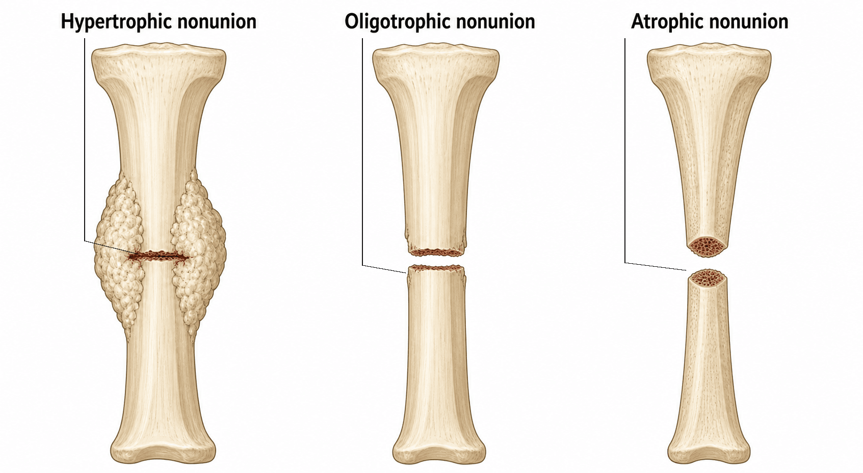
Oligotrophic is the trap. It shows no callus on X-ray and looks avascular, but the bone scan is positive: the biology is there, the fragments are simply too unstable or too poorly apposed to bridge, and it is treated as a hypertrophic nonunion.
Vascular (hypertrophic) nonunions
- Appearance
- Abundant hypertrophic callus, wide bone ends
- Cause
- Good blood supply but insufficient stability
- Treatment
- Stability (exchange nail, compression plate)
- Appearance
- Mildly hypertrophic, moderate callus
- Cause
- Slightly unstable
- Treatment
- Stability
- Appearance
- No callus on X-ray but vascular (bone scan positive)
- Cause
- Major instability or poor apposition
- Treatment
- Stability
Avascular (atrophic) nonunions
- Appearance
- One side healed, intermediate fragment avascular
- Cause
- Devascularised wedge fragment
- Treatment
- Biology plus stability
- Appearance
- Necrotic intermediate fragments
- Cause
- Multiple avascular fragments
- Treatment
- Biology plus stability
- Appearance
- Bone loss beyond critical size
- Cause
- Bone loss from injury or infection
- Treatment
- Masquelet or bone transport
- Appearance
- Rounded, sclerotic bone ends, sealed canal
- Cause
- Complete loss of biological activity
- Treatment
- Biology plus stability
Clinical Presentation and Assessment
History. The complaint is persistent pain at the fracture site, especially with weight-bearing, and start-up pain is characteristic. Ask about function: an inability to progress weight-bearing or activity level. Constitutional symptoms, fevers and sweats, are a screen for infection.
The background. Take a smoking history, a medication history (steroids, NSAIDs) and a dietary one. Review the operative notes of any previous surgery for complications, infection or a poor reduction.
Examination. Pain at the fracture site is the clinical hallmark. Motion there is gross instability, a false joint or pseudarthrosis, and a sinus or drainage means active infection. Look beyond the fracture too: angular deformity, muscle atrophy and joint stiffness each change the plan.
- Significance
- Clinical hallmark of nonunion
- Action
- Confirms diagnosis
- Significance
- Gross instability (false joint/pseudarthrosis)
- Action
- Surgical stabilisation required
- Significance
- Active infection
- Action
- Staged surgical management
- Significance
- Malunion component
- Action
- Deformity analysis required
- Significance
- Chronic disuse
- Action
- Rehabilitation planning
- Significance
- Secondary contracture
- Action
- May need arthrolysis
Every nonunion is infected until proven otherwise, and infection eradication is the priority before any reconstruction. Any history of wound drainage is highly predictive of infection. The screen is:
- Bloods: WCC, CRP (the most sensitive) and ESR (tracks the chronic course)
- Biopsy: the gold standard - obtain 5 or more samples from the nonunion site at surgery for culture
- Nuclear medicine: where the question is septic versus aseptic, the combined scan in the imaging table below
Investigations
Each modality answers a different question: the radiograph asks whether the fracture is bridging and the implant intact, the CT asks it precisely, and nuclear medicine and MRI ask whether the nonunion is infected.
- Role
- First line
- Key Findings
- Fracture line persistence, callus assessment (Weber-Cech), implant failure
- Role
- Detailed assessment
- Key Findings
- Fine detail of bridging bone, rotational profile, sequestrum
- Role
- Distinguish septic vs aseptic
- Key Findings
- Combined WBC/bone marrow scan is the gold standard nuclear test
- Role
- Infection/soft tissue
- Key Findings
- High sensitivity for osteomyelitis, oedema, abscess (requires MARS sequences for metal)
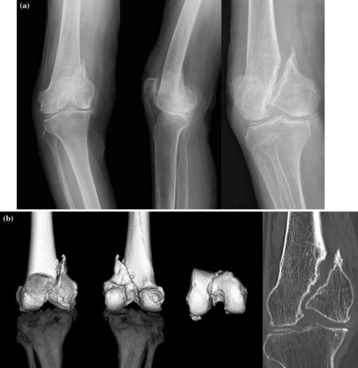
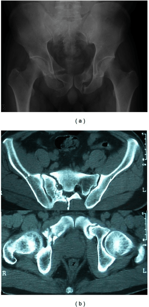
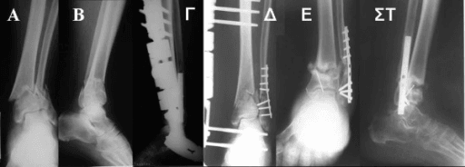
Radiographs. Four views: AP, lateral and obliques, plus the joints above and below. Look for persistence of the fracture line (a visible gap), the callus pattern (hypertrophic versus atrophic) and implant failure: broken screws, a bent plate, a fractured nail.
Bridging bone on at least 3 of the 4 cortices on orthogonal views is the usual radiographic definition of union. Note that it is three of four, not all four, and it is the criterion used by the nonunion series on this page. CT often over-calls nonunion because of metal and partial-volume artefact, so clinical correlation (pain, tenderness, motion) is vital. Always assess the rotational profile if malunion is suspected.
Management Algorithm
The personality of the nonunion. Treatment depends on answering five questions, in this order.
- Is it infected? If yes, staged management: debridement, antibiotics, then reconstruction
- Is it hypertrophic? It has biology but needs stability (fix it stiffer)
- Is it atrophic? It has no biology, so it needs stability and biology (graft plus fixation)
- Is there a deformity? It needs correction (osteotomy) plus fixation, planned around the CORA
- Is there a defect? Bone loss over 3-4cm needs the Masquelet technique or bone transport
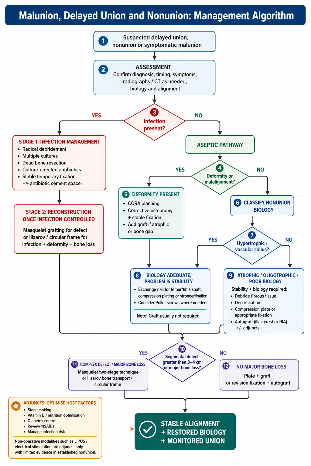
A limited role. Once a nonunion is established (over 9 months) non-operative treatment is primarily adjunctive, and the adjuncts are weak. LIPUS (low-intensity pulsed ultrasound) has conflicting evidence: the TRUST trial (BMJ 2016) showed no benefit in fresh tibial fractures, and the evidence for established nonunions is weak. Electrical stimulation, capacitive coupling or pulsed electromagnetic fields, has weak evidence too.
Metabolic optimisation is the other adjunct: vitamin D replacement, smoking cessation and protein supplementation.
Non-negotiable for elective reconstruction. Most surgeons will not operate electively on active smokers for nonunion surgery. Refer patients to structured cessation support (behavioural counselling plus nicotine-replacement therapy).
When bone transport or lengthening is used, examiners expect the biology and the practical numbers of distraction osteogenesis:
- The corticotomy is low-energy, preserving the periosteal and endosteal blood supply, and it is the controlled distraction of this cut that stimulates new bone (regenerate) in the gap.
- The three phases: (1) Latency, a wait of about 5-7 days after corticotomy before distraction, to let the early callus and haematoma organise; (2) Distraction, lengthening at a rate of about 1 mm/day, delivered as a rhythm of about 0.25 mm four times daily (more frequent smaller increments give better regenerate than one big daily turn); (3) Consolidation, in which the regenerate mineralises, classically taking roughly 1-2 months per cm gained (the high "healing index").
- The regenerate zones (histology): a central radiolucent fibrous interzone (longitudinally oriented collagen and proliferating cells) flanked by two mineralisation fronts maturing into new cortical bone. Distraction osteogenesis is intramembranous ossification, not endochondral.
- Getting rate or rhythm wrong: distracting too fast thins the regenerate (poor or failed bone, even a cyst); too slow causes premature consolidation of the corticotomy.
Surgical Technique
Setup. Supine on a traction table for the femur, or supine on a radiolucent table for the tibia, with the image intensifier positioned for AP and lateral views. Make sure the same brand of nailing system is available, to remove the old nail.
Procedure
- Incision: use the previous incision over the nail entry point
- Expose the nail: identify the end cap or locking screws
- Remove hardware: remove the end cap and locking screws
- Extract the nail: use the appropriate extraction device
- Assess the canal: pass a flexible reamer to assess the nonunion site
- Over-ream: ream to 1-2mm larger than the previous reamer size
- Collect the reaming debris: this is the autograft
- Insert a larger nail: 1-2mm greater diameter than the previous nail
- Lock the nail: static or dynamic depending on stability, static initially; it may be dynamised later
- Poller screws: consider them if the canal is wide or unstable
What matters. Do not strip the soft tissue at the nonunion site. The larger nail is critical, because the increase in stiffness is the mechanical half of the operation. Blocking screws improve alignment where the fracture extends into the metaphysis.
Complications
- Incidence
- 5-15%
- Prevention/Management
- Address both biology and stability; consider revision technique
- Incidence
- 3-5%
- Prevention/Management
- Staged approach; adequate debridement; culture-directed antibiotics
- Incidence
- Up to 30%
- Prevention/Management
- Consider RIA as alternative; limit harvest volume
- Incidence
- Rare
- Prevention/Management
- High index of suspicion with acute correction or lengthening
- Incidence
- 1-3%
- Prevention/Management
- Peroneal nerve at risk with valgus correction; radial nerve with humeral plating
- Incidence
- 5-10%
- Prevention/Management
- Accurate CORA planning; intraoperative assessment
- Incidence
- 3-5%
- Prevention/Management
- Adequate plate length; protected weight bearing
The iliac crest. Chronic donor-site pain occurs at the rate in the table. Haematoma occurs in 5-10%. The nerves at risk are the lateral femoral cutaneous nerve anteriorly and the cluneal nerves posteriorly. Fracture of the crest is rare but reported with a large harvest.
Reducing it. Consider RIA for large graft volumes. Limit an anterior crest harvest to the inner table, use the posterior approach for larger volumes with less pain, and close the periosteum to reconstruct the contour.
Postoperative Care and Rehabilitation
- Touch weight bearing typically
- Wound surveillance
- VTE prophylaxis
- Continue smoking cessation
- Optimise nutrition
- X-ray assessment for callus
- Progress weight bearing as callus forms
- Range of motion exercises for adjacent joints
- Consider dynamisation of nail if delayed healing
- Serial radiographic assessment
- Full weight bearing once bridging callus visible
- Progressive strengthening
- Return to light activities
- CT if union uncertain on X-ray
- Address any residual stiffness
- Hardware removal if symptomatic (after solid union)
- Return to sport or manual work
The principles behind the timeline. Continued abstinence from smoking is vital for healing, and nutrition means protein and calcium supplementation with vitamin D replacement if deficient. Weight bearing is tailored to the stability of the fixation and the healing response. For nails, dynamisation, removing the locking screws, is considered at 3-6 months if healing is slow.
Outcomes and Prognosis
Union rates by treatment. The union rate and the time to union differ by technique, and the frame is the slow one.
- Union Rate
- Over 90%
- Time to Union
- 4-6 months
- Union Rate
- 85-90%
- Time to Union
- 6-9 months
- Union Rate
- 85-95%
- Time to Union
- 4-6 months
- Union Rate
- 80-90%
- Time to Union
- 6-12 months
- Union Rate
- 80-90%
- Time to Union
- Prolonged (roughly 1-2 months per cm)
Prognosis. The favourable nonunion is hypertrophic, uninfected, in a non-smoker who is well nourished and has had a single previous operation. The unfavourable one is atrophic, with active or previous infection, continued smoking, multiple previous surgeries, a large bone defect, or an immunocompromised host.
Under 5% of nonunions ultimately require amputation. This is reserved for cases with uncontrollable infection, severe limb shortening, or patient preference after multiple failed procedures.
Guidelines, Registries & Global Practice
Global Epidemiology (evidence-based):
The most robust contemporary data come from the Zura et al. inception cohort of 309,330 fractures across 18 bones (JAMA Surgery 2016, PMID 27603155), which reported an overall nonunion rate of 4.9%, consistent with the widely quoted 5-10% range for diaphyseal long-bone fractures. The highest-risk anatomical sites were the scaphoid, the tibia plus fibula, and the femur. The tibial shaft remains the prototypical problem fracture worldwide owing to its subcutaneous position, watershed blood supply and frequent open injury.
- Figure
- 4.9%
- Source
- Zura 2016 (PMID 27603155)
- Figure
- 5-10%
- Source
- Zura 2016; consistent across series
- Figure
- 2.32 (95% CI 1.76-3.06)
- Source
- Scolaro 2014 (PMID 24740664)
- Figure
- 1.66
- Source
- Zura 2016 (PMID 27603155)
- Figure
- 1.40
- Source
- Zura 2016 (PMID 27603155)
Smoking is the single most important modifiable risk factor globally (adjusted OR 2.32; Scolaro 2014). Cessation support should be offered to every patient with delayed or nonunion before elective reconstruction.
Guideline & Society Guidance (side-by-side):
- Position on nonunion / fracture healing
- Systematic biology-versus-mechanics assessment; autograft remains gold standard; rhBMP-2 reserved for selected open tibial fractures
- Evidence level
- Based on Level I RCTs (BESTT, SPRINT)
- Position on nonunion / fracture healing
- Senior decision-making, early specialist referral for nonunion, structured MDT for limb reconstruction; does not endorse LIPUS for routine healing
- Evidence level
- Guideline (GRADE-based)
- Position on nonunion / fracture healing
- Open-fracture and limb-reconstruction standards: combined orthoplastic care, debridement and early soft-tissue cover to prevent nonunion
- Evidence level
- Consensus standard
- Position on nonunion / fracture healing
- Diamond Concept framework (scaffold, cells, signals, stability, vascularity); strain theory guides fixation
- Evidence level
- Expert/educational consensus
- Position on nonunion / fracture healing
- Endorses induced-membrane (Masquelet) and distraction osteogenesis for segmental defects; emphasises infection exclusion
- Evidence level
- Narrative/consensus
There is no dedicated international nonunion implant registry; high-quality evidence is trial-driven. Practice is anchored by the SPRINT RCT (reamed nailing reduces reoperation in closed tibial fractures; PMID 19047701), the BESTT RCT (rhBMP-2 in open tibial fractures; PMID 12473698) and the TRUST RCT (LIPUS ineffective; PMID 27797787). National joint replacement registries (AOANJRR, NJR, AJRR) capture arthroplasty but not diaphyseal nonunion outcomes.
Exchange nailing predominates for hypertrophic shaft nonunions across high-income systems. The Masquelet/induced-membrane technique and Ilizarov bone transport are both used for segmental defects, with choice driven by surgeon expertise, defect size and infection status; distraction osteogenesis is relatively more common in centres with established limb-reconstruction units. rhBMP-2 use varies widely with cost and regulatory access.
- Adult daily-smoking prevalence in high-income populations is around 10-11%, and is higher in regional and remote communities where trauma rates are also elevated, reinforcing the need for routine cessation counselling before elective reconstruction.
- The Reamer-Irrigator-Aspirator (RIA) is used at major trauma centres internationally for large-volume autograft harvest with lower donor-site morbidity than iliac crest.
Be prepared to discuss the systematic approach to nonunion: infection screen, classification (Weber-Cech), treatment selection (biology versus mechanics), and patient optimisation (especially smoking cessation). Know exchange nailing technique and the Masquelet technique for defects, and be able to quote the landmark trials (SPRINT, BESTT, TRUST) and the contemporary nonunion rate of approximately 5% (Zura 2016).
MCQ Practice Points
Q: Smoking increases nonunion risk by approximately how much? A: About 2.3-fold - Scolaro's adjusted odds ratio was 2.32 (95% CI 1.76-3.06) across 19 cohort studies. Carbon monoxide causes tissue hypoxia and nicotine inhibits neovascularisation, and this is the single most important modifiable risk factor for failure to unite. The discriminating detail: in that same review, healing time in smokers (30.2 vs 24.1 weeks) did not differ significantly (p = 0.18), so a stem offering "smokers take significantly longer to heal" is testing whether you know the union finding from the non-significant one.
Q: An elephant foot appearance on X-ray represents which type of nonunion? A: Hypertrophic (vascular) nonunion. This has good blood supply and abundant callus but lacks stability. Treatment is mechanical - exchange nail or compression plate. No graft needed.
Q: What does an atrophic nonunion require that hypertrophic does not? A: Biological augmentation (bone graft). Atrophic nonunion has no biological activity (sealed canal, no callus). It needs graft PLUS stability. Hypertrophic needs stability only.
Q: What is the key technical principle of exchange nailing? A: Over-ream by 1-2mm and insert a nail 1-2mm larger diameter. This provides biological effect (reaming autograft) and mechanical effect (larger stiffer nail).
Q: According to Perren strain theory, what strain is required for primary bone healing? A: Under 2% strain. 2-10% strain produces secondary healing with callus. Over 10% strain produces only fibrous tissue.
Clinical Decision Scenarios
Practise clinical reasoning and management decisions out loud
“A 35-year-old male presents with persistent pain 9 months after intramedullary nailing for a closed tibial shaft fracture. He is a smoker. X-rays show a visible fracture line with minimal callus. The nail appears intact.”
“A 45-year-old woman presents with a tibial nonunion 12 months after a Grade IIIB open fracture treated with external fixation and flap coverage. X-rays show atrophic bone ends with a 4cm segmental defect. CRP is normal.”
“A 28-year-old woman presents 18 months after a distal femoral fracture with a united but malunited fracture. She has 15 degrees of varus deformity and 2cm of shortening. She complains of medial knee pain and difficulty with stairs.”
DEFINITIONS
- Delayed union: Not healed by 3-6 months but progressing
- Nonunion: Over 9 months with no progress for 3 months
- Malunion: Healed in unacceptable position (ARTS analysis)
INFECTION SCREEN
- Every nonunion is infected until proven otherwise
- Bloods: CRP (most sensitive), ESR, WCC
- Biopsy: 5+ samples at surgery for culture
- Nuclear medicine: Combined WBC/marrow scan
WEBER-CECH CLASSIFICATION
- Hypertrophic (Elephant foot): Good biology, needs STABILITY
- Oligotrophic: Appears avascular but bone scan positive - treat as hypertrophic
- Atrophic: No biology, sealed canal, needs GRAFT + stability
TREATMENT ALGORITHM
- Infected: Staged debridement then reconstruction
- Hypertrophic: Exchange nail or compression plate (stability only)
- Atrophic: Plate + graft (biology + stability)
- Defect over 4cm: Masquelet or bone transport
EXCHANGE NAILING
- Indication: Aseptic hypertrophic femur/tibia shaft nonunion
- Over-ream 1-2mm larger than previous
- Insert nail 1-2mm larger diameter
- Success rate over 90%
KEY NUMBERS
- Smoking risk: 2.3x increased nonunion
- Primary healing strain: Under 2%
- Secondary healing strain: 2-10%
- ICBG chronic pain: Up to 30%
Evidence Base
Zura et al. - Epidemiology of Fracture Nonunion in 18 Human Bones
- Inception cohort of 309,330 fractures across 18 bones in a US payer database
- Overall nonunion rate 4.9%
- Highest-risk sites: scaphoid, tibia plus fibula, and femur
- Independent risk factors included open fracture (OR 1.66), diabetes (OR 1.40), smoking (OR 1.20) and NSAID plus opioid use (OR 1.84)
TRUST Investigators (Busse et al.) - LIPUS for Operatively Managed Tibial Fractures
- Blinded, sham-controlled RCT of 501 operatively managed tibial fractures across 43 centres
- No difference in time to radiographic healing (hazard ratio 1.07, 95% CI 0.86 to 1.34)
- No improvement in SF-36 physical component or other functional measures at 1 year
SPRINT Investigators (Bhandari et al.) - Reamed vs Unreamed Tibial Nailing
- Multicentre blinded RCT of 1319 tibial shaft fractures, 1226 (93%) followed to one year
- NO overall difference in the composite reoperation outcome (relative risk 0.90, 95% CI 0.71 to 1.15)
- In closed fractures reamed nailing reduced the composite outcome from 17% to 11% (relative risk 0.67, 95% CI 0.47 to 0.96, p = 0.03) - but the authors state this was largely due to differences in DYNAMISATION, not union
- In open fractures the point estimate favoured UNREAMED nailing (relative risk 1.27, 95% CI 0.91 to 1.78)
- Only 57 of 1226 patients (4.6%) needed implant exchange or grafting for nonunion
BESTT Study (Govender et al.) - rhBMP-2 for Open Tibial Fractures
- RCT of 450 open tibial fractures, all nailed, comparing standard care against rhBMP-2 on an absorbable collagen sponge laid over the fracture AT DEFINITIVE WOUND CLOSURE
- Only the HIGHER 1.50 mg/mL dose (12 mg total) worked: 44% reduction in secondary intervention, relative risk 0.56 (95% CI 0.40 to 0.78), p = 0.0005 - the 0.75 mg/mL arm did not separate from control
- Faster fracture healing (p = 0.0022) and wound healing (83% vs 65% healed at six weeks)
- Fewer infections specifically in Gustilo-Anderson type-III injuries (p = 0.0219)
Scolaro et al. - Cigarette Smoking Increases Complications Following Fracture
- Systematic review of 19 cohort studies (7 prospective, 12 retrospective) drawn from 7,110 records
- Adjusted odds ratio of nonunion in smokers 2.32 (95% CI 1.76 to 3.06), p less than 0.001 - this is the figure this page quotes as 2.3x
- Higher nonunion in smokers with tibial fractures (OR 2.16, 95% CI 1.55 to 3.01) and open fractures (OR 1.95, 95% CI 1.3 to 2.9)
- Mean healing time 30.2 vs 24.1 weeks - but this did NOT reach significance (p = 0.18), and neither did superficial (p = 0.13) or deep (p = 0.33) infection
Swanson, Brinker et al. - Systematic Exchange Nailing for Aseptic Femoral Nonunion
- Retrospective cohort of 50 aseptic femoral nonunions in 49 patients, presenting with a nail in situ an average of 25 months after the original fixation
- Systematic protocol: exchange nail at least 2 mm larger, DIFFERENT MANUFACTURER, static interlocking, correction of any metabolic or endocrine abnormality, and secondary dynamisation if progress was slow
- All 50 (100%) united; average time to union 7 months, range 3 to 26 months
Calori et al. - Validation of the Non-Union Scoring System in 300 Long Bone Non-Unions
- 300 long bone nonunions scored with NUSS and treated by the score-matched 'Ladder Strategy'
- Union was essentially FLAT across severity classes: 60/69 (86.9%) in class I, 102/117 (87.1%) in class II and 69/84 (82.1%) in class III
- Mean radiographic healing 8.78, 9.02 and 9.53 months across the three classes - a difference of under a month between the mildest and the worst
- Complication rate 5.5% (15 of 270)