Metacarpophalangeal Joint Degenerative and Inflammatory Arthritis
Classifications
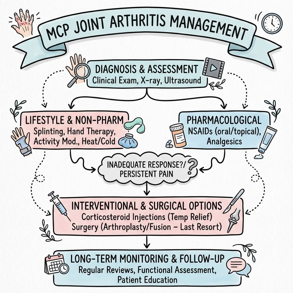
Critical Must-Knows
- Mechanism: Rheumatoid chronic synovitis stretches the volar plate and radial collateral ligament, causing volar subluxation and ulnar drift with ulnar extensor-tendon displacement
- Management: Optimise DMARDs (methotrexate first-line) and biologics first; operate only for established deformity with functional loss after 3-6 months of failed medical therapy
- Digit-specific surgery: silicone arthroplasty for fingers (digits 2-5), arthrodesis for the thumb MCP (pinch stability); soft-tissue rebalancing is integral to arthroplasty
Clinical Pearls
- "Silicone is a flexible spacer, not a load-bearing joint replacement; fibrous encapsulation provides stability
- "Arthroplasty without extensor centralisation and radial collateral reconstruction will recur in ulnar drift
- "Thumb MCP is fused at 10-15 degrees flexion in neutral rotation; test pinch intra-operatively before fixation
Clinical Imaging
Imaging Gallery


Critical MCP Arthritis Exam Points
Rheumatoid MCP Pathomechanics
Synovitis stretches volar plate and collateral ligaments. MCP subluxes volarly and ulnarly. Extensor tendons displace ulnarly into valleys between MCPs. Results in ulnar drift deformity. Know mechanism: VUSEX (Volar, Ulnar, Synovitis, Extensor, X-ray).
Silicone Arthroplasty Principles
Silicone spacer arthroplasty is standard for MCP arthritis (digits 2-5). NOT a load-bearing hinge - acts as flexible spacer. Fibrous encapsulation provides stability. Restores alignment, preserves 30-40 degrees motion. 10-15 year survivorship. Adjunct soft tissue balancing essential.
Thumb MCP Management
Arthrodesis preferred for thumb MCP arthritis. Provides stable lateral key pinch and pulp pinch. Fusion position: 10-15 degrees flexion, neutral rotation. Arthroplasty risks instability with high pinch loads. Plate fixation preferred over K-wires. 95% fusion rate.
Soft Tissue Balancing
Correct ALL pathology, not just bone. Synovectomy for active rheumatoid disease, extensor tendon centralization (reposition over MCP center), radial collateral ligament reconstruction (correct ulnar drift), intrinsic release if tight. Arthroplasty alone WILL fail without soft tissue balancing.
At a Glance
MCP joint arthritis is predominantly rheumatoid (90% of RA patients affected), presenting with the classic volar subluxation and ulnar drift deformity caused by chronic synovitis stretching the volar plate and collateral ligaments. Surgical management differs by digit: silicone arthroplasty for digits 2-5 (acts as flexible spacer, not load-bearing, provides 30-40° motion arc with 10-15 year survivorship), while arthrodesis is preferred for the thumb MCP (10-15° fusion position for stable pinch). Critical principle: soft tissue balancing is essential - synovectomy, extensor centralisation, radial collateral ligament reconstruction, and intrinsic release must accompany arthroplasty or it will fail.
VUSEXRheumatoid MCP Deformity Mechanism
| V | V - Volar subluxation (proximal phalanx subluxes volarly) |
| U | U - Ulnar drift (fingers deviate ulnarly from ligament laxity) |
| S | S - Synovitis (primary pathology - chronic inflammation) |
| E | E - Extensor displacement (tendons displace ulnarly into valleys) |
| X | X - X-ray shows subluxation (diagnostic confirmation) |
| V | V - Volar subluxation (proximal phalanx subluxes volarly) | E | E - Extensor displacement (tendons displace ulnarly into valleys) |
| U | U - Ulnar drift (fingers deviate ulnarly from ligament laxity) | X | X - X-ray shows subluxation (diagnostic confirmation) |
| S | S - Synovitis (primary pathology - chronic inflammation) |
Hook:VUSEX captures the rheumatoid MCP mechanism - examiners expect you to describe this sequence!
SPACERSilicone Arthroplasty Principles
| S | S - Silicone elastomer (flexible, not load-bearing) | C | C - Centralize extensors (rebalance over MCP center) |
| P | P - Preserves motion (30-40 degrees flexion arc) | E | E - Encapsulation (fibrous capsule forms around implant) |
| A | A - Alignment restored (corrects ulnar drift and volar subluxation) | R | R - Release intrinsics (tight ulnar intrinsics must be released) |
| S | S - Silicone elastomer (flexible, not load-bearing) | A | A - Alignment restored (corrects ulnar drift and volar subluxation) | E | E - Encapsulation (fibrous capsule forms around implant) |
| P | P - Preserves motion (30-40 degrees flexion arc) | C | C - Centralize extensors (rebalance over MCP center) | R | R - Release intrinsics (tight ulnar intrinsics must be released) |
Hook:SPACER - silicone acts as a spacer, not a true joint replacement!
CASCADEMCP Arthrodesis Fusion Positions
| C | C - Cascading flexion (ulnar digits progressively more flexed) | A | A - Avoid excessive flexion or hyperextension |
| A | A - Angle for thumb: 10-15 degrees flexion | D | D - Digits 2-5: Index 25-30, Middle 35-40, Ring 40-45, Small 45-50 |
| S | S - Small finger most flexed: 45-50 degrees | E | E - Essential for grip function (mimics normal hand cascade) |
| C | C - Check pinch position intraoperatively |
| C | C - Cascading flexion (ulnar digits progressively more flexed) | C | C - Check pinch position intraoperatively | E | E - Essential for grip function (mimics normal hand cascade) |
| A | A - Angle for thumb: 10-15 degrees flexion | A | A - Avoid excessive flexion or hyperextension | ||
| S | S - Small finger most flexed: 45-50 degrees | D | D - Digits 2-5: Index 25-30, Middle 35-40, Ring 40-45, Small 45-50 |
Hook:CASCADE reminds you of the cascading flexion pattern from radial to ulnar!
Overview and Epidemiology
Why MCP Arthritis Matters in Exams
MCP joint arthritis is predominantly rheumatoid. Examiners expect detailed knowledge of rheumatoid pathomechanics (synovitis leading to volar subluxation and ulnar drift), surgical decision-making (silicone arthroplasty for digits 2-5, arthrodesis for thumb), and adjunct soft tissue procedures (extensor centralization, ligament reconstruction). This is a pattern recognition and surgical planning topic.
MCP Joint Arthritis is inflammation and degeneration of the metacarpophalangeal joints, presenting as pain, stiffness, deformity, and functional impairment.
Epidemiology
Rheumatoid Arthritis (Most Common)
Prevalence:
- 90% of RA patients develop MCP involvement
- Bilateral and symmetric distribution
- Female greater than male (3:1 ratio)
- Peak onset 40-60 years of age
- Progression over years to decades
Natural History:
- Early: synovitis, pain, morning stiffness
- Moderate: ulnar drift, volar subluxation
- Late: severe deformity, extensor tendon displacement, functional disability
Post-Traumatic and Other Causes
Post-Traumatic Arthritis:
- Following MCP fracture with articular involvement
- Chronic MCP instability from collateral ligament injury
- Prior MCP dislocation
- Usually unilateral, single digit
Primary Osteoarthritis:
- Rare at MCP (unlike CMC-1, DIP, PIP joints)
- More common in manual laborers
- Typically less severe than rheumatoid
Other Inflammatory:
- Psoriatic arthritis (seronegative, DIP and MCP)
- Crystalline arthropathy (gout, pseudogout)
Risk Factors
Rheumatoid Arthritis:
- Autoimmune predisposition (RF positive, anti-CCP antibodies)
- Genetic factors (HLA-DR4, family history)
- Female sex, smoking history
- Environmental triggers (infections, hormonal changes)
Post-Traumatic:
- Intra-articular MCP fracture (especially volar plate avulsion)
- Chronic MCP instability (collateral ligament injury)
- Dorsal MCP dislocation with articular damage
- Inadequate initial treatment of MCP injuries
Occupational:
- Repetitive gripping and pinching (manual laborers)
- Vibratory tool use
- Heavy manual work
Anatomy and Biomechanics
MCP Joint Architecture
Cam and Post Configuration:
- Metacarpal head: Cam-shaped (eccentric condyle)
- Wider volarly than dorsally (approximately 20-30% larger volar diameter)
- Proximal phalanx base: Shallow concave (post)
- Articular mismatch: Allows increased ROM but inherent instability
Collateral Ligaments:
- Proper collateral ligament: Origin at metacarpal head dorsal to axis of rotation, inserts on proximal phalanx base
- Accessory collateral ligament: Origin at metacarpal, inserts on volar plate
- Function: Lax in extension (allows lateral deviation), tight in flexion (stabilizes joint)
- In rheumatoid: Stretched by synovitis, leading to instability
Volar Plate:
- Thick fibrocartilaginous structure on palmar aspect
- Prevents MCP hyperextension
- Attachment: Weak proximally (allows volar subluxation in RA), strong distally to proximal phalanx
- In rheumatoid: Stretched, allowing volar subluxation of proximal phalanx
Extensor Mechanism:
- Extensor digitorum communis: Inserts on proximal phalanx base via extensor hood
- Sagittal bands: Stabilize extensor tendon over MCP center (radial and ulnar bands)
- In rheumatoid: Sagittal bands attenuate, extensor displaces ulnarly into valley between MCP heads
Biomechanics of Normal MCP Function
Range of Motion:
- Flexion: 80-90 degrees (digits 2-5), 50-60 degrees (thumb)
- Extension: 0-20 degrees hyperextension (normal variation)
- Radial-ulnar deviation: 10-20 degrees in extension (lax collaterals), minimal in flexion
Stability:
- Bony congruity: Minimal (cam and post mismatch)
- Static stabilizers: Collateral ligaments, volar plate, joint capsule
- Dynamic stabilizers: Intrinsic muscles (lumbricals, interossei), extensor tendons
Load Transmission:
- Power grip: High compressive loads across MCPs (up to 5-10 times grip force)
- Precision pinch: Index and thumb MCPs experience high loads
- Implication: Thumb MCP requires arthrodesis for pinch stability (arthroplasty fails under load)
Pathophysiology and Deformity Mechanisms
Rheumatoid MCP Deformity Mechanism (VUSEX)
Volar subluxation and Ulnar drift result from Synovitis causing capsular stretch, with Extensor tendon displacement ulnarly, confirmed on X-ray. This is the pathomechanical sequence examiners expect you to recite.
Rheumatoid Arthritis MCP Deformity
Sequential Pathomechanics:
-
Synovitis (Primary Event):
- Chronic inflammation of MCP synovium
- Pannus formation (invasive synovial tissue)
- Release of inflammatory cytokines (TNF-alpha, IL-1, IL-6)
- Enzymatic degradation of cartilage (matrix metalloproteinases)
-
Capsular and Ligament Stretch:
- Synovial hypertrophy distends joint capsule
- Radial collateral ligament stretches (allows ulnar drift)
- Volar plate stretches proximally (allows volar subluxation)
- Sagittal bands attenuate (allows extensor tendon displacement)
-
Volar Subluxation:
- Proximal phalanx subluxes volarly on metacarpal head
- Visible step-off at MCP dorsally
- Loss of normal MCP contour
- Worsens with grip activities (force vector pulls phalanx volarly)
-
Ulnar Drift:
- Radial collateral ligament laxity allows ulnar deviation
- Ulnar intrinsic muscles (ulnar lumbricals, interossei) pull digits ulnarly
- Wrist radial deviation compounds MCP ulnar drift (Z-collapse deformity)
- Gravity and grip forces perpetuate ulnar deviation
-
Extensor Tendon Displacement:
- Sagittal band attenuation allows extensor to displace ulnarly
- Extensor falls into valley between MCP heads
- Acts as ulnar deviator instead of pure extensor
- Creates extensor lag and perpetuates ulnar drift
-
Progressive Deformity:
- Biomechanical imbalance worsens with hand use
- Deformity becomes fixed (contracture)
- Articular cartilage erosion from abnormal loading
- End-stage: mutilating arthropathy
Nalebuff Rheumatoid Hand Classification:
| Type | Deformity Pattern | Mechanism | Treatment Consideration |
|---|---|---|---|
| Type I | Swan-neck (MCP flex, PIP hyperextension, DIP flexion) | Intrinsic tightness, PIP volar plate laxity, FDS weakness | Address MCP and PIP (may need PIP fusion or reconstruction) |
| Type II | Boutonniere (MCP hyperextension, PIP flexion, DIP hyperextension) | Central slip rupture at PIP, lateral band volar displacement | PIP central slip reconstruction or fusion |
| Type III | MCP ulnar drift with swan-neck | Combined Type I and MCP pathology | MCP arthroplasty with PIP management |
| Type IV | Severe MCP volar subluxation | End-stage rheumatoid destruction | MCP arthroplasty or arthrodesis (if bone stock poor) |
Type I (swan-neck with MCP flexion deformity) is the most common pattern requiring MCP arthroplasty.
Post-Traumatic Arthritis Mechanism
Direct Cartilage Injury:
- Intra-articular fracture of metacarpal head or proximal phalanx base
- Articular step-off greater than 2mm leads to abnormal load distribution
- Focal cartilage loss at impact site
- Secondary degenerative changes over months to years
Chronic Instability:
- Collateral ligament injury (acute or chronic)
- Recurrent MCP subluxation/dislocation
- Abnormal joint kinematics cause cartilage wear
- Progressive arthrosis
Stiffness-Related:
- Prolonged immobilization after MCP injury
- Adhesions and capsular contracture
- Decreased joint motion leads to cartilage nutrition impairment
- Degenerative changes from stiffness
Primary Osteoarthritis (Rare)
Mechanism:
- Idiopathic cartilage degeneration
- Repetitive microtrauma in manual laborers
- Genetic predisposition (rare in MCPs compared to DIP/PIP/CMC-1)
- Progressive: cartilage loss, subchondral sclerosis, osteophyte formation
Why rare at MCP?
- MCP joint is congruous and mobile (less focal stress compared to DIP/PIP)
- CMC-1 (thumb base) is much more common site for primary OA
Clinical Presentation and Assessment
History
Rheumatoid Arthritis:
- Bilateral symmetric hand pain and stiffness
- Morning stiffness greater than 1 hour (classic, improves with activity)
- Progressive ulnar drift and visible deformity
- Difficulty with power grip (holding objects) and precision pinch
- Known RA diagnosis with systemic involvement (other joints, lungs, heart)
- DMARD treatment history (methotrexate, biologics)
- Duration of hand symptoms (months to years)
Post-Traumatic:
- History of MCP trauma (fracture, dislocation, ligament injury)
- Unilateral, single digit pain and stiffness
- Reduced ROM compared to contralateral side
- Pain with gripping activities
- Delayed onset (months to years after injury)
Primary OA:
- Insidious onset, gradual progression
- Usually older age (greater than 60 years)
- Occupational history (manual labor, repetitive gripping)
- Less systemic symptoms than RA
Examination
Inspection:
- Ulnar drift: Fingers deviate ulnarly at MCPs (pathognomonic for rheumatoid)
- Volar subluxation: Proximal phalanx displaced volarly, dorsal step-off at MCP
- Swelling: Boggy synovitis at MCP joints (active RA), fusiform swelling
- Deformity: Swan-neck (MCP flexion, PIP hyperextension, DIP flexion) or boutonniere at IP joints
- Extensor tendon position: Displaced ulnarly into valleys between MCP heads
- Skin: Rheumatoid nodules (extensor surface, olecranon, MCP), thinning, fragility
- Z-collapse: Wrist radial deviation with MCP ulnar drift
Palpation:
- MCP joint line tenderness (dorsal palpation)
- Synovial thickening (boggy, compressible, warm in active inflammation)
- Collateral ligament stability (radial and ulnar stress testing at MCP)
- Volar plate (test for hyperextension laxity)
Range of Motion:
- Active MCP flexion-extension: Normal 0-90 degrees, reduced in arthritis
- Passive ROM: Compare to active (capsular tightness vs extensor lag)
- Extensor lag: Inability to fully extend MCP actively (extensor displacement/weakness)
- Compare to contralateral hand
Special Tests:
- Intrinsic tightness test: With MCP extended, attempt PIP flexion. If intrinsics tight, PIP flexion is limited. With MCP flexed (relaxes intrinsics), PIP flexion should improve.
- Extensor lag: Active extension deficit compared to passive extension (indicates extensor tendon pathology)
- Collateral ligament stability: Radial and ulnar stress at MCP in flexion (normally tight) and extension (normally lax). Excessive laxity suggests ligament attenuation.
- Grip strength: Dynamometer testing (compare to contralateral, age-matched norms)
- Pinch strength: Key pinch and pulp pinch (assess thumb MCP stability)
Functional Assessment:
- Power grip: Holding objects, jar opening
- Precision pinch: Writing, buttoning
- ADLs: Dressing, eating, hygiene
- Work demands: Manual labor vs sedentary
Examination findings guide surgical planning and inform patient expectations.
Differential Diagnosis of MCP Joint Arthritis
Distinguishing Causes of MCP Joint Arthritis
| Condition | Distribution | Serology / Labs | Radiographic Hallmark | Discriminating Clue |
|---|---|---|---|---|
| Rheumatoid arthritis | Bilateral, symmetric, MCP and wrist; spares DIP | RF positive 70-80%, anti-CCP positive 70% (specificity over 95%) | Periarticular osteopenia, marginal erosions, ulnar drift, volar subluxation | Symmetric MCP synovitis with ulnar drift and morning stiffness over 1 hour |
| Psoriatic arthritis | Asymmetric, ray pattern, DIP and MCP | RF negative, anti-CCP negative (seronegative) | Pencil-in-cup deformity, periostitis, osteolysis, ankylosis | Skin/nail psoriasis, dactylitis (sausage digit), DIP involvement |
| Primary osteoarthritis | Index and middle MCP, often in manual workers | Inflammatory markers normal, seronegative | Osteophytes, subchondral sclerosis, preserved bone density | Rare at MCP; suspect haemochromatosis if index/middle MCP OA in a younger man |
| Post-traumatic arthritis | Unilateral, single digit | Seronegative, normal markers | Focal joint-space loss, sclerosis, malunion/articular step-off | Antecedent fracture, dislocation or collateral ligament injury |
| Crystalline arthropathy (gout/CPPD) | Mono- or oligoarticular, episodic | Urate may be raised; aspirate shows crystals | Tophi/erosions with overhanging edges (gout); chondrocalcinosis (CPPD) | Acute hot swollen joint; negatively birefringent needles (gout) vs positively birefringent rhomboids (CPPD) |
| Haemochromatosis arthropathy | Second and third MCP characteristically | Raised ferritin and transferrin saturation | Hook-like osteophytes on metacarpal heads, chondrocalcinosis | Square hand-grip pain; screen iron studies in atypical MCP OA |
Non-Operative Management
Conservative Treatment Goals:
- Reduce inflammation and pain
- Preserve function and ROM
- Slow disease progression (rheumatoid)
- Delay or avoid surgery
- Optimize medical management before considering surgical intervention
Disease-Modifying Therapy (Rheumatoid)
DMARDs (Disease-Modifying Anti-Rheumatic Drugs):
First-Line:
- Methotrexate (MTX): 10-25mg weekly (oral or subcutaneous)
- Mechanism: Folate antagonist, anti-inflammatory
- Efficacy: 60-70% response rate
- Monitoring: CBC, LFTs, renal function (every 8-12 weeks)
- Side effects: Nausea, hepatotoxicity, bone marrow suppression, teratogenic
- Supplement: Folic acid 1mg daily (reduces side effects)
Alternative/Combination:
- Sulfasalazine: 2-3g daily (divided doses)
- Efficacy: Moderate (less than MTX)
- Use: Combination with MTX or if MTX intolerant
- Hydroxychloroquine: 200-400mg daily
- Efficacy: Mild disease
- Monitoring: Ophthalmology (retinal toxicity, rare)
- Leflunomide: 10-20mg daily
- Efficacy: Similar to MTX
- Use: MTX alternative
Triple Therapy: MTX + sulfasalazine + hydroxychloroquine (moderate-severe RA, as effective as some biologics)
Biologic DMARDs (Moderate-Severe RA)
Anti-TNF Agents:
- Adalimumab (Humira): 40mg subcutaneous every 2 weeks
- Etanercept (Enbrel): 50mg subcutaneous weekly
- Infliximab (Remicade): 3-10mg/kg IV every 8 weeks (with MTX)
- Mechanism: Inhibit TNF-alpha (key inflammatory cytokine)
- Efficacy: 60-70% ACR20 response (20% improvement)
- Risks: Infections (TB reactivation, screen PPD), malignancy (lymphoma, skin cancer)
IL-6 Inhibitors:
- Tocilizumab (Actemra): 8mg/kg IV every 4 weeks or 162mg subcutaneous weekly
- Sarilumab (Kevzara): 200mg subcutaneous every 2 weeks
- Mechanism: Block IL-6 receptor
- Efficacy: Similar to anti-TNF
JAK Inhibitors (Newer):
- Tofacitinib (Xeljanz): 5mg oral twice daily
- Baricitinib (Olumiant): 2mg oral daily
- Mechanism: Inhibit Janus kinase (intracellular signaling)
- Efficacy: Similar to biologics, oral administration (advantage)
- Risks: Infections, thrombosis (black box warning)
B-Cell Depletion:
- Rituximab (Rituxan): 1000mg IV x2 (day 0 and 14), repeat every 6 months
- Mechanism: Depletes CD20+ B cells
- Use: Failed anti-TNF
T-Cell Costimulation Blockade:
- Abatacept (Orencia): IV or subcutaneous
- Mechanism: Blocks T-cell activation
Indication for Biologics: Moderate-severe RA uncontrolled on MTX monotherapy, high disease activity, erosive disease.
Perioperative Management: Hold anti-TNF for 1-2 half-lives pre-op (e.g., adalimumab hold 2-4 weeks), restart when wound healed (2 weeks post-op). Coordinate with rheumatology.
Anti-Inflammatory Medications
NSAIDs:
- Ibuprofen: 400-800mg three times daily
- Naproxen: 500mg twice daily
- Celecoxib (COX-2 selective): 200mg daily or twice daily
- Mechanism: Inhibit cyclooxygenase, reduce prostaglandin synthesis
- Efficacy: Symptomatic relief (pain, stiffness), do NOT modify disease progression
- Risks: GI bleeding (PPI co-prescription if high risk), renal impairment, cardiovascular events (especially COX-2 inhibitors)
- Monitoring: Renal function, CBC
Corticosteroids:
- Low-dose oral prednisone: 5-10mg daily
- Use: Acute flares, bridge therapy while starting DMARDs (takes 8-12 weeks for DMARD effect)
- Minimize long-term use: Osteoporosis, infection risk, glucose intolerance, Cushing syndrome
- Taper: Gradual taper when DMARDs effective
- Intra-articular corticosteroid injections: See next tab
Medical management is first-line for rheumatoid MCP arthritis. Surgery reserved for failed medical management with persistent symptoms and functional impairment.
Indications for Surgical Management:
- Failed conservative management (DMARD/biologic therapy, injections, splinting) for at least 3-6 months
- Persistent pain affecting ADLs and quality of life
- Progressive deformity affecting function (ulnar drift limiting grip, volar subluxation)
- Severe ulnar drift or volar subluxation (cosmetic and functional concerns)
- Extensor tendon rupture or displacement (mechanical dysfunction)
- Patient desire for improved alignment and function
- Larsen Grade III-V radiographic changes (severe erosions, subluxation)
Surgical Management
Silicone MCP Arthroplasty
Historical Context:
- Introduced by Alfred Swanson in 1960s-1970s
- Revolutionized rheumatoid hand surgery
- Flexible spacer concept (not load-bearing hinge)
- Fibrous encapsulation provides stability
Indications:
- Rheumatoid MCP arthritis with ulnar drift and/or volar subluxation
- Post-traumatic MCP arthritis (digits 2-5)
- Failed conservative management (DMARDs, splinting, injections for greater than 3-6 months)
- Desire to preserve motion (vs arthrodesis)
- Larsen Grade III-V radiographic changes
- Functional impairment (difficulty with grip, ADLs)
Contraindications:
- Active infection (absolute)
- Inadequate soft tissue coverage (exposed bone, compromised skin)
- Severe bone loss (insufficient bone stock for implant stems)
- Thumb MCP (arthrodesis preferred)
- Active rheumatoid flare (defer until controlled)
- Non-compliant patient (will not adhere to post-op splinting protocol)
Implant Types:
- Swanson silicone implant: Original design, hinge with stems
- Sutter silicone implant: Similar to Swanson
- NeuFlex: Newer silicone design with titanium grommets (reinforced)
- Pyrocarbon (surface replacement): Not silicone, requires intact bone stock, higher revision rate, NOT standard
- Silicone is standard: Flexible elastomer, NOT load-bearing, acts as spacer
Silicone Implant Mechanism:
- NOT a true joint replacement: Does not replicate normal joint biomechanics
- Acts as flexible spacer maintaining joint space
- Fibrous capsule forms around implant (encapsulation) - provides stability
- Allows motion through implant flexion (elastomer property)
- Not load-bearing: Cannot withstand high compressive loads (hence thumb MCP contraindication)
Surgical Technique
Pre-operative Planning:
- Bilateral hand x-rays (PA, lateral, oblique)
- Assess bone stock (degree of erosion, metacarpal head destruction)
- Measure implant size from x-ray (templating)
- Coordinate with rheumatology (DMARD perioperative management)
- Optimize medical management (control active synovitis)
- Educate patient on post-op splinting commitment (6 weeks full-time)
Patient Position:
- Supine, arm on radiolucent hand table
- Pneumatic tourniquet (upper arm, 250mmHg)
- Exsanguination with Esmarch or elevation
Incision:
- Longitudinal dorsal incision centered over MCP joint
- 3-4cm length
- Multiple MCPs: Can use single longitudinal incision (index through small) or separate incisions for each digit
- Avoid excessive skin undermining (preserve venous drainage)
Exposure:
- Subcutaneous dissection: Preserve dorsal veins and cutaneous nerves
- Identify extensor tendon: Extensor digitorum communis (EDC) overlying MCP
- Develop plane between extensor and joint capsule
- Elevate extensor mechanism radially: Preserves sagittal bands if intact, or elevate as a flap if ruptured
- Longitudinal capsulotomy: Open capsule to expose MCP joint
Synovectomy (if rheumatoid):
- Complete synovectomy: Remove all hypertrophic, inflamed synovium from MCP joint
- Use rongeur, curette, or electrocautery
- Critical to remove as much pannus as possible (reduces recurrent synovitis)
Bone Preparation:
- Resect metacarpal head: Oscillating saw, perpendicular cut to metacarpal shaft
- Amount: Resect minimal bone (approximately 5-10mm), preserve length
- Angle: Perpendicular to shaft (not angled)
- Remove osteophytes, smooth edges
- Ream intramedullary canals:
- Metacarpal canal (hand reamers or burr)
- Proximal phalanx canal
- Goal: Snug fit for implant stems (avoid over-reaming)
- Trial implant sizing: Insert trial implant, assess fit (should be snug, not loose)
- Select definitive implant size: Match to trial size
- Insert silicone implant: Stems into metacarpal and proximal phalanx canals
- Avoid over-stuffing: Too large implant causes stiffness, implant fracture
- Avoid under-sizing: Too small implant allows recurrent deformity
Soft Tissue Balancing (CRITICAL):
1. Extensor Tendon Centralization:
- Rationale: Extensor tendon has displaced ulnarly into valley, acts as ulnar deviator
- Technique: Reposition extensor tendon over center of MCP
- Suture to radial capsule: 3-0 non-absorbable suture (Ethibond, Ti-Cron), secure extensor to radial side
- Alternative: Radial sagittal band reconstruction (if sagittal band ruptured)
2. Radial Collateral Ligament Reconstruction:
- Rationale: Radial collateral ligament is stretched/incompetent, allows ulnar drift
- Technique: Reef (plicate) radial collateral ligament, or
- Reconstruct: Use radial capsule, suture to metacarpal neck (radial side)
- Goal: Tighten radial structures, resist ulnar drift
3. Intrinsic Release (if tight):
- Test intrinsic tightness: With MCP extended, attempt PIP flexion (limited if intrinsics tight)
- Release ulnar intrinsics: Release ulnar interosseous from proximal phalanx (ulnar side)
- Preservation: Preserve radial intrinsics (counteract ulnar drift)
4. Crossed Intrinsic Transfer (advanced, selective):
- Indication: Severe recurrent ulnar drift despite ligament reconstruction
- Technique: Transfer ulnar intrinsic to radial side (e.g., transfer ulnar lateral band to radial side)
- Rarely performed: Reserve for severe, recurrent cases
Without soft tissue balancing, arthroplasty WILL fail (recurrent ulnar drift).
Closure:
- Capsule: Close capsule over implant (2-0 absorbable suture, Vicryl)
- Extensor mechanism: Ensure extensor centralized, close sagittal bands if opened
- Subcutaneous: 3-0 absorbable
- Skin: 4-0 or 5-0 nylon, running or interrupted
Dressing:
- Non-adherent dressing (Xeroform), gauze
- Immediate application of dynamic MCP extension splint (see post-op protocol)
Post-operative Protocol
Immobilization:
- Dynamic MCP extension outrigger splint:
- Custom-fabricated by certified hand therapist
- Wrist in 20-30 degrees extension, MCPs in extension (0 degrees) with radial deviation
- Elastic bands pull MCPs into extension and radial deviation
- Allows controlled passive flexion, blocks ulnar deviation
- Wear schedule:
- Weeks 0-6: Full-time (23 hours/day, remove for hygiene only)
- Weeks 6-12: Night-time only
- Months 3-6: Night-time as needed
Rehabilitation:
- Immediate (Day 1-2): Passive ROM with dynamic splint (flexion allowed, extension assisted by splint)
- Week 1: Hand therapy begins, passive ROM exercises (therapist-guided)
- Weeks 2-6: Progressive passive ROM, gentle active ROM within splint
- Week 6: Remove dynamic splint during day, begin active ROM exercises
- Weeks 6-12: Progressive active ROM, strengthening begins (gentle resistance)
- Week 12: Progress to full strengthening (power grip, resistance training)
Critical: Patient compliance with dynamic splinting is key to preventing recurrent ulnar drift.
Outcomes
Range of Motion:
- Expected: 30-40 degrees MCP flexion arc (0-40 degrees typical)
- Not normal: Silicone does not restore full ROM (normal 0-90 degrees)
- Functional: 30-40 degrees sufficient for most ADLs and grip
Alignment:
- Ulnar drift correction: 80-85% maintain correction long-term (if soft tissue balanced)
- Volar subluxation correction: 85-90% maintain reduction
Pain Relief:
- Significant improvement: 80-90% of patients
- Mechanism: Removes painful synovium, stabilizes joint, improves alignment
Survivorship:
- 10 years: 80-90% survival (implant in situ, functioning)
- 15 years: 60-70% survival
- Failure modes: Implant fracture (5-10%), subsidence (5%), recurrent deformity (10-15%)
Patient Satisfaction:
- High: 85-90% satisfied with pain relief and alignment
- Expectations: Counsel that this is spacer, not normal joint (limited ROM)
Complications:
- Implant fracture: 5-10% (see Complications section)
- Recurrent ulnar drift: 10-15% (inadequate soft tissue balancing)
- Stiffness: 20-30% (less than expected ROM, adhesions)
- Subsidence: 5% (implant sinks into bone)
- Infection: Less than 2%
- Squeaking: Occasional patient complaint (silicone friction)
Silicone MCP arthroplasty is highly successful for rheumatoid MCP arthritis when combined with meticulous soft tissue balancing and post-operative dynamic splinting.
Complications and Their Management
Complications of MCP Arthritis Surgery
| Complication | Incidence | Prevention | Management |
|---|---|---|---|
| Implant fracture | 5-10% | Proper implant sizing, avoid over-stuffing | Observe if asymptomatic; revise if painful or unstable |
| Recurrent ulnar drift | 10-15% | Meticulous soft tissue balancing, dynamic splinting | Revision with aggressive soft tissue balancing, crossed intrinsic transfer |
| Stiffness | 20-30% | Early passive motion, intensive hand therapy | Dynamic splinting, manipulation (rare), tenolysis (if severe adhesions) |
| Infection | Less than 2% | Sterile technique, perioperative antibiotics | Early: Wash out, antibiotics. Late: Implant removal, antibiotics, staged fusion |
| Subsidence | 5% | Proper implant sizing, avoid over-reaming | Observe if mild; revise if severe and symptomatic |
| Nonunion (arthrodesis) | 5-10% | Rigid fixation (plate), smoking cessation, bone graft if poor quality | Revision fusion with bone graft and plate fixation |
Implant Fracture (Silicone Arthroplasty)
Mechanism:
- Fatigue failure from cyclic loading (silicone is flexible but not indestructible)
- Occurs over years (typically 5-10 years post-op)
- Often asymptomatic: Fibrous capsule (encapsulation) maintains some stability even after fracture
Presentation:
- Many patients asymptomatic (incidental finding on x-ray)
- Some have return of pain, instability, or crepitus
- Palpable fracture (rare)
Imaging:
- X-ray: Visible break in silicone implant (radiolucent line through implant)
Management:
- If asymptomatic: Observation (no intervention needed)
- If symptomatic (pain, instability): Revision arthroplasty (remove fractured implant, insert new) OR arthrodesis (if poor bone stock)
Prevention:
- Proper implant sizing (avoid over-stuffing, which increases stress)
- Soft tissue balancing (reduces abnormal forces on implant)
Recurrent Ulnar Drift
Mechanism:
- Inadequate soft tissue balancing at index surgery (extensor not centralized, radial collateral ligament not reconstructed)
- Patient non-compliance with post-operative dynamic splinting
- Persistent ulnar intrinsic tightness
Presentation:
- Progressive ulnar deviation of fingers at MCPs (months to years post-op)
- Return of pre-operative deformity
- May be associated with extensor lag
Management:
- Early (mild): Dynamic extension splinting, hand therapy
- Established (moderate-severe): Revision surgery with aggressive soft tissue balancing:
- Extensor tendon centralization (more aggressive suturing to radial capsule)
- Radial collateral ligament reconstruction (tighten radial structures)
- Release ulnar intrinsics
- Crossed intrinsic transfer: Transfer ulnar lateral band to radial side (creates active radial deviation force)
Prevention:
- Meticulous soft tissue balancing at index surgery (non-negotiable)
- Post-operative dynamic extension splinting (6 weeks full-time, patient compliance critical)
- Hand therapy supervision
Stiffness
Mechanism:
- Adhesions between implant and surrounding tissues
- Capsular contracture
- Inadequate hand therapy or patient non-compliance
- Over-stuffing (implant too large)
Presentation:
- Limited MCP ROM (less than expected 30-40 degrees)
- Difficulty with fist making
- Functional impairment
Management:
- Primary prevention: Early passive ROM (Day 1-2 post-op), intensive hand therapy
- Established stiffness:
- Dynamic splinting (flexion splint to improve flexion, extension splint to improve extension)
- Gentle manipulation by therapist
- Manipulation under anesthesia (rare, risk of implant fracture)
- Tenolysis (surgical release of adhesions, rarely needed)
Prevention:
- Immediate post-operative passive ROM with dynamic splint
- Intensive hand therapy (weekly sessions for first 3 months)
- Patient education and compliance
Infection
Incidence:
- Less than 2% (rare)
Timing:
- Early (less than 6 weeks): Surgical site infection
- Late (greater than 6 weeks): Hematogenous seeding (rare)
Presentation:
- Pain, swelling, erythema, warmth at MCP
- Drainage from incision
- Systemic: Fever, malaise (uncommon)
Diagnosis:
- Clinical diagnosis
- Labs: Elevated WBC, ESR, CRP
- Joint aspiration: Synovial fluid WBC greater than 50,000 (highly suggestive), positive culture
- X-ray: Usually normal acutely, may show implant loosening if chronic
Management:
- Early infection (less than 3 weeks, acute):
- Surgical wash-out (incision and drainage, debridement)
- Retain implant if well-fixed (attempt salvage)
- IV antibiotics (6 weeks, culture-directed)
- Success rate: 50-70% (implant salvage)
- Late infection or failed salvage:
- Implant removal (definitive)
- IV antibiotics (6 weeks)
- Staged arthrodesis: After infection cleared (3-6 months), perform fusion
- Prevention:
- Perioperative antibiotics (cefazolin 1-2g IV within 1 hour of incision)
- Sterile technique
- Coordinate DMARD management with rheumatology (hold biologics perioperatively to reduce infection risk)
Infection is rare but devastating (often requires implant removal and fusion).
Outcomes and Evidence
- Prospective study at three referral centres (USA and England), 70 surgical and 93 non-surgical rheumatoid patients with severe ulnar drift and/or extensor lag
- At one year, mean overall Michigan Hand Outcomes Questionnaire (MHQ) score improved significantly in the surgical group but did not change in the non-surgical group
- Ulnar deviation and extensor lag improved significantly after silicone arthroplasty
- Grip and pinch strength and Arthritis Impact Measurement Scales showed no significant change
- Non-surgical patients had better baseline MHQ scores and did not deteriorate over one year
- 208 silicone MCP arthroplasties in 52 hands of 36 rheumatoid patients, mean 14 years follow-up
- Mean MCP arc of motion 30 degrees pre-op, 46 degrees early post-op, declining to 36 degrees at final follow-up
- Mean ulnar drift corrected from 26 degrees to less than 5 degrees early, recurring to 16 degrees at final follow-up
- 130 implants (63%) fractured and a further 45 (22%) deformed by final follow-up; fracture associated with increased ulnar drift
- Only 38% of hands rated satisfactory and only 27% pain-free at final follow-up
- 56 thumb MCP fusions (12 tension-band wiring, 44 plate-and-screw), mean follow-up 32 months
- Overall union rate 95%; mean fusion angle 12.8 degrees (plate) and 16.5 degrees (tension band)
- Twelve complications overall, the majority in the plate-and-screw group
- Use of fully locked plates specifically increased the rate of delayed or non-union
- Authors do not recommend routine use of locked plates for thumb MCP fusion
- Foundational description of reconstructive priorities for the rheumatoid wrist, MCP joint and thumb
- Established indications distinguishing arthrodesis from arthroplasty in the rheumatoid hand
- Companion paper (Nalebuff and Millender, same volume) defined the four-type swan-neck classification guiding procedure selection
- Framework remains the international reference for communicating rheumatoid hand deformity patterns
- 5-year prospective cohort of 170 rheumatoid patients (73 surgical, 97 non-surgical)
- Significantly higher upper-extremity (MHQ) outcomes in the surgical group maintained at 5 years
- Observed surgical revision rate 5.5% over 5 years
- Incremental cost-effectiveness was favourable and robust to revision rates up to about 6%
- Short-term functional gains after silicone MCP arthroplasty are sustained to 5 years at relatively low cost
- Reviews implant arthroplasty of the MCP, PIP and trapeziometacarpal joints across implant materials
- Hinged silicone prosthesis remains, in many cases, the gold standard despite deformity-correction loss, implant fracture and synovitis
- Alternative metal-plastic and pyrocarbon implants evolved but survivorship and reoperation rates remain a concern
- Implant arthroplasty predictably relieves pain with high satisfaction but historically high complication rates
- Global age-standardised prevalence and incidence of rheumatoid arthritis rose significantly from 1990 to 2021
- Age-standardised disability-adjusted life-year (DALY) rate fell over the same period
- Females consistently showed higher rates across all metrics; burden correlates positively with socio-demographic index
- Mendelian randomisation confirmed a causal effect of smoking on rheumatoid arthritis
- Incidence is projected to rise moderately through 2050, with marked regional variation (highest in Andean Latin America)
Exam Viva Scenarios
Use these scenarios to practise clinical reasoning and management decisions
Scenario 1: Rheumatoid MCP Arthritis
"A 55-year-old woman with rheumatoid arthritis presents with progressive ulnar drift of all four fingers bilaterally. She has failed DMARD therapy (methotrexate and adalimumab) and has persistent pain with gripping. X-rays show Larsen Grade III changes with volar subluxation and marginal erosions at MCPs. Describe your management."
Scenario 2: Thumb MCP Arthritis
"A 60-year-old man with post-traumatic arthritis of the thumb MCP joint (old intra-articular fracture 10 years ago) has severe pain with lateral key pinch and difficulty with jar opening. He has failed conservative management including NSAIDs, activity modification, and corticosteroid injections. X-rays show joint space loss, subchondral sclerosis, and osteophytes. What is your surgical plan?"
Scenario 3: Failed MCP Arthroplasty
"A 58-year-old woman had silicone MCP arthroplasty of the index and middle fingers 8 years ago for rheumatoid arthritis. She now presents with return of pain, recurrent ulnar drift of both digits, and difficulty with grip. X-ray shows fractured silicone implant in the index MCP with some bone resorption around the implant. The middle finger implant is intact but digits have drifted ulnarly. What is your approach to this failed arthroplasty?"
MCQ Practice Points
Clinical Pearl
Q: What is the typical deformity pattern in rheumatoid MCP joint arthritis?
A: Ulnar drift and volar subluxation of the proximal phalanges. Mechanism: Radial collateral ligament attenuation, extensor tendon ulnar subluxation, intrinsic muscle imbalance. Associated with radial deviation at wrist (zig-zag deformity). Sagittal band rupture allows extensor tendon ulnar displacement.
Clinical Pearl
Q: What is the preferred surgical treatment for rheumatoid MCP arthritis?
A: Silicone MCP arthroplasty (Swanson design) remains gold standard. Provides pain relief and improved appearance. ROM typically 30-40° post-op. Requires soft tissue balancing including extensor tendon centralization, intrinsic release, and collateral ligament reconstruction. Contraindicated in manual laborers.
Clinical Pearl
Q: What differentiates osteoarthritis from rheumatoid arthritis at the MCP joint?
A: OA: Index/middle finger MCP involvement, osteophytes, subchondral sclerosis, preserved bone density. RA: Symmetric polyarticular involvement, periarticular osteopenia, marginal erosions, soft tissue swelling, ulnar drift. RA rarely affects DIP (contrast with OA which commonly affects DIP).
Clinical Pearl
Q: What is the role of MCP arthrodesis versus arthroplasty?
A: Arthrodesis preferred for: thumb MCP (requires stability for pinch), single-digit involvement, young laborers, post-traumatic arthritis. Arthroplasty preferred for: Multiple digit RA involvement (maintains finger cascade motion). Arthrodesis position: Index 25°, middle 30°, ring 35°, small 40° flexion.
Clinical Pearl
Q: What soft tissue procedure is essential during MCP arthroplasty for rheumatoid arthritis?
A: Extensor tendon centralization - the extensor tendons must be relocated from their ulnarly subluxed position over the MCP joint center. Techniques include: radial sagittal band repair, crossed intrinsic transfer, juncturae release. Without centralization, ulnar drift recurs post-operatively.
Guidelines, Registries & Global Practice
OrthoVellum is a global resource; the principles below apply to any board (FRCS, FRACS, EBOT, ABOS, DNB) and the key regional differences a candidate may be examined on are flagged explicitly.
Global Epidemiology
According to PubMed (Ma et al, GBD 2021 DOI), the global age-standardised prevalence and incidence of rheumatoid arthritis (the dominant cause of MCP arthritis) rose significantly from 1990 to 2021, with the disability burden (DALY rate) falling as medical management improved. Disease is consistently commoner in women, correlates with higher socio-demographic index, and is causally linked to smoking. Roughly 90% of patients with established rheumatoid disease develop MCP involvement. Primary osteoarthritis of the finger MCP joints is rare relative to the trapeziometacarpal, DIP and PIP joints.
Major Guidelines, Side by Side
There is no single dedicated surgical guideline for MCP arthritis; practice is framed by rheumatology disease-control guidelines (which determine when, and whether, surgery is needed) and by hand-surgery consensus. Recommendations are broadly concordant across regions.
| Body (region) | Position relevant to MCP arthritis | Evidence basis |
|---|---|---|
| EULAR (Europe) | Treat-to-target with conventional synthetic DMARDs (methotrexate anchor) first-line, escalating to biologic or targeted-synthetic DMARDs; tight inflammatory control reduces the deformity that drives surgery | High-level consensus, regularly updated |
| ACR (USA) | Methotrexate-first, treat-to-target strategy aligned with EULAR; biologic/targeted DMARDs for inadequate response | Systematic-review-based consensus |
| NICE / BSR (UK) | Early combination/methotrexate-based DMARD therapy with treat-to-target; biologics restricted to moderate-to-severe disease failing conventional DMARDs | Health-technology appraisal |
| AAOS / ASSH (USA hand surgery) | Silicone MCP arthroplasty for symptomatic rheumatoid MCP destruction with ulnar drift/volar subluxation failing medical management; arthrodesis preferred for the thumb MCP and for high-demand or bone-deficient digits | Cohort and comparative evidence |
| BSSH / BOA (UK hand surgery) | Equivalent: silicone arthroplasty for finger MCP rheumatoid destruction, fusion for the thumb MCP; soft-tissue rebalancing integral | Cohort-based consensus |
Practical convergence: medical control of synovitis is first-line everywhere; surgery is reserved for established deformity with functional loss after optimised DMARD/biologic therapy. The arthrodesis-for-thumb / arthroplasty-for-fingers division is universal.
Registry and Comparative Evidence
Unlike hip and knee replacement, small-joint hand implants are not comprehensively tracked by the major national arthroplasty registries (NJR, AJRR, AOANJRR, Swedish/SHAR, Norwegian, NZJR), so the evidence base is institutional cohorts and comparative series rather than registry survivorship:
- Durability: at 5 years, patient-reported gains are maintained and cost-effective (Squitieri/Chung, PMID 25909303); by 14 years, motion, alignment and satisfaction decline substantially, with about 63% implant fracture (Goldfarb and Stern, PMID 14563791).
- Implant choice: silicone remains the default; pyrocarbon and metal-plastic surface replacements have not shown superior survivorship and carry comparable or higher reoperation rates (Srnec/Wagner/Rizzo, PMID 28869061).
- Thumb fusion: union approaches 95% with either tension-band or plate fixation; all-locked-screw plates raise non-union rates (Lutsky, PMID 29989437).
Global Practice Variation
- Falling surgical demand: effective treat-to-target DMARD/biologic therapy has markedly reduced the incidence of severe rheumatoid hand deformity in high-resource settings, so silicone MCP arthroplasty is performed far less often than in the pre-biologic era.
- Resource-limited settings: where early DMARDs and biologics are less accessible, candidates still present with advanced, fixed deformity, and reconstructive arthroplasty/arthrodesis retains a larger role; cost favours silicone over pyrocarbon implants.
- Perioperative DMARD management: broadly harmonised internationally — continue methotrexate through surgery in most cases, time biologics around the dosing cycle and restart once the wound is healed, and give stress-dose corticosteroid cover for patients on chronic glucocorticoids. Decisions are made jointly with rheumatology.
- Antibiotic prophylaxis: a single dose of a first-generation cephalosporin within 60 minutes of incision is standard worldwide, with a glycopeptide or clindamycin alternative for beta-lactam allergy.
Counselling and Consent (universal)
- Document failed conservative management (optimised DMARD/biologic therapy, injections, splinting) over 3 to 6 months.
- Set realistic expectations: silicone arthroplasty restores roughly 30 to 40 degrees of motion (not a normal joint), with implant fracture in 5 to 10% (often asymptomatic) and recurrent drift in 10 to 15% if soft tissues are not balanced.
- Emphasise that dynamic extension splinting for 6 weeks and certified hand-therapy supervision are integral to success, not optional.
- Counsel smoking cessation — it impairs fusion and raises infection risk — and document it.
MCP JOINT ARTHRITIS
Clinical summary
Etiology and Epidemiology
- •Rheumatoid arthritis: 90% of RA patients have MCP involvement (most common cause)
- •Post-traumatic: Following MCP fracture, dislocation, ligament injury (unilateral, single digit)
- •Primary OA: Rare at MCP (unlike CMC-1, DIP, PIP)
- •Psoriatic/crystalline: Seronegative spondyloarthropathy, gout, pseudogout
- •Female greater than male 3:1 (RA), peak onset 40-60 years
Rheumatoid MCP Pathomechanics (VUSEX)
- •Volar subluxation: Proximal phalanx subluxes volarly from volar plate stretch
- •Ulnar drift: Radial collateral ligament laxity, ulnar intrinsics pull ulnarly
- •Synovitis: Primary event - chronic inflammation stretches capsule and ligaments
- •Extensor displacement: Tendons displace ulnarly into valleys (sagittal band attenuation)
- •X-ray: Shows volar subluxation (lateral view) and ulnar deviation (PA view)
Nalebuff Rheumatoid Hand Classification
- •Type I: Swan-neck (MCP flexion, PIP hyperextension, DIP flexion) - most common
- •Type II: Boutonniere (MCP hyperextension, PIP flexion, DIP hyperextension)
- •Type III: Combined MCP ulnar drift with swan-neck deformity
- •Type IV: Severe MCP volar subluxation (end-stage rheumatoid destruction)
Imaging and Larsen Grading
- •Larsen Grade I: Periarticular swelling, osteopenia, no erosions (non-operative)
- •Larsen Grade II: Erosions, joint space narrowing less than 50%
- •Larsen Grade III-V: Severe erosions, narrowing greater than 50%, subluxation (SURGICAL)
- •PA and lateral hand x-rays: Assess ulnar drift, volar subluxation, erosions
- •Post-traumatic: Joint space narrowing, subchondral sclerosis, osteophytes
Non-Operative Management
- •DMARDs: Methotrexate first-line (10-25mg weekly), sulfasalazine, leflunomide
- •Biologics: Anti-TNF (adalimumab, etanercept), IL-6 inhibitors (tocilizumab), JAK inhibitors (moderate-severe RA)
- •NSAIDs: Symptomatic relief only, do NOT modify disease
- •Injections: Corticosteroid (triamcinolone 20mg), 50-70% relief for 3-6 months
- •Splinting: MCP ulnar drift orthosis (slows progression, does NOT reverse deformity)
Silicone MCP Arthroplasty (Digits 2-5)
- •Indications: Rheumatoid MCP arthritis, digits 2-5 (NOT thumb), failed medical management
- •Implant: Silicone elastomer spacer (Swanson, Sutter, NeuFlex), NOT load-bearing
- •Technique: Resect MC head, ream canals, insert implant with stems into MC and phalanx
- •Adjuncts (ESSENTIAL): Synovectomy, extensor centralization, radial collateral ligament reconstruction, intrinsic release
- •Post-op: Dynamic MCP extension splint 6 weeks full-time (CRITICAL for preventing recurrent drift)
- •Outcomes: 30-40 degrees ROM, 80-90% at 10 years survivorship, 85% pain relief
MCP Arthrodesis (Preferred for Thumb)
- •Indications: Thumb MCP arthritis (post-traumatic or RA), post-traumatic digits 2-5 with bone loss, failed arthroplasty
- •Fusion position: Thumb 10-15 degrees flexion, Index 25-30, Middle 35-40, Ring 40-45, Small 45-50 (cascading flexion)
- •Fixation: Dorsal locking plate preferred (rigid, 95% fusion rate), K-wires alternative
- •Rationale for thumb: Pinch stability essential (high loads 5-10x pinch force), arthroplasty risks instability
- •Outcomes: 95% fusion rate, excellent pain relief, stable pinch, high satisfaction
Soft Tissue Adjunct Procedures
- •Extensor tendon centralization: Reposition extensor over MCP center, suture to radial capsule
- •Radial collateral ligament reconstruction: Reef radial structures, correct ulnar drift
- •Ulnar intrinsic release: Release tight ulnar interosseous from proximal phalanx
- •Crossed intrinsic transfer: Transfer ulnar lateral band to radial side (severe recurrent drift)
- •WITHOUT soft tissue balancing, arthroplasty WILL fail (recurrent ulnar drift)
Post-operative Protocol
- •Dynamic MCP extension splint: 6 weeks full-time, then night splinting 3 months
- •Immediate passive ROM: Day 1-2 with dynamic splint (flexion allowed, extension assisted)
- •Active ROM: Week 6 (after splint removed during day)
- •Strengthening: Week 12 (gentle grip, progressive resistance)
- •Hand therapy: Certified hand therapist essential for splint fabrication and supervision
Complications
- •Implant fracture: 5-10% (often asymptomatic due to fibrous encapsulation, observe if pain-free)
- •Recurrent ulnar drift: 10-15% (inadequate soft tissue balancing at index surgery)
- •Stiffness: 20-30% (hand therapy essential, dynamic splinting, tenolysis if severe)
- •Subsidence: 5% (implant sinks into bone, observe if mild)
- •Infection: Less than 2% (early: wash-out, antibiotics; late: implant removal, staged fusion)
- •Nonunion (arthrodesis): 5-10% (revision with bone graft and rigid plate)
Thumb vs Fingers Decision
- •Thumb MCP: Arthrodesis preferred (pinch stability, 10-15 degrees flexion)
- •Digits 2-5: Silicone arthroplasty preferred (motion needed for grip function)
- •DO NOT fuse multiple MCPs bilaterally (eliminates grip function)
- •Thumb arthroplasty risks instability under pinch loads (silicone not load-bearing)
Exam Pearls (High Yield)
- •VUSEX: Volar subluxation, Ulnar drift, Synovitis, Extensor displacement, X-ray (rheumatoid mechanism)
- •SPACER: Silicone acts as spacer, not load-bearing hinge (Preserves motion, Alignment, Centralize extensors, Encapsulation, Release intrinsics)
- •CASCADE: Cascading MCP fusion angles (Thumb 10-15, Index 25-30, Middle 35-40, Ring 40-45, Small 45-50)
- •Silicone arthroplasty REQUIRES soft tissue balancing (extensor centralization, ligament reconstruction, intrinsic release)
- •Dynamic MCP extension splinting for 6 weeks is CRITICAL (prevents recurrent ulnar drift)