Traumatic and Stress-Related Injuries
- Zone 2 (Jones) is a vascular watershed area prone to nonunion
- Zone 1 (Pseudo-Jones) heals well with protected weight-bearing
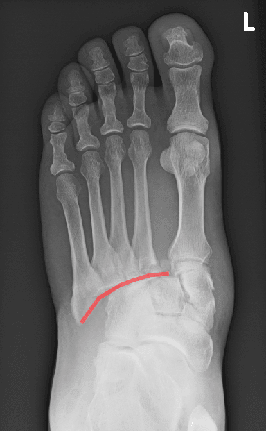
- Lisfranc injury must be excluded with weight-bearing views if subtle
- 1st Metatarsal requires zero displacement tolerance due to load bearing
- Smoking significantly increases the risk of Jones fracture nonunion
- “Jones fracture entry point: 'High and Inside' (High-Dorsal, Inside-Medial)
- “Fleck sign: Pathognomonic for Lisfranc avulsion (Base of 2nd MT)
- “Stress fractures: 2nd MT (Good) vs 5th MT (Poor) prognosis
- “Early fixation in athletes improves time to union and return to play
Metatarsal Fractures
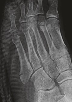
The exam favourite is the Zone 2 (Jones) fracture of the 5th metatarsal. It is a vascular watershed area (metaphyseal-diaphyseal junction) and prone to nonunion. Do not confuse it with Zone 1 (Pseudo-Jones) which is a tuberosity avulsion and heals universally. Zone 3 is a diaphyseal stress fracture and has the highest nonunion rate.
Overview and Epidemiology
Metatarsal fractures are common foot injuries, representing approximately 35% of all foot fractures. The fifth metatarsal is the most frequently injured, accounting for nearly 70% of metatarsal fractures. These injuries range from simple avulsion fractures to complex stress reactions and traumatic Lisfranc disruptions.
- 5th Metatarsal: Most common (Zones 1-3). Zone 1 (Avulsion) is the most frequent.
- Central Metatarsals (2-4): Often fractured together. Isolated fractures are rare and should raise suspicion of Lisfranc involvement.
- 1st Metatarsal: Least common but most critical for weight-bearing (carries 1/3 of body weight).
- Stress Fractures: "March fracture" classically involves the 2nd or 3rd metatarsal shaft. 5th metatarsal stress fractures (Zone 3) are high-risk.
Anatomy and Biomechanics
The foot's structural integrity depends on the metatarsals' role in the longitudinal and transverse arches.
Structural Anatomy
- The Columns:
- Medial Column: 1st Metatarsal + Medial Cuneiform (Flexible).
- Middle Column: 2nd/3rd Metatarsals + Middle/Lateral Cuneiforms (Rigid, "Keystone").
- Lateral Column: 4th/5th Metatarsals + Cuboid (Mobile).
- Ligamentous Support: The Lisfranc ligament (oblique) connects the medial cuneiform to the base of the 2nd metatarsal. There is no ligamentous connection between the 1st and 2nd metatarsal bases.
The 5th metatarsal base has a dual blood supply:
- Metaphyseal Arteries: Supply the tuberosity (Zone 1).
- Nutrient Artery: Enters the mid-diaphysis and travels proximally. The Junction: The area between these two supplies (Zone 2 - Jones fracture site) is a vascular watershed area, leading to high nonunion rates. [1]
Biomechanics
- Weight Distribution: During gait, the 1st metatarsal takes double the load of the lesser metatarsals.
- Load Sharing: The metatarsal heads follow a parabolic curve. Shortening of one metatarsal (e.g., malunion) leads to transfer metatarsalgia under adjacent heads.
The Metatarsal Parabola: Why Length and Alignment Matter
The anatomy section notes the metatarsal heads follow a parabolic curve and that shortening causes transfer metatarsalgia — this is the principle behind the topic's repeated insistence on anatomic reduction, especially of the first metatarsal.
- The normal parabola. The metatarsal heads form a smooth, gently curved arc: the second metatarsal is usually the longest, the first and third are slightly shorter and roughly similar, and the fourth and fifth are progressively shorter. This distributes forefoot load evenly across the heads.
- What malreduction does. Shortening of a metatarsal unloads its own head and transfers load to the adjacent heads; plantar (dorsiflexion-apex) angulation drives a head plantarward so it bears excess pressure. Either produces a transfer lesion — a painful plantar callosity, metatarsalgia, and sometimes a secondary stress fracture under the overloaded neighbour.
- The first metatarsal is unforgiving. Because it carries roughly a third of forefoot load, it has zero tolerance for shortening or malalignment — hence anatomic ORIF for any displacement, and the warning that a dorsal malunion causes chronic metatarsalgia and can accelerate hallux rigidus.
- Practical rule. Restore length and the plantar plane, not just the coronal alignment, when fixing metatarsal fractures; residual shortening of more than a few millimetres, or plantar angulation, is the mechanism behind late transfer metatarsalgia — later managed with metatarsal pads/orthotics or a Weil shortening osteotomy of the overloaded ray.
Q: Why is anatomic reduction (especially of the first metatarsal) so important in metatarsal fractures? A: The metatarsal heads sit on a parabola that shares forefoot load. Shortening or plantar malunion of one ray shifts load to its neighbours, causing transfer metatarsalgia (painful callosity, even a secondary stress fracture). The first metatarsal carries about a third of the load, so it tolerates no shortening/malalignment — restore length and the plantar plane, not just coronal alignment.
Classification Systems
Divides the 5th metatarsal base into three zones based on anatomy and healing potential.
| Grade/Type | Description | Management |
|---|---|---|
| Pseudo-Jones | Tuberosity avulsion. Involves the cancellous bone. Heals universally. | Boot and weight-bear as tolerated. Excellent prognosis. |
| Jones Fracture | Metaphyseal-diaphyseal junction. Extends into the 4th-5th intermetatarsal joint. Watershed area. | NWB cast 6-8 weeks OR intramedullary screw fixation (athletes). |
| Stress Fracture | Proximal diaphyseal fracture. Distal to the 4th-5th intermetatarsal joint. Very poor biology. | Intramedullary screw fixation strongly recommended. High nonunion risk. |
The Torg Classification: Chronicity of the 5th Metatarsal Base Fracture
The Lawrence-Botte zones describe where a proximal 5th metatarsal fracture sits; the Torg classification (cited in the evidence base) describes its chronicity and healing potential on the radiograph — the two are complementary, and both are examinable.
- Torg Type I — acute. A sharp, narrow fracture line with no intramedullary sclerosis and no periosteal reaction. Best healing potential; many unite in a non-weight-bearing cast.
- Torg Type II — delayed union. A widened fracture line with some intramedullary sclerosis and evidence of prior periosteal reaction — a fracture that has struggled to heal.
- Torg Type III — established nonunion. Complete obliteration of the medullary canal by sclerotic bone. Needs operative treatment: curettage/drilling of the sclerotic canal with bone grafting and/or a larger intramedullary screw.
- Why it matters. Radiographic chronicity, not the millimetre of location, drives prognosis — sclerosis or canal obliteration predicts poorer healing and pushes toward surgery, whether the fracture is labelled a "Jones" or a proximal diaphyseal fracture (Chuckpaiwong). Detailed Jones-fracture management is developed in the jones-fractures topic.
Q: How does the Torg classification of a proximal 5th metatarsal fracture guide treatment? A: Type I (acute) = narrow line, no sclerosis → can heal in a non-weight-bearing cast. Type II (delayed union) = widened line with intramedullary sclerosis → often needs fixation. Type III (nonunion) = the medullary canal is obliterated by sclerotic bone → operative curettage/drilling, bone graft and a larger IM screw. Sclerosis/canal obliteration, not exact location, is the key adverse sign.
Clinical Assessment
A high degree of clinical suspicion for Lisfranc injury is required for all midfoot trauma.
- Mechanism: Direct blow (crush) vs. Indirect twisting (Lisfranc).
- Pain: Inability to weight-bear is a significant indicator of instability.
- Training History: Recent increase in load (military recruits, marathon runners) for stress fractures.
Investigations
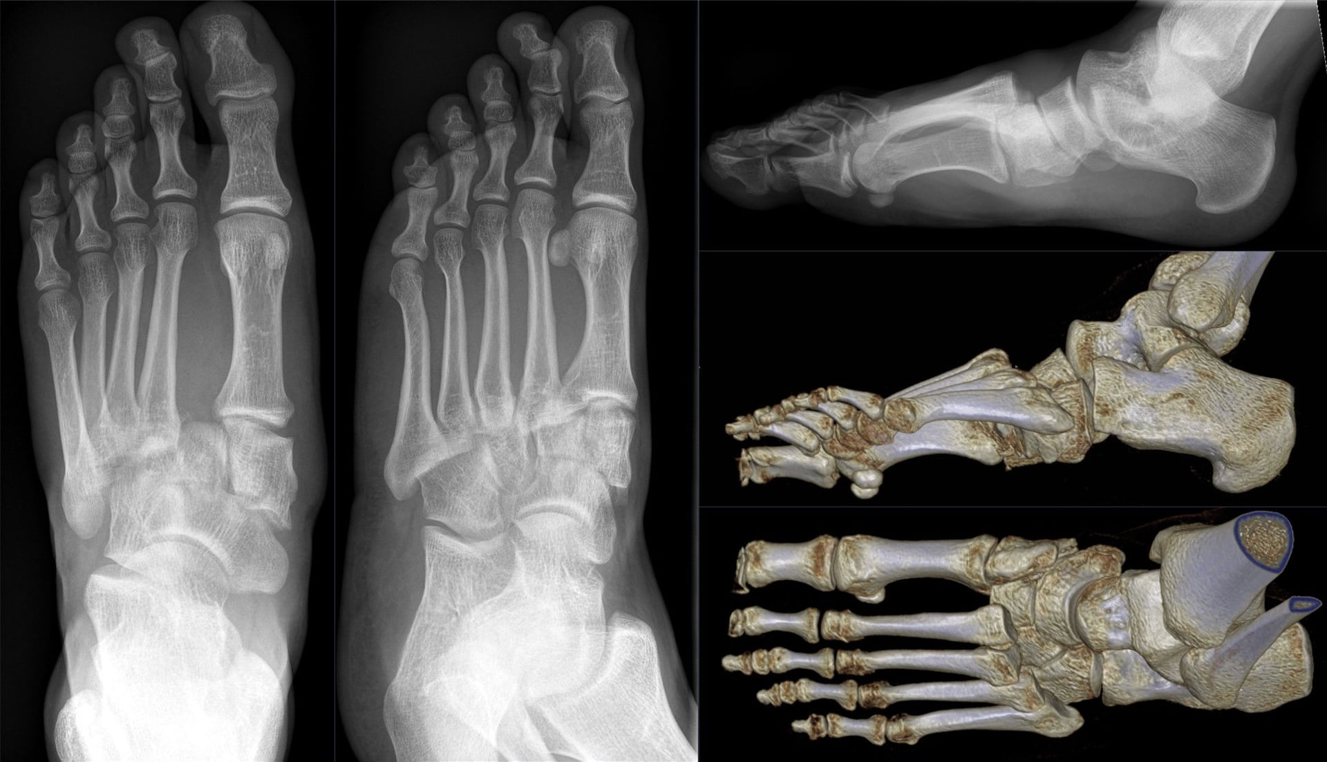
Radiographic assessment is the gold standard, but subtle injuries may require advanced imaging.
Standard Views AP, Lateral, and 30° Oblique views. Lisfranc Signs:
- Gap greater than 2mm between 1st/2nd MT bases.
- Fleck Sign: Bony avulsion in the 1st intermetatarsal space.
Stability Assessment Essential for subtle Lisfranc injuries. If painful, consider stress radiographs under anesthesia.
Pre-op Planning Superior for identifying small avulsion fractures (Fleck sign) and assessing articular involvement.
Stress Fractures The most sensitive test for early stress reactions (bone marrow edema) before cortical changes appear on X-ray.
Differential Diagnosis
Forefoot and midfoot pain has several mimics. Key discriminators below.
- Discriminating Features
- Trauma history, focal bony tenderness, deformity
- Best Test
- Plain radiographs (AP/oblique/lateral)
- Discriminating Features
- Insidious overuse pain, normal early X-ray, training spike
- Best Test
- MRI (marrow oedema) or delayed callus on X-ray
- Discriminating Features
- Plantar ecchymosis, midfoot instability, gap over 2mm
- Best Test
- Weight-bearing/stress radiographs, CT
- Discriminating Features
- 2nd MT head, adolescent/young female, AVN flattening
- Best Test
- Radiograph/MRI of MT head
- Discriminating Features
- Burning interdigital pain, Mulder click, no bony tenderness
- Best Test
- Clinical, ultrasound/MRI
- Discriminating Features
- Dorsal MTP pain, positive drawer, no fracture line
- Best Test
- Clinical exam, MRI/ultrasound
- Discriminating Features
- Smooth corticated bone lateral to cuboid, asymptomatic
- Best Test
- Compare with contralateral foot
Management Algorithm
- Zone 1: Symptomatic weight-bearing in boot.
- Zone 2 (Jones): NWB cast (6-8 weeks) OR Screw fixation in athletes.
- Zone 3 (Stress): Aggressive surgical fixation recommended.
Surgical Technique
- Positioning: Patient supine with a bolster under the ipsilateral hip to rotate the foot medially.
- Imaging: Enables optimal lateral foot C-arm view.
- Entry Point: Establish a 'High and Inside' (dorsomedial) entry point on the tuberosity.
- Significance: Avoids splitting the lateral cortex and aligns with the intramedullary canal.
- Guidewire: Pass a 1.6mm or 2.0mm guidewire down the intramedullary canal.
- Confirmation: Confirm central placement on AP and Lateral views.
- Drilling: Drill over the wire cautiously.
- Screw: Insert a 4.5mm - 5.5mm partially threaded screw.
- Compression: Confirm compression across the watershed zone on C-arm.
- Screw Selection: Use the largest diameter solid screw (4.5-5.5mm).
- Thread Engagement: Ensure all threads cross the fracture site.
- Length: Screw tip should reach within 5mm of the distal MT head.
- Bone Graft: Consider for delayed unions (greater than 6 months).
- Undersized screw: Leads to persistent nonunion.
- Lateral entry point: Risks splitting the lateral cortex.
- Eccentric wire: Leads to distal cortical perforation.
- Sural Nerve: Avoid excessive lateral dissection.
Complications
Metatarsal fractures, particularly the 5th, are prone to specific long-term issues.
- Risk Site / Cause
- Zone 2/3 (5th MT)
- Management / Sign
- ORIF + Bone Graft (ICBG)
- Risk Site / Cause
- Malunion (Plantar angulation)
- Management / Sign
- Transfer lesion to adjacent heads
- Risk Site / Cause
- Lateral approach to 5th MT base
- Management / Sign
- Numbness on lateral foot border
- Risk Site / Cause
- Pain over screw head
- Management / Sign
- Hardware removal after union
- Risk Site / Cause
- Premature return to sport
- Management / Sign
- Occurs in 10-15% of athletes
- Risk Site / Cause
- High-energy crush injury
- Management / Sign
- RARE but emergent fasciotomy required
- Risk Site / Cause
- Lisfranc injury (50% rate)
- Management / Sign
- May require fusion at 2-5 years
- Risk Site / Cause
- Prolonged immobilization
- Management / Sign
- Early mobilization, physio, pain clinic
Detailed Complication Management
Nonunion (Zone 2/3 - Fifth Metatarsal)
- Incidence: 30-50% without surgery for Jones fractures.
- Risk Factors:
- Smoking (most significant).
- Delayed presentation (>6 weeks).
- Premature weight-bearing.
- Undersized screw fixation (<4.5mm).
- Management:
- Revision: Larger diameter screw (5.5mm+).
- Biology: Debridement + Bone Graft (ICBG/BMAC).
- Stimulation: External bone stimulator.
Postoperative Care
Recovery depends on the stability of the construct and the biology of the fracture.
Protection Splint/Backslab. Strictly Non-Weight Bearing. Elevation to manage edema.
Mobilization Transition to a CAM walking boot. Gentle ROM of toes. Weight-bearing status per surgeon/topic protocol.
Rehabilitation Gradual weight-bearing as tolerated. Physical therapy for intrinsic foot muscle strengthening.
Outcomes and Prognosis
Overall prognosis for metatarsal fractures is good, provided structural alignment is maintained.
Excellent Predictable healing within 6-10 weeks. Functional return to baseline is standard.
Guarded Highest rate of secondary intervention (up to 30%). Return to high-impact sport may take 4-6 months if nonunion develops.
Long-term Prognosis
- Arthritis: Post-traumatic arthritis is common following Lisfranc injuries, even with anatomical reduction.
- Deformity: Malunion (plantar angulation) leads to chronic metatarsalgia.
Guidelines, Registries & Global Practice
Global epidemiology:
- Metatarsal fractures account for roughly 5-6% of all fractures and about 35% of foot fractures worldwide; the fifth metatarsal is involved in the majority (commonly quoted ~70% of metatarsal fractures).
- Stress (fatigue) fractures cluster in military recruits, distance runners, dancers and footballers. The 2nd/3rd metatarsal shaft ("march fracture") is the classic fatigue site; proximal 5th metatarsal stress fractures are the high-risk subset.
- Bone stress injuries frequently signal an underlying metabolic problem — screen for the Relative Energy Deficiency in Sport (RED-S) / female athlete triad, low vitamin D, and (in recurrent or atypical cases) coeliac disease.
Side-by-side guidance:
- Position on 5th MT base / Jones fractures
- Nonoperative for Zone 1 avulsions; intramedullary screw favoured for Zone 2/3 and for athletes seeking faster return.
- Position on 5th MT base / Jones fractures
- Early IM screw for competitive athletes; solid, largest-diameter screw to reduce refracture.
- Position on 5th MT base / Jones fractures
- Tuberosity (Zone 1) and most acute Jones fractures managed nonoperatively (boot, weight-bear as tolerated); surgery for displacement, nonunion or high-demand athletes.
- Position on 5th MT base / Jones fractures
- Lag-screw or plate ORIF for displaced 1st and central shaft fractures; IM screw for proximal 5th MT; anatomic restoration of length/alignment to prevent transfer metatarsalgia.
Across societies, anatomic reduction is mandatory; primary arthrodesis is favoured for purely ligamentous injuries (Ly & Coetzee Level I evidence), while ORIF remains standard for bony / combined patterns.
- Well-resourced settings: MRI for occult stress fractures, weight-bearing and stress radiographs (or weight-bearing CT) for subtle Lisfranc injuries, and early IM screw fixation for athletes to compress return-to-play timelines.
- Limited-resource settings: Plain radiographs and clinical examination drive decisions; NWB casting is a wholly acceptable primary treatment for acute Jones and tuberosity fractures, reserving surgery for nonunion, displacement or unequivocal instability. Smoking cessation and nutritional optimisation are universally available, high-yield interventions.
Controversies & Areas of Uncertainty
Level I evidence supports early screw fixation in athletes, but the optimal strategy for sedentary or low-demand patients is debated — many heal with NWB casting, avoiding hardware and refracture risk.
Larger, solid screws are widely recommended to reduce refracture, but high-quality comparative data on the ideal diameter, headed vs headless, and the added value of bone graft or biologics remain limited.
Primary arthrodesis is favoured for purely ligamentous injuries, but for combined bony-ligamentous patterns the choice between fusion and ORIF (and timing of routine hardware removal) is still individualised.
Early protected weight-bearing after fixation may speed recovery without compromising union, but cautious surgeons still use prolonged NWB in watershed-zone fractures; consensus thresholds are lacking.
Essential Mnemonics
1235th Metatarsal Zones
Proximal to Distal
Hook:1-2-3: The further down the bone (distal), the worse it heals
MMLLisfranc Stability
The 3 Columns
Hook:M-M-L: Medial, Middle (the key), Lateral
HIJones Entry Point
Screw Position
Hook:Stay 'HI' (High and Inside) to avoid the lateral cortex
MCQ Practice Points
Q: What are the three zones of 5th metatarsal base fractures and their significance?
A: Zone 1 (tuberosity avulsion): Peroneus brevis insertion, excellent healing, weight-bear as tolerated. Zone 2 (Jones fracture): Metaphyseal-diaphyseal junction, poor blood supply, high nonunion risk - consider early fixation in athletes. Zone 3 (diaphyseal stress): Chronic, requires intramedullary screw fixation.
Q: What associated injury must be excluded with any metatarsal fracture?
A: Lisfranc injury - tarsometatarsal joint complex disruption. Check for: 2nd MT base alignment with middle cuneiform on AP, medial border of 4th MT aligns with medial cuboid on oblique. Fleck sign (avulsion between 1st/2nd MT bases) pathognomonic. Weight-bearing radiographs if subtle. Missed injury leads to painful flatfoot.
Q: What are the indications for surgical fixation of metatarsal shaft fractures?
A: Surgical indications: greater than 10° sagittal angulation, greater than 3-4mm shortening, displacement greater than 3-4mm in transverse plane, multiple metatarsal fractures (disrupts transverse arch), open fractures, compartment syndrome. Central metatarsals (2-4) tolerate less displacement than 1st or 5th due to load distribution.
Q: What is the mechanism and treatment of first metatarsal stress fractures?
A: First MT stress fractures occur in runners with forefoot varus or hypermobility. Located at proximal metaphysis or diaphysis. Treatment: Activity modification, rigid-soled shoe/boot for 6-8 weeks. Surgical fixation for nonunion or elite athletes. Risk factors include vitamin D deficiency, female athlete triad, training errors.
Q: What is the "March fracture" and its typical location?
A: March fracture = stress fracture of metatarsal shaft, classically 2nd or 3rd metatarsal neck/shaft. Named for military recruits. Caused by repetitive stress exceeding bone remodeling capacity. X-ray may be negative initially; periosteal reaction or callus appears 2-3 weeks later. MRI/bone scan for early diagnosis if needed.
At a Glance
- Detail
- 5th Metatarsal (70%)
- Detail
- 1st Metatarsal (Load bearing)
- Detail
- Zone 2/3 (Watershed nonunion)
- Detail
- Plantar ecchymosis
- Detail
- Fleck sign (Base of 2nd MT)
- Detail
- Gap >2mm between 1st/2nd MT
Exam Cheat Sheet
5th MT Base Zones
- Zone 1: Avulsion → Boot and weight-bear as tolerated
- Zone 2: Jones → Watershed, NWB Cast or Screw (Athlete)
- Zone 3: Stress → Diaphyseal, High risk of nonunion
Lisfranc Pearls
- Plantar ecchymosis (Pathognomonic sign)
- Gap greater than 2mm between 1st/2nd MT bases
- Fleck sign is a pathognomonic avulsion fragment
Management Limits
- 1st MT: Zero displacement tolerance
- 2-4 MT: less than 10° angulation, less than 3mm shortening
- 5th MT: Zone 2 always requires non-operative protection or ORIF
Clinical Decision Scenarios
Practise clinical reasoning and management decisions out loud
“A 19-year-old elite basketball player presents with sudden lateral foot pain after a pivot. X-ray shows a transverse fracture at the 5th MT base, 2cm distal to the tuberosity tip. How would you proceed?”
Evidence Base
Early Screw Fixation vs Casting (RCT)
- RCT, 37 acute Jones fractures (19 screw vs 18 cast)
- Cast group: 44% treatment failure (5 nonunions, 1 delayed, 2 refractures)
- Screw: median union 7.5 wks and return to sport 8.0 wks vs 14.5 and 15.0 wks for cast
Surgery vs Nonop in Athletes (Meta-analysis)
- 22 studies, 646 Jones fractures in athletes
- Return to play: 98.8% with IM screw vs 71.6% nonoperative; union 97.3% vs 71.4%
- Overall refracture rate 10.2%; time to RTP 9.6 wks (screw) vs 13.1 wks (nonop)
Torg Classification (Landmark)
- 46 fractures distal to tuberosity stratified into acute, delayed union and nonunion
- Acute fractures: 14/15 united in ~7 wks with NWB toe-to-knee cast vs 4/10 with weight-bearing methods
- Sclerosis / canal obliteration defines chronic disease needing surgery