Robbing Peter to Pay Paul
- Converts a high-level injury (long regeneration time) to a low-level injury (short time).
- Requires a viable motor end plate (must be done generally within 12 months).
- Donor nerve must be expendable or redundant.
- Donor and Recipient must be synergistic for easier retraining (e.g., wrist flexor to finger extensor).
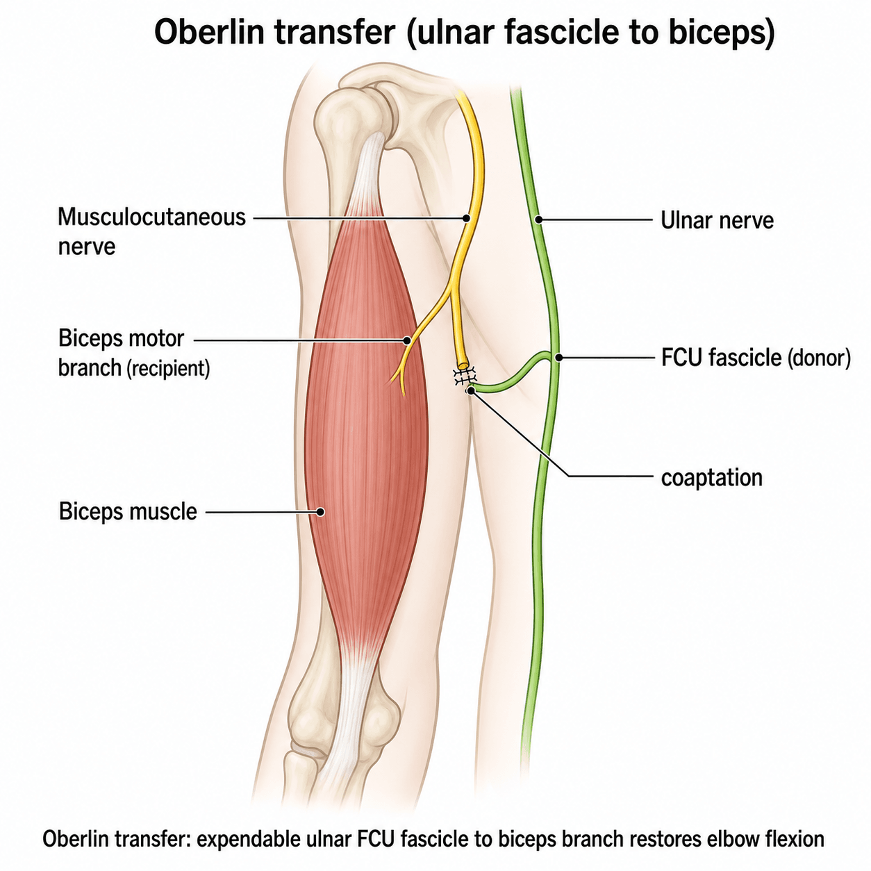
- “Oberlin Transfer restores Elbow Flexion (Ulnar fascicle to Biceps).
- “Somsak Transfer restores Triceps (Intercostal to Radial).
- “AIN to Ulnar reduces the risk of Claw Hand in high ulnar palsy.
Overview
Nerve Transfer (Neurotization): The surgical coaptation of a healthy, expendable donor nerve (proximal to the injury) to a denervated recipient nerve (distal to the injury) to restore function.
Nerve transfer has revolutionised the management of brachial plexus injuries (BPI) and high peripheral nerve injuries.
This page covers the transfer (neurotization) option specifically. The broader reconstructive menu it sits within - direct epineurial/fascicular repair, grafts and conduits, and when to choose each - is on the Nerve Repairs page, the regeneration biology and the motor-end-plate time window that governs the timing on the Nerve Injury and Regeneration page, and the structure and conduction basic science on Nerve Anatomy and Physiology; the four are companion reads.
Principles: Distance, Time and the Donor
Why a transfer works. Axons regenerate at about 1mm/day. If the injury is 30cm from the muscle, they take around 300 days to reach it; transferring a donor distally cuts that distance to under 5cm, about 50 days. A high-level injury with a long regeneration time is converted into a low-level injury with a short one.
Time is muscle. Motor end plates degenerate irreversibly at 12-18 months after denervation, and a nerve transfer must be performed before that window closes.
Transfer versus graft. A graft bridges the injury with sensory nerve, the sural being the standard for long gaps with the medial antebrachial cutaneous and saphenous as alternatives, and it needs two coaptations, proximal and distal, with the entire length still to regenerate. A transfer takes an expendable motor branch, makes one coaptation close to the target and leaves a short distance. The trade is numbness at the graft donor site against minor weakness in the transfer donor's distribution.
- Nerve Graft
- Sural (Sensory only)
- Nerve Transfer
- Expendable Motor Branch
- Nerve Graft
- Two (Proximal & Distal)
- Nerve Transfer
- One (Distal)
- Nerve Graft
- Long (entire length)
- Nerve Transfer
- Short (close to target)
- Nerve Graft
- Good for sensation, variable for motor
- Nerve Transfer
- Excellent for specific motor targets
- Nerve Graft
- Numbness
- Nerve Transfer
- Weakness (minor)
The ideal donor. The donor is chosen for what it can give and what it can afford to lose:
- Powerful: a high axon count (MCA greater than 1000)
- Expendable: its loss is acceptable
- Reach: long enough to reach the target
- Synergistic function, which makes retraining easier
- Independent excursion
- Calibre: a size match with the recipient
- Timing: available early
When to transfer. The indications are the situations in which repair or graft cannot deliver axons to a living muscle in time:
- An impossible repair, typically a root avulsion, where there is no proximal stump to graft from
- A very proximal injury with a viable distal target, where the transfer protects the motor end plates from the long wait
- Enhancing function by "baby-sitting" a muscle while the native axons regenerate down the main trunk
Classification
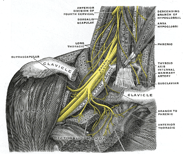
Transfers are classified by where the donor comes from.
Intra-plexus transfers use roots or trunks adjacent to the injury: with a C5 rupture and a C6 avulsion, the C5 stump (if available) or the medial pectoral nerve. The donor integrates within the same limb, but the supraclavicular plexus has to be explored to reach it, and that is a danger zone.
Extra-plexus transfers bring axons in from outside the plexus altogether:
- Intercostal nerves (3rd-6th) for elbow flexion or the shoulder
- Spinal accessory (XI) for the shoulder, into the suprascapular nerve
- Phrenic nerve, sometimes used, at the risk of hemidiaphragm paralysis
- Contralateral C7 in total plexus avulsion, crossed over the neck; it requires a vascularised nerve graft (usually ulnar) to reach
Distal transfers target nerve branches close to the muscle, the Oberlin transfer being the example.
Clinical Assessment
Checking the donor. The donor muscle must be at least MRC grade 4, preferably 5. Make sure that taking it will not leave an unacceptable deficit, for example where other muscles are already weak, and check that the patient can activate it easily.
Mapping the targets. The examination is organised around the three functions a transfer can restore:
- Shoulder: deltoid and rotator cuff, for the axillary and suprascapular targets. The suprascapular nerve drives supraspinatus and infraspinatus, abduction and external rotation, and spinal accessory to suprascapular is the workhorse of shoulder reanimation
- Elbow: biceps and brachialis, for the musculocutaneous target. An ulnar FCU fascicle to the biceps branch is the Oberlin transfer, a median FCR fascicle to the brachialis branch is Mackinnon's addition, and the double fascicular transfer yields the best results
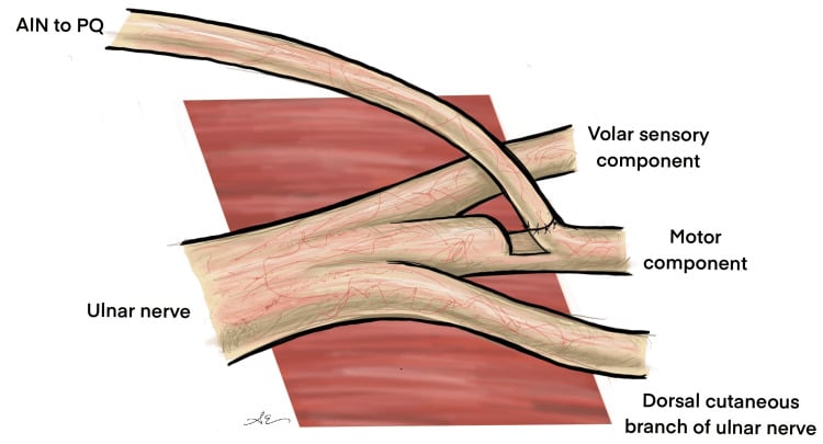
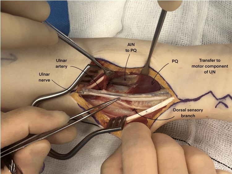
- Hand: the intrinsics, supplied by the ulnar motor branch. The anterior interosseous nerve (pronator quadratus branch) transfer prevents clawing and restores power pinch, and is a standard for high ulnar nerve injuries
Investigations
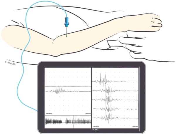
EMG and NCS. Electrodiagnostics confirm both halves of the operation. Motor unit potentials in the donor muscle confirm healthy axons; fibrillations and positive sharp waves in the recipient muscle confirm denervation and the need for a target. It is crucial to verify that the "expendable" donor is actually working properly before it is cut.
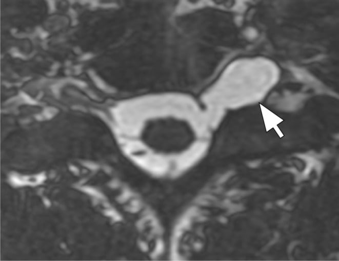
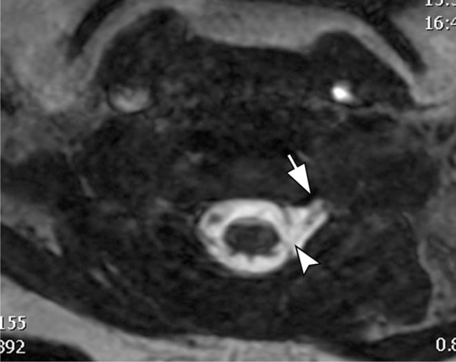
MRI of the plexus defines the anatomy of the injury, avulsion versus rupture. A pseudomeningocele indicates that the root has been pulled from the cord, so an extra-plexus transfer is required. CT myelography is the gold standard, but MRI is less invasive.
Choosing the Reconstruction: Differential of Options
When the clinical picture is set, the viva test is choosing between competing reconstructive strategies. Distinguish them by indication, not just by name.
- Best Indication
- Sharp, clean transection seen acutely
- Key Limitation
- Useless once a gap or avulsion is present
- Best Indication
- Rupture with a healthy proximal stump
- Key Limitation
- Two coaptations and long regeneration distance; inferior to transfer for proximal motor targets
- Best Indication
- Avulsion or very proximal injury with viable distal target and end-plates
- Key Limitation
- Needs an expendable synergistic donor and a still-receptive motor end plate (under about 12 months)
- Best Indication
- Late presentation (over 12 months) or failed transfer
- Key Limitation
- Major procedure; outcomes below a healthy native muscle
- Best Indication
- Local donor muscles available, motor end plate already lost
- Key Limitation
- Borrows existing function; limited independent control
The same donor-expendability, synergy, distance and timing principles apply outside the upper limb, and examiners may push you here. For foot drop from a proximal common or deep peroneal palsy, a branch of the tibial nerve (for example to a redundant head of gastrocnemius or soleus) can be transferred to the motor branch of tibialis anterior or the deep peroneal nerve to restore dorsiflexion when the lesion is too proximal to graft and the target end plate is still viable. Results are less reliable than upper-limb transfers, and a tibialis posterior tendon transfer to the dorsum of the foot remains the standard, more dependable alternative.
Nerve transfers are also described in spinal-cord injury, using supralesional donors to restore elbow extension, grasp or pinch, and in facial reanimation (masseteric or cross-face transfers), all sharing the same rules: wherever an expendable synergistic motor donor lies close to a viable but denervated target within the end-plate window, a transfer can convert a hopeless proximal lesion into a short distal repair.
Treatment
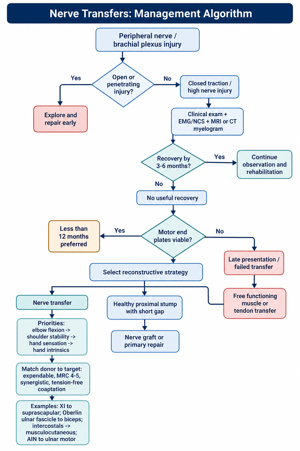
Diagnosis (day 0 to 3 weeks). Confirm the level of the injury and rule out penetrating trauma, which is explored early. For a closed injury the question is whether to wait and watch. Mechanism is key: high-velocity traction usually means avulsion.
Decision (3-6 months). If there are no signs of recovery, clinical or on EMG, by 3-6 months, the patient needs surgery.
Execution. The pattern of injury sets the plan. In Erb's palsy (C5/6) the shoulder is reanimated (XI to suprascapular) and the elbow (Oberlin). In a total palsy (C5-T1) elbow flexion (intercostal to musculocutaneous) and shoulder stability are prioritised. The function priority hierarchy:
- Elbow flexion
- Shoulder stability
- Hand sensation
- Hand intrinsics
Surgical Technique
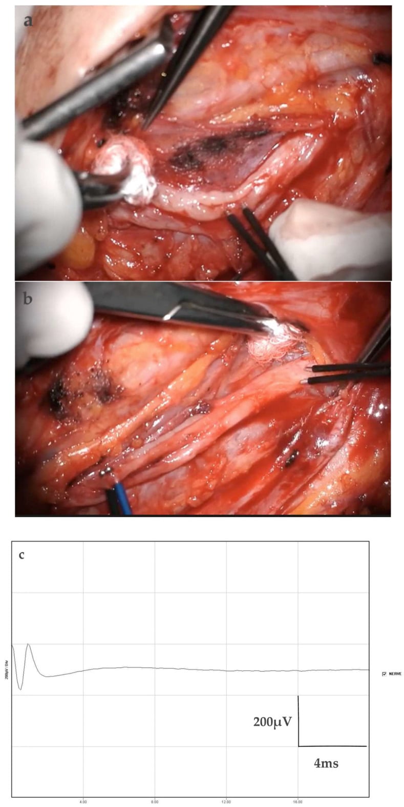
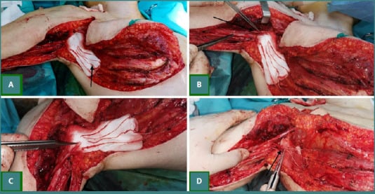
What every transfer has in common. The cords and their motor branches are identified against the axillary vessels. The donor fascicle is stimulated to confirm it is expendable, then coapted tension-free, end-to-end (or supercharged end-to-side), as close to the recipient's motor entry point as possible to minimise the reinnervation distance.
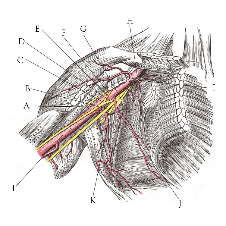
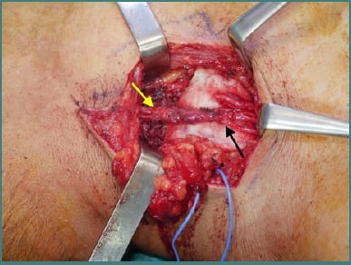
Spinal accessory to suprascapular. Through a dorsal approach, locate XI distal to its trapezius innervation and the suprascapular nerve in the suprascapular notch, release the ligament and repair directly. Taking XI distal to its important trapezius branches preserves proximal trapezius innervation and limits donor shoulder-girdle weakness.
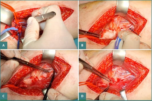
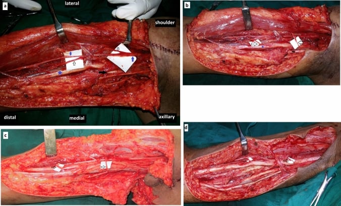
Radial (triceps) to axillary. Through a posterior or axillary approach, the radial nerve's branch to the long head of triceps is transferred to the anterior division of the axillary nerve, for deltoid, which restores abduction. The anterior axillary division is exposed and confirmed, an expendable triceps branch is selected with stimulation, divided distally and coapted to the recipient proximally. Paired with spinal accessory to suprascapular, it reinnervates both deltoid and rotator cuff.
Complications
Donor morbidity. Weakness in the donor distribution, trapezius weakness for example, is usually transient or well compensated. Each transfer has its own version:
- Oberlin: FCU weakness is rarely clinically significant because of redundancy with FCR; ulnar nerve injury during fascicle identification is rare but catastrophic
- Spinal accessory to suprascapular: trapezius weakness may impair overhead activities; shoulder shrug weakness is usually well tolerated
- Intercostal: chest wall pain (usually temporary); rare pneumothorax during harvest; breathing-elbow synchrony takes months to overcome
- AIN to ulnar motor: mild pronation weakness is usually not clinically apparent; the intrinsics may fail to recover despite a successful coaptation
Failure and co-contraction. The transfer may produce no reinnervation despite surgery, or the patient may have difficulty isolating the movement. Where sensory transfers are used there is sensory loss, and sensory re-education is vital to overcome cortical confusion.
The patient's part. Patients must learn that activating the donor now produces the recipient action, and that is a long-term commitment rather than a quick fix, so motivation is a key selection criterion. Pain management is essential: some patients develop chronic neuropathic pain requiring specialist input.
Postoperative Care
A sling or splint protects the repair. No tension on the coaptation.
The patient activates the donor, "breathe" for intercostals and "flex wrist" for Oberlin, with biofeedback providing visual cues that link the donor action to the recipient effect. This phase requires intense physiotherapy and patient motivation.
The brain adapts and the movement becomes natural. Plasticity allows the patient to eventually just "flex elbow" without thinking "flex wrist".
Reading the first signs. Triggered nascent motor-unit potentials on needle EMG can appear before visible movement. Their presence should prompt focused donor-recipient activation and biofeedback rather than prolonged protection; serial examination still determines whether the new units mature into useful MRC power.
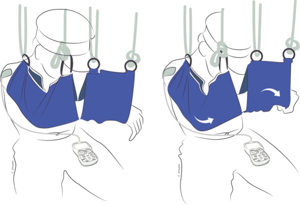
Early motor retraining. After a double fascicular transfer, suspension slings remove gravity while the patient deliberately couples the donor command with elbow flexion, and EMG biofeedback makes weak recipient activation visible. Assistance is progressively reduced as strength emerges, followed by donor de-activation and task-specific endurance work.
Prognosis
Upper trunk (Erb's). Good prognosis; the shoulder and elbow are usually recoverable. Abduction recovery is generally 80-90% of normal range, and elbow flexion by Oberlin transfer is very reliable, with greater than 90% success.
Total plexus. Poor prognosis. The goal is a "helper hand" or just elbow flexion, and hand function is unlikely to be significantly restored. Pain relief is a major goal, with DREZ lesions for avulsion pain.
Time. Earlier is better: results degrade significantly after 6-9 months, and the "Goldilocks" period is 3-6 months. After 12 months a free functioning muscle transfer (gracilis) is the only option for motor recovery, with tendon transfers as an alternative if local muscles are available. Always have a Plan B for salvage if the transfer fails. Pre-operative counselling is crucial to manage expectations.
Guidelines, Registries & Global Practice
Global Epidemiology:
- Traumatic adult brachial plexus injury is overwhelmingly a disease of young men: most series report a male-to-female ratio of roughly 4-9 to 1 with peak incidence in the late teens to mid-30s.
- High-energy motorcycle and road-traffic trauma is the dominant mechanism worldwide, accounting for the majority of supraclavicular avulsion injuries in most published cohorts.
- The burden is disproportionately high in regions with large motorcycle populations (parts of South and Southeast Asia, Latin America), which is also where several of the landmark distal-transfer techniques were developed and refined.
Side-by-Side Society / Group Practice (no formal RCT-based guideline exists):
- Common international position
- Sharp/penetrating injury: explore early (ideally within days). Closed injury with no recovery: refer by 3 months, operate by 3-6 months.
- Common international position
- CT myelography remains the reference for root avulsion; MR neurography is increasingly used and is less invasive. Baseline EMG/NCS deferred to about 3-4 weeks.
- Common international position
- Strong convergence (supported by Garg et al.) toward dual nerve transfer over long interposition grafts for shoulder and elbow.
- Common international position
- Reserve donor nerves for free functioning muscle transfer rather than late nerve reconstruction (supported by Hoang/Seruya).
- Unlike arthroplasty, there is no large international implant-style registry for nerve transfers; evidence is pooled from high-volume centre series and systematic reviews.
- Dedicated multidisciplinary peripheral-nerve and brachial-plexus services (combining plastic, orthopaedic, hand and neurosurgery with specialist hand therapy) consistently report better and more reproducible outcomes than ad hoc care.
- High-resource settings: routine microsurgical fascicular transfers, intra-operative nerve stimulation, MR neurography, and structured biofeedback rehabilitation over 12-24 months.
- Limited-resource settings: greater reliance on extra-plexus donors (intercostal, spinal accessory) and on tendon transfers or free muscle transfer where microsurgical theatre time, nerve stimulators or long-term therapy are scarce; later presentation is common, shifting the balance toward muscle transfer.
Controversies & Areas of Uncertainty
Double fascicular transfer reinnervates brachialis as well as biceps and series report high M4+ rates, but a single Oberlin transfer already gives reliable flexion with even lower donor burden. Whether the second coaptation is always worth the extra median-nerve dissection remains debated.
Reverse end-to-side transfers (for example distal AIN into the side of the ulnar motor branch) are used to "baby-sit" muscle while native axons regenerate down the main trunk. How much functional motor recovery genuinely comes from the donor versus the regenerating native axons is still uncertain.
Contralateral C7 can provide donor axons in total avulsion, but the risk to the healthy limb and the modest, mainly sensory/median functional gain mean its role is contested and centre-dependent.
The "12-month" end-plate window is a teaching rule, not a hard cut-off; reinnervation potential declines progressively and varies with patient age and injury level. Earlier surgery is consistently better, but the exact point of futility is individual.
MCQ Practice Points
Q: Which fascicle of the Ulnar nerve is used in the Oberlin transfer? A: The fascicle supplying the Flexor Carpi Ulnaris (FCU).
Q: What is the maximum time window for successful motor nerve transfer? A: Generally 12-18 months before motor end plate fibrosis.
Q: What is the risk of using the Phrenic nerve as a donor? A: Hemidiaphragm paralysis (Reduced Vital Capacity).
Q: What is the Oberlin transfer and what does it restore? A: Transfer of Ulnar FCU fascicle to Biceps motor branch. Restores elbow flexion in C5/C6 injuries.
Q: What is the purpose of AIN to Ulnar motor transfer? A: Prevents clawing and restores pinch in high ulnar nerve injuries. Reinnervates intrinsic muscles before motor end plate fibrosis.
Q: What is the rate of nerve regeneration after repair? A: Approximately 1mm per day (1 inch per month). This determines the urgency of distal transfers for long injuries.
Viva Scenarios
Practise clinical reasoning and management decisions out loud
“A 25-year-old male motorcyclist presents with a C5/C6 avulsion injury 4 months post-accident. No recovery. Examination shows deltoid/biceps paralysis. Hand is normal.”
“Patient with a high ulnar nerve transection at the axilla. 6 months post-injury. Primary repair was done but prognosis is guarded.”
“A 22-year-old male sustained a motorcycle accident with complete C5-T1 avulsion injury. MRI shows pseudomeningoceles at all levels. He presents at 5 months with a flail arm.”
Principles
- Donor Expendability
- Recipient Viability
- Proximity (Distal target)
- Synergy (Easier rehab)
Common Transfers
- Oberlin: Ulnar to Biceps (Elbow flexion)
- Somsak: Intercostal to MC (Elbow flexion)
- XI to SSN: Shoulder reanimation
- AIN to Ulnar: Intrinsic salvage
Timing
- Early: 3-6 months (ideal)
- Late: Greater than 12 months (Muscle transfer required)
- Motor end plate fibrosis by 18 months
- Regeneration: 1mm/day
Evidence Base
Nerve-transfer literature is dominated by retrospective Level III-IV case series and pooled systematic reviews; no randomised controlled trial has compared transfer against graft. Outcomes are reported on the Medical Research Council (MRC) motor scale, where M3 means movement against gravity and M4 means movement against resistance. All statistics below are taken directly from the cited papers.
Oberlin Transfer (original description)
- First description: transfer of part of the ulnar nerve to the biceps motor branch in 4 patients with C5-C6 avulsion
- Approximately 10 percent of the ulnar nerve bulk harvested and sutured directly to the biceps branch
- Restored elbow flexion with no significant impairment of hand function
Oberlin Transfer (consolidating series)
- 18 patients (8 with C5-C6, 10 with C5-C6-C7 paralysis)
- 7 of 8 C5-C6 patients recovered useful elbow flexion after the transfer alone
- Results were poorer and often needed a supplementary Steindler when surgery was delayed beyond a few months
Double Fascicular Transfer (Mackinnon technique)
- 6 patients: expendable fascicles from BOTH ulnar and median nerves transferred to the biceps and brachialis branches
- Mean recovery of elbow flexion was MRC grade 4 plus
- No motor or sensory donor deficit; pinch and grip unchanged or improved
Double Fascicular Transfer (larger series)
- 29 patients; 28 (97 percent) regained elbow flexion
- 8 reached M5, 15 reached M4, 4 reached M3
- No functional deficit in the ulnar or median donor distributions
Nerve Transfers vs Nerve Grafting (upper plexus)
- Systematic review of 31 studies in traumatic upper-trunk (C5-C6) palsy
- M4+ elbow flexion: 247 of 299 (83 percent) after transfer vs 32 of 57 (56 percent) after grafting
- Dual transfer for shoulder gave M4+ abduction in 74 percent vs 46 percent for grafting
Triceps-to-Axillary (Leechavengvongs / Somsak group)
- Anatomic feasibility study in 36 cadaveric shoulder girdles
- Nerve to the long head of triceps reaches the anterior branch of the axillary nerve via a posterior approach without grafting
- Donor carries roughly 1,233 axons against approximately 2,704 in the axillary anterior branch
Intercostal-to-Biceps Transfer
- 17 patients with avulsion injury; intercostal neurotisation of biceps, mean 5-year follow-up
- 8 of 17 (47 percent) achieved good or excellent elbow flexion
- Results improved when surgery was done under 5 months and in patients under 50 years
Distal Median-to-Ulnar (AIN) Transfer
- Technical description of AIN to deep motor branch of ulnar nerve to restore intrinsic function
- Combined with median third-webspace sensory transfer for ulnar volar sensation
- Reduces denervation time for distal intrinsics in proximal (Sunderland IV-V) ulnar injury
Nerve Reconstruction vs Free Muscle Transfer (late cases)
- Systematic review, 103 patients presenting at 12 months or later
- Upper-trunk M3+ elbow flexion: 53 percent after nerve reconstruction vs 100 percent after free functional muscle transfer
- For total plexus injuries, free muscle transfer outperformed nerve reconstruction (78 vs 37 percent M3+)