Atrophic vs Hypertrophic | Stability | Biology
- Nonunion: Fracture that will not heal without intervention (9 months + no progress for 3 months)
- Hypertrophic = mechanical failure (unstable) → improve stability
- Atrophic = biological failure (avascular) → improve biology
- Infection must be excluded in all nonunions
- Diamond concept: Cells + scaffolds + growth factors + stability
- “Elephant foot = most callus, easiest to treat (just stabilise)
- “Horse hoof = less callus but still biological activity
- “Atrophic = avascular ends need resection, grafting
- “Smokers have 2x higher nonunion risk
Definition and Classification
Definition. A nonunion is a fracture that will not heal without intervention. It is often defined, following the FDA, as at least 9 months from injury with no radiographic progress for 3 consecutive months.
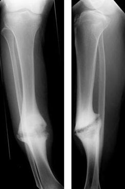
The Weber-Cech classification. The radiograph sorts a nonunion by the biology of its bone ends, and the category says what has failed.
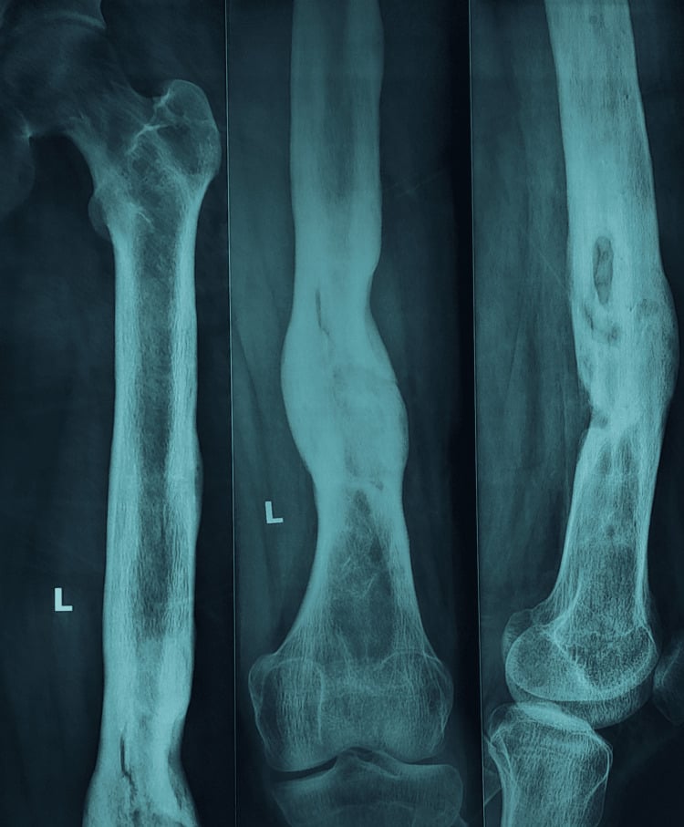
- Hypertrophic ("elephant foot"), viable: abundant callus, very vascular
- Slightly hypertrophic ("horse hoof"), viable: less callus, but still some biological activity
- Oligotrophic, viable: minimal callus, the bone ends present but inactive
- Atrophic, non-viable (avascular): no callus and avascular ends, sometimes comminuted or with bone loss
What the pattern means. A hypertrophic nonunion is a mechanical failure. The biology is intact, as the callus shows, but the fracture is unstable, so the answer is more stability and it may not need bone graft. An atrophic nonunion is a biological failure of avascular ends: the dead ends are resected, the biology improved with graft, and stability provided as well. Whatever the pattern, infection must be excluded in every nonunion.
Pathophysiology
Normal healing. Fracture healing proceeds through overlapping phases:
- Inflammation (days 1-7): haematoma, inflammatory cells, cytokines (IL-1, IL-6, TNF-α)
- Soft callus (weeks 1-4): chondrogenesis, fibrous callus
- Hard callus (weeks 4-12): woven bone replaces cartilage by endochondral ossification
- Remodelling (months to years): lamellar bone replaces woven bone and the cortex is restored
Nonunion occurs when mechanical or biological factors disrupt that cascade.
Mechanical failure, the hypertrophic pattern. Three factors are at work: excessive motion at the fracture, which prevents bridging; inadequate fixation, such as an undersized nail, loose screws or a failed plate; and distraction, since a gap of more than 2mm impairs bridging. The biology is intact, but instability prevents consolidation.
Biological failure, the atrophic pattern. The biological factors:
- Vascular disruption: severe soft-tissue injury (Gustilo IIIB/C), periosteal stripping at surgery
- Avascular bone: the scaphoid waist, the intracapsular femoral neck, the talar body
- Infection: low-grade biofilm infection suppresses osteoblast function
- Host and metabolic factors: age and comorbidity, diabetes, smoking (nicotine vasoconstriction), malnutrition, vitamin D deficiency
- Medication: NSAIDs inhibit the COX-2 needed for bone healing; corticosteroids impair osteoblast function
Local causes. Soft tissue interposed in the fracture, and an inadequate reduction that leaves a gap or malalignment, are further causes of nonunion.
The diamond concept. Giannoudis and colleagues (2007) proposed that healing requires all four of these elements to be optimised:
- Osteogenic cells: MSCs and osteoprogenitor cells from periosteum, bone marrow and the circulation
- An osteoconductive scaffold: bone graft matrix, collagen, HA/TCP ceramics
- Osteoinductive signals: BMPs, PDGF, TGF-β, VEGF, PRP
- Mechanical stability: absolute or relative, depending on the healing mechanism
Vascularity, the blood supply that brings oxygen, nutrients and cells, was added later as a fifth element. An atrophic nonunion fails on biology (elements 1-3 and 5); a hypertrophic nonunion fails on mechanics (element 4).
Infection. Bacteria, especially Staphylococcus epidermidis and Propionibacterium acnes, form a biofilm on implants and bone that protects them from antibiotics and the immune system. Bacterial toxins and inflammatory cytokines then inhibit osteoblast function. The result is an infected nonunion, which will not heal until the infection is eradicated, and up to 10% of nonunions are infected with occult, low-grade organisms.
Clinical Presentation
History. The patient presents months to years after the original fracture, with persistent pain at the fracture site despite what should have been adequate healing time. Pain on weight-bearing or activity is mechanical and suggests instability; constant pain or night pain suggests infection or severe instability. Function is lost: the patient cannot return to work or manage daily activities, still uses crutches or a walker, and has reduced movement in the adjacent joints. Ask about previous failed treatment, since bone grafting or revision fixation may already have been tried.
Risk factors. Ask specifically about:
- Smoking: 2-3 times the nonunion risk and dose dependent (OR 2.50, 95% CI 1.73-3.61; Xu 2021)
- NSAIDs, particularly high-dose, prolonged use after the fracture
- Diabetes, especially if poorly controlled (HbA1c over 8%)
- Malnutrition (low albumin, low vitamin D)
- Osteoporosis or metabolic bone disease
- Chronic corticosteroid use (over 7.5mg prednisolone daily)
- Severe soft-tissue injury at the original fracture (Gustilo III open fracture)
Look. Muscle atrophy from chronic disuse, scars from previous surgery, swelling or erythema from infection, and malalignment in varus, valgus or rotation. A sinus tract, if present, is pathognomonic of infection.
Feel. Tenderness at the nonunion site, a palpable gap or instability if the implants have failed, and warmth from infection.
Move. A healed fracture should be stable, so look for abnormal motion at the site and for pain on axial loading, bending or rotation. Check the adjacent joints for stiffness, from compensation and prolonged immobilisation.
Neurovascular. Look for nerve injury from previous surgery or chronic hardware irritation, and assess the vascular status, since a chronic injury may carry vascular compromise.
Red flags for infection. Any nonunion with persistent pain, raised CRP or ESR, or sinus drainage should be cultured. The flags:
- A draining sinus
- Persistent pain despite apparently stable fixation
- Multiple failed surgeries without union
- Raised inflammatory markers (CRP, ESR) at presentation
- A previous open fracture (Gustilo II/III)
Investigations
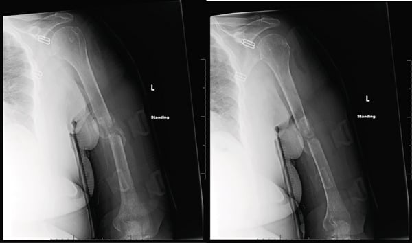
Radiographs. Orthogonal views, AP and lateral at a minimum. Read them for:
- Callus: hypertrophic, oligotrophic or atrophic
- The fracture line: a persistent gap, sclerosis of the bone ends
- Hardware: loose screws (lucency around the threads), broken plates or screws, a nail backing out
- Alignment: varus or valgus, angulation, rotation, shortening
- Bone stock: comminution, bone loss, osteopenia
CT is the best test of bone healing, more sensitive than radiographs for cortical bridging. 3D reconstruction helps plan surgery by showing the deformity, bone stock and implant position, and in infected cases CT identifies sequestrum or bone loss.
MRI assesses infection, through marrow oedema, fluid collections, sinus tracts, soft-tissue abscess and sometimes sequestrum, and the vascularity of the bone ends from their signal characteristics. It shows bony detail less well than CT.
Nuclear medicine is not routinely needed. A Tc-99m MDP bone scan has high sensitivity but low specificity, because uptake is increased at a nonunion whether or not it is infected; an In-111 white-cell scan or FDG-PET is more specific for infection. Reserve them for difficult cases in which infection is suspected but cultures are negative.
Bloods for infection. A raised CRP suggests infection, although CRP may be mildly elevated in a chronic nonunion without infection. ESR is less specific, but persistent elevation is concerning. The white cell count is usually normal in chronic low-grade infection.
Tissue culture is the gold standard.
- Tissue by CT-guided biopsy or aspiration of the nonunion site
- At least 5 tissue specimens, not swabs
- Prolonged culture (14 days) to catch slow-growing organisms (Propionibacterium, Cutibacterium)
Metabolic and nutritional status. Screen with:
- 25-OH vitamin D: target over 75 nmol/L for optimal bone healing
- Calcium, phosphate, alkaline phosphatase and PTH: metabolic bone disease
- Albumin and pre-albumin: nutritional status, with low albumin meaning poor healing
- HbA1c in diabetics: target under 7% for healing
- Thyroid function (TSH, free T4): hyperthyroidism impairs healing
Specialised tests, if indicated. DEXA for bone mineral density; bone turnover markers, CTX for resorption and P1NP for formation, which remain research tools; and genetic testing in rare cases (osteogenesis imperfecta, hypophosphatasia).
Work-up and Differential Diagnosis
Infection first. Exclude it in every nonunion, with the bloods (CRP, ESR, WCC), aspiration or biopsy for prolonged culture, and imaging described above. An infected nonunion is treated differently, by the staged pathway under Management.
Modifiable factors. Address each of them:
- Smoking: advise cessation. The four-week preoperative cessation figure is evidence for fewer wound infections, not for a lower nonunion rate
- Nutrition: optimise protein and vitamins
- Diabetes: control it (HbA1c)
- NSAIDs: avoid them, since they may impair healing
The differential. A painful fracture that is not progressing is not always a simple nonunion. The pattern on imaging and the inflammatory markers point to the diagnosis, and each entity is treated very differently.
- Callus / Radiograph
- Abundant callus (elephant foot / horse hoof), persistent line
- Biology vs Mechanics
- Mechanical failure, biology intact
- Infection markers
- Usually normal
- Key action
- Increase stability (exchange nail, compression plate)
- Callus / Radiograph
- Minimal callus, viable but quiet bone ends
- Biology vs Mechanics
- Mixed - often inadequate reduction/gap
- Infection markers
- Usually normal
- Key action
- Optimise reduction/stability +/- graft
- Callus / Radiograph
- No callus, tapered avascular sclerotic ends, possible gap
- Biology vs Mechanics
- Biological failure
- Infection markers
- Usually normal
- Key action
- Resect ends to bleeding bone + autograft + stable fixation
- Callus / Radiograph
- Lucency around implants, sequestrum, periosteal reaction
- Biology vs Mechanics
- Biology suppressed by biofilm
- Infection markers
- CRP/ESR often raised; may be normal
- Key action
- Deep tissue culture, staged debridement + antibiotics
- Callus / Radiograph
- Progressive callus, healing slower than expected
- Biology vs Mechanics
- Healing trajectory intact
- Infection markers
- Normal
- Key action
- Protect, optimise biology, observe
- Callus / Radiograph
- Lytic or blastic lesion, cortical destruction, soft-tissue mass
- Biology vs Mechanics
- Underlying lesion prevents healing
- Infection markers
- Variable
- Key action
- Staging, biopsy before any fixation
Measuring Union Objectively: the RUST and RUSH Scores
The problem they solve. The definition of nonunion rests on "no radiographic progression", yet union and progression are subjective and poorly reproducible between observers. RUST and RUSH make radiographic healing objective and reproducible, and give the otherwise arbitrary 9-month and 3-month definition a measurable anchor.
RUST (Radiographic Union Score for Tibial fractures). Each of the four cortices on two orthogonal radiographs is scored: anterior and posterior on the lateral view, medial and lateral on the AP.
- 1: fracture line visible, no callus
- 2: callus present, fracture line still visible
- 3: bridging callus, fracture line gone
The four scores sum to a total from 4 (no healing) to 12 (complete union). A modified RUST adds a fourth grade (remodelled, line absent) for finer discrimination.
RUSH (Radiographic Union Score for Hip) is the analogous cortical-and-trabecular score, validated for femoral neck fractures.
Reading the score. A rising score across serial films means progression. A low or plateauing score, such as a RUST that stops climbing well below 10 over three months, supports the diagnosis of nonunion and the need for intervention. The scores also serve as trial endpoints, as in the TRUST trial of LIPUS.
The criteria for union. Radiographic bridging, typically 3 of 4 cortices bridged or a high RUST, combined with clinical union: no pain on weight-bearing or on stressing the fracture, and the ability to bear weight.
Management
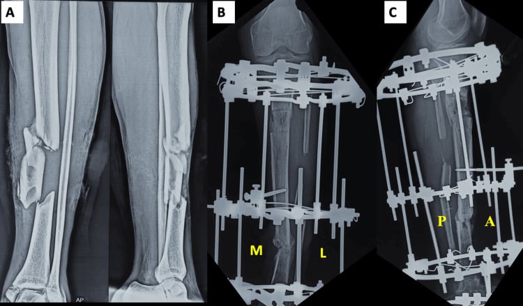
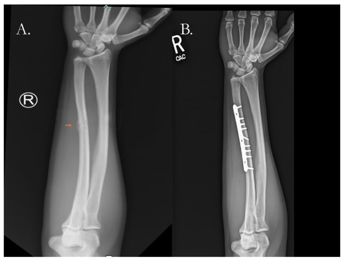
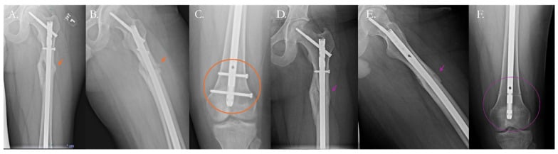
The problem is instability; the biology is good. Improve the stability:
- Compression plating: a DCP in compression mode
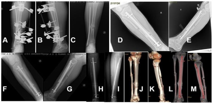
- Exchange nailing: a larger, stiffer nail
- Additional fixation: an added locking plate, cerclage
Bone graft may not be needed, because the biology is sufficient.
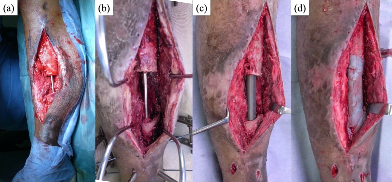
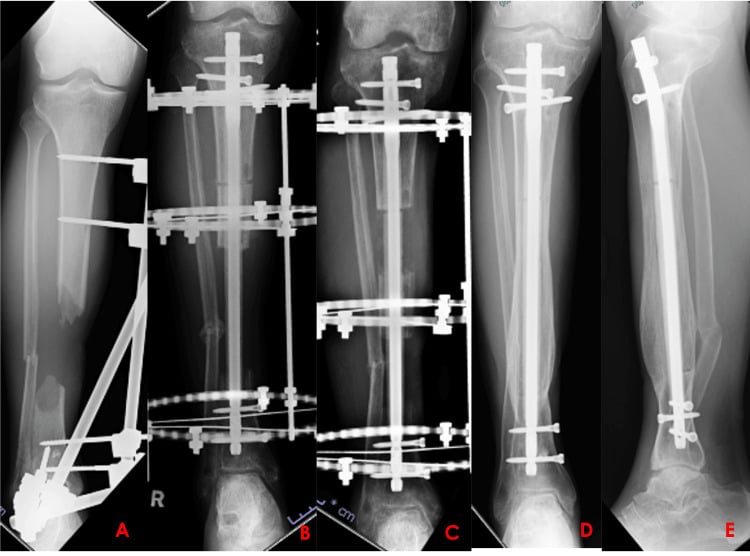
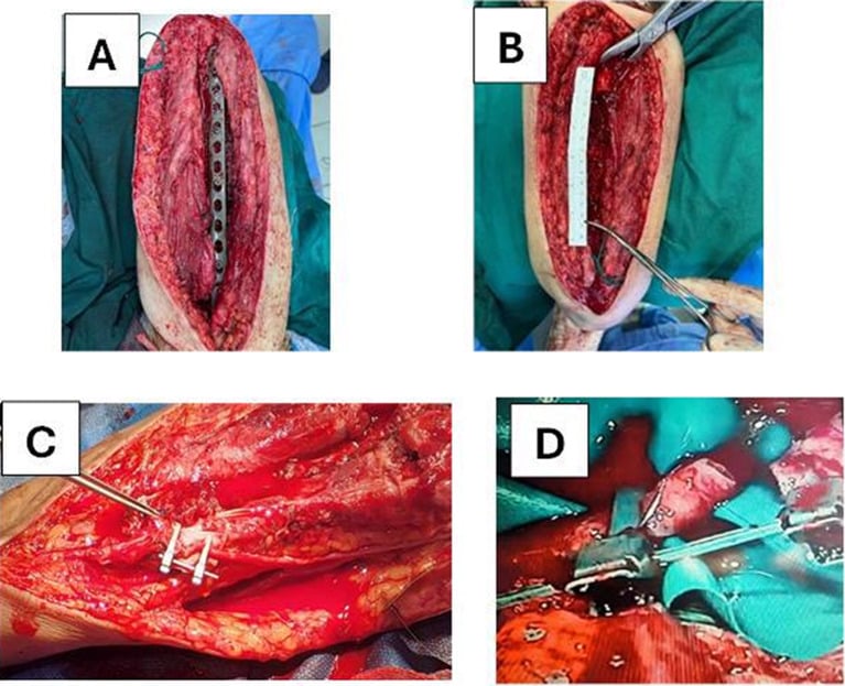
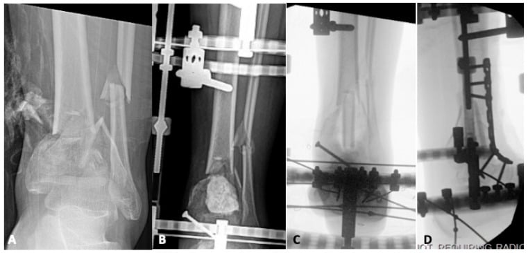
When reconstruction fails. Amputation is indicated for:
- Multiple failed reconstructions (persistent nonunion after 2-3 surgeries)
- Chronic infection not controllable with limb salvage
- Severe soft-tissue loss with inadequate coverage
- A neuropathic limb (insensate foot, severe nerve injury)
- Patient preference after informed consent, since some patients choose amputation over prolonged unsuccessful limb salvage
It may provide pain relief better than the chronic pain of an unstable nonunion, a faster return to function because modern prosthetics enable good mobility, and an end to repeated surgery.
The Reamer-Irrigator-Aspirator (RIA) Autograft
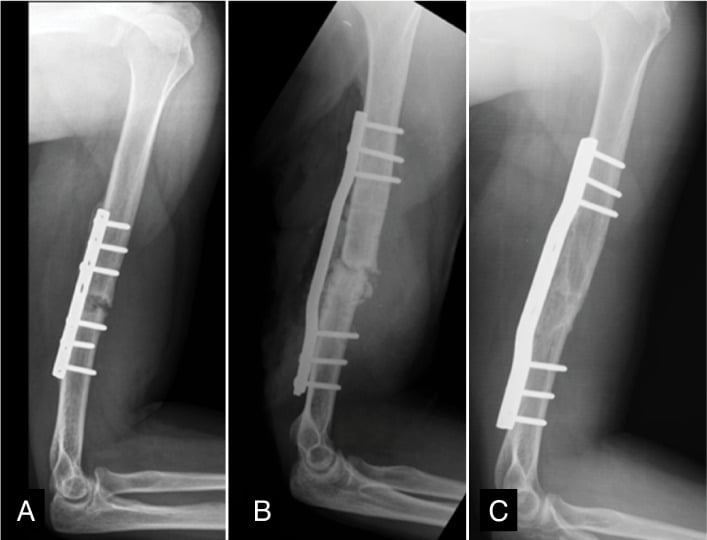
Reamer-irrigator-aspirator (RIA) graft is the answer to "where do I get enough autograft for a large atrophic or segmental defect?", and its blood loss is the reason that answer is not free.
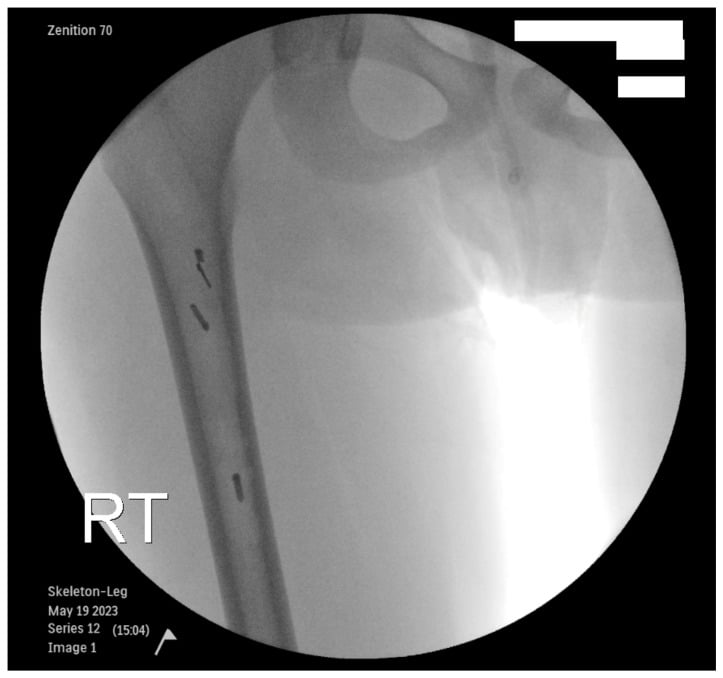
What it is. A single-pass intramedullary reaming system that reams the femoral (or tibial) canal while irrigating and aspirating, collecting the reamings in an in-line filter. It harvests a large volume of autologous cancellous bone and marrow from inside the long bone. The contralateral femur is the usual donor for a femoral or tibial recipient, and the donor canal must be intact and adequately sized.
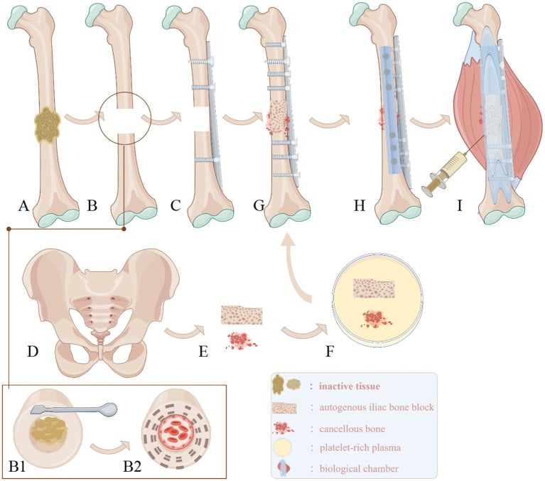
Why it matters in nonunion. Iliac crest bone graft yields a limited volume and carries significant donor-site pain. A large atrophic or segmental defect, or the graft needed to fill a Masquelet membrane, may require more than one iliac crest can provide. RIA delivers a large volume, often 40-90 mL, of biologically rich graft containing osteogenic cells, an osteoconductive matrix and osteoinductive growth factors, three arms of the diamond concept, with generally less chronic donor-site pain than iliac crest harvest.
The trade-offs. Blood loss of roughly 500-800 mL, and a risk of iatrogenic fracture, cortical perforation or eccentric over-reaming of the donor bone.
Where it sits. RIA is a harvest technique, not a different graft: the product is autograft, so it complements rather than replaces the diamond-concept biology. General graft selection is covered in the bone-grafts topic; for the large volume of autograft needed in atrophic or segmental nonunion, RIA is the workhorse.
Complications
Of the nonunion itself. The patient lives with chronic pain, mechanical or neuropathic, and loss of limb function, to the point of being unable to work or dependent in daily activities. The limb wastes and weakens, and the psychological cost is depression, anxiety and a reduced quality of life. Abnormal loading, and stiffness from prolonged immobilisation, lead to degenerative change in the adjacent joints.
Deformity. Malalignment in varus, valgus, rotation or shortening, and a limb-length discrepancy that can reach several centimetres in an atrophic nonunion with bone resorption.
Infection. Biofilm on the implants, a chronic draining sinus and osteomyelitis.
At operation. Revision surgery carries its own intraoperative risks:
- Bleeding, particularly with takedown of hypertrophic callus and with RIA harvest
- Iatrogenic nerve injury: the radial nerve in plating, the peroneal nerve in tibial work
- Vascular injury, dissecting through scarred tissue planes
- Fracture during hardware removal or reaming
After operation, early.
- Infection: surgical site infection 2-5%, deep infection requiring implant removal 1-2%
- Wound dehiscence, from a poor soft-tissue envelope or tension on closure
- Compartment syndrome, particularly in the lower limb after extensive surgery
- DVT and PE, with prolonged surgery and re-operation as risk factors
After operation, late.
- Re-nonunion, the graft failing to incorporate: 5-15% even with appropriate treatment
- Donor-site morbidity: chronic iliac crest pain in 5-10%, haematoma, infection, and nerve injury (lateral femoral cutaneous nerve numbness in 10%)
- Hardware failure: plate breakage, screw loosening, more likely if the biology is inadequate
- Malunion: alignment may be suboptimal despite union
- CRPS (reflex sympathetic dystrophy): chronic pain, stiffness, vasomotor change (1-2%)
By technique. Each reconstruction has its own complications.
- Exchange nailing: femoral or tibial fracture during reaming or nail insertion (1-2%), nail malposition, cortical perforation with reaming
- Bone grafting: resorption without incorporation, particularly of large structural grafts; fracture through the graft site if loaded prematurely
- Bone transport (Ilizarov): pin-tract infection, almost universal, with 20-30% needing antibiotics; peroneal nerve palsy in 5-10% of tibial transports; knee and ankle contractures from prolonged external fixation; refracture after frame removal (5-10%); treatment lasting 12-24 months, a significant burden on the patient; equinus in tibial transport if the frame is malpositioned
- Vascularised fibula: flap failure from arterial or venous thrombosis (5-10%); donor-site ankle instability, sural nerve numbness and gait disturbance; stress fracture of the graft before consolidation; it requires microsurgical expertise, which is not widely available
- rhBMP-2: heterotopic ossification, swelling and cost (see Controversies), and a theoretical concern about carcinogenicity with supraphysiological doses that has not been proven
Guidelines, Registries & Global Practice
Global Epidemiology
- Tibial shaft fractures carry the highest long-bone nonunion rate; pooled prevalence is approximately 7% (Tian et al, 2020), rising sharply with open, high-energy and Gustilo IIIB/IIIC injuries.
- Smoking roughly doubles nonunion risk worldwide (OR ~2.5); diabetes, NSAID use, BMI over 40, age over 60 and infection are consistent global risk factors.
- In limited-resource settings, delayed presentation, open injuries from road traffic trauma, and constrained access to implants and microsurgery shift the burden toward established and infected nonunion.
Guidelines & Society Positions, Side by Side
- Emphasis
- Diamond concept - analyse stability and biology together
- Practical recommendation
- Match construct to fracture personality; reamed exchange nailing for diaphyseal aseptic nonunion; staged management for infection
- Emphasis
- Open fracture and bone-infection pathways
- Practical recommendation
- Combined orthoplastic care, early specialist transfer, multidisciplinary management of infected nonunion
- Emphasis
- Adjuncts and biologics evidence appraisal
- Practical recommendation
- Biophysical stimulation (LIPUS/PEMF) not supported as a substitute for surgical correction of stability/biology
- Emphasis
- Defect reconstruction strategy
- Practical recommendation
- Induced membrane (Masquelet) and distraction osteogenesis as complementary options selected by defect size, infection and host
Where guidance genuinely converges: exclude infection in every nonunion, correct the dominant deficit (mechanics vs biology), optimise modifiable host factors, and refer complex segmental or infected cases to units with limb-reconstruction and orthoplastic capability.
Registry & Outcome Evidence
- Arthroplasty-style national registries (NJR, AJRR, AOANJRR, Swedish/Norwegian) do not track diaphyseal nonunion directly, but trauma and fracture-outcome datasets consistently identify open tibial fractures, infection and smoking as the dominant drivers of revision for nonunion.
- Reported union rates: reamed exchange nailing ~90% in selected aseptic tibial nonunion; induced membrane technique ~84-90% across long bones and tibia-specific series.
Practice Variation by Resource Setting
- High-resource: routine deep-tissue culture with prolonged incubation, RIA autograft, antibiotic-loaded cement spacers, rhBMP-2 as off-label salvage, microvascular free fibula, and circular frames available.
- Limited-resource: reliance on iliac crest autograft, conventional external fixation and bone transport over costly biologics; emphasis on infection control, soft-tissue coverage and modifiable-factor optimisation. Antibiotic cement spacers (gentamicin/vancomycin-loaded PMMA) remain a low-cost, widely applicable standard for staged infected-nonunion care.
- Antibiotic principles (global): empiric cover then de-escalation guided by deep-tissue culture and sensitivities, typically around 6 weeks of targeted therapy before definitive reconstruction in infected nonunion, in conjunction with infectious-diseases input.
Related pages: Fracture Healing for the biology this page describes failing, and Malunion and Delayed Union for the two diagnoses most often confused with nonunion at the point of decision; Open Fracture Management and Osteomyelitis Pathophysiology for the infected nonunion that must be excluded before any biological reconstruction; Masquelet Induced Membrane Technique, Bone Transport Techniques and Ilizarov External Fixation for the segmental defect that exceeds what grafting can bridge; and Tibial Shaft Fractures and Femoral Shaft Fractures for the index injuries behind most of the series cited here.
Controversies & Areas of Uncertainty
Nonunion management carries several genuinely unresolved debates that examiners use to separate safe from outstanding candidates.
Definition and timing. The "9 months plus 3 months without progression" FDA wording is a regulatory construct rather than a biological truth. Many surgeons now diagnose nonunion functionally, when union is not expected without intervention, whatever the calendar says, and increasingly use the RUST and RUSH scores to quantify progression.
Reamed versus unreamed exchange nailing. Reaming delivers local autograft and allows a larger, stiffer nail, but its thermal and vascular cost in a compromised diaphysis is debated. The benefit is clearest in hypertrophic patterns and least convincing in atrophic biology.
rhBMP-2: efficacy against harm and cost. Despite the BESTT data, routine use in long-bone nonunion is off-label, since FDA approval covers only the tibial shaft and ALIF spinal fusion. It is expensive and often not covered, and it is associated with heterotopic ossification (ectopic bone in the soft tissues, up to 30% in some series) and inflammatory swelling. Enthusiasm has cooled, and many units reserve it for salvage or graft-volume problems rather than first-line use.
Masquelet versus distraction osteogenesis (bone transport). For segmental loss there is no high-quality head-to-head trial. Masquelet offers a shorter, more predictable timeline; bone transport avoids a large graft harvest and corrects length and deformity, at the price of prolonged frame time and a high pin-site burden. The choice remains surgeon- and patient-specific.
Biophysical adjuncts. Low-intensity pulsed ultrasound (LIPUS) and pulsed electromagnetic fields are widely marketed, but large pragmatic trials, such as TRUST in tibial fractures, showed no clinically important benefit. They are no substitute for addressing stability and biology.
When to stop limb salvage. The threshold for converting to amputation after repeated failed reconstructions is value-laden. With modern prosthetics and shared decision-making, amputation is a legitimate and sometimes superior functional choice, not a failure.
Essential Mnemonics
SCGMSDiamond Concept (Enhanced)
Hook:SCGMS = Diamond Concept for healing (Stability, Cells, Growth, Matrix, Supply)!
HABTreatment Selection
Hook:HAB = Hypertrophic fix, Atrophic graft, Big defect reconstruct!
MCQ Practice Points
Q: What distinguishes hypertrophic from atrophic nonunion?
A: Hypertrophic nonunion: Abundant callus ("elephant foot" or "horse hoof"), adequate blood supply, lacks mechanical stability. Treatment: improved fixation alone. Atrophic nonunion: No callus, avascular bone ends, requires biological stimulation. Treatment: bone graft + stable fixation. Radiographic appearance guides treatment strategy.
Q: What are the four components of the Diamond Concept for nonunion treatment?
A: The Diamond Concept requires: 1) Osteogenic cells (mesenchymal stem cells), 2) Osteoconductive scaffold (bone graft matrix), 3) Osteoinductive factors (BMPs, growth factors), 4) Mechanical stability (adequate fixation). All four elements must be optimized for successful union. Fifth element added: vascularity.
Q: What is the definition of delayed union versus nonunion?
A: Delayed union: Fracture not healed by expected time but showing progressive healing signs (typically 3-6 months depending on location). Nonunion: Fracture that will not heal without intervention - typically defined as no radiographic progression over 3 consecutive months or failure to unite by 9 months. FDA definition: 9 months without healing.
Q: What investigation is essential before treating an apparent nonunion?
A: Infection must be excluded before treating any nonunion. Investigations: ESR, CRP, WBC count, and tissue cultures (not swabs). Consider CT-guided biopsy for deep infections. Infected nonunion requires debridement, antibiotics, and staged reconstruction. Up to 10% of nonunions are occultly infected.
Q: What is the role of exchange nailing in tibial shaft nonunion?
A: Exchange nailing (reaming + larger diameter nail) achieves 70-90% union rates in hypertrophic tibial nonunions. Mechanism: Reaming provides local bone graft, improves nail-cortex contact, and increases stability. Best for hypertrophic nonunions. May add dynamization or bone graft augmentation for atrophic patterns.
Exam Viva Scenarios
Practise clinical reasoning and management decisions out loud
“A patient has a tibial shaft fracture that was treated with IM nailing 12 months ago. X-rays show no healing and abundant callus (hypertrophic). How do you manage?”
“A 45-year-old man sustained an open femoral shaft fracture in a motor vehicle accident 15 months ago. He was initially treated with debridement and intramedullary nailing. X-rays at 15 months show no healing with a 6cm bone defect and atrophic bone ends with no callus formation. The nail appears loose with some backing out proximally. He has persistent thigh pain and cannot weight-bear. CRP is 8 (normal), ESR 15 (normal). How do you assess this patient and what are your treatment options for this challenging nonunion?”
“A 52-year-old diabetic woman presents with persistent tibial nonunion 18 months after initial plating and bone grafting for a closed tibial fracture. She has already undergone one revision surgery 6 months ago where the plate was changed to a longer plate with additional bone graft, but there is still no healing. She complains of ongoing pain and occasional serous discharge from one of the old screw holes. X-rays show atrophic nonunion with lucency around several screws. CRP is 25, ESR 45. The referring surgeon is planning another attempt at plating with BMP augmentation and asks for your opinion. What is your assessment and how would you proceed?”
Classification
- Hypertrophic: Abundant callus, unstable
- Atrophic: No callus, avascular
- Exclude infection in all cases
Hypertrophic Treatment
- Problem: Instability
- Solution: Increase stability
- Exchange nail, compression plate
Atrophic Treatment
- Problem: Biology (avascular)
- Solution: Resect ends + bone graft
- Autograft, BMP, stable fixation
Modifiable Factors
- Smoking cessation
- Optimize nutrition
- Avoid NSAIDs, control diabetes
Evidence Base
Diamond Concept of Fracture Healing - Foundational Framework
- Proposed expanding the classic tissue-engineering triangle (cells, scaffold, growth factors) into a four-element 'diamond' by adding mechanical stability
- Argues the mechanical environment is consistently underestimated and must be given equal weight to biology
- Conceptual basis for the modern strategy of analysing every nonunion as a cell, scaffold, signal and stability problem
- Vascularity later incorporated as a fifth contributing element
Exchange Nailing for Aseptic Tibial Shaft Nonunion
- Prospective series of 25 consecutive tibial shaft aseptic nonunions previously nailed, treated by reamed exchange nailing
- 24 of 25 (96%) united at a mean of 4 months (range 3-6 months)
- The single failure united after a subsequent cancellous bone graft
- No wound infection or malunion; selection required under 1 cm shortening and no segmental defect
rhBMP-2 for Open Tibial Fractures - BESTT RCT
- Prospective, randomised, controlled, single-blind multicentre trial of 450 acute open tibial shaft fractures treated with IM nailing
- 1.50 mg/mL rhBMP-2 reduced the risk of secondary intervention for delayed/nonunion by 44% versus standard care (RR 0.56, 95% CI 0.40-0.78, p=0.0005)
- Fewer invasive secondary procedures (bone grafting, nail exchange), faster fracture and wound healing
- Fewer infections in Gustilo type III injuries with the higher dose
Masquelet (Induced Membrane) Technique - Systematic Review
- Systematic review of 17 studies (427 patients) of the induced membrane technique in long bones, defect length 0.6-26 cm
- Bone union achieved in 89.7% and infection eradicated in 91.1% of cases
- Persistent infection or nonunion requiring further surgery in 18%; deep surgical site infection 4.4%
- Need for reintervention correlated with poorer union; infected indications carried higher complication risk
Masquelet Technique for Tibial Segmental Defects - Meta-analysis
- Random-effects meta-analysis of 30 studies, 643 tibiae with segmental bone loss
- Pooled union rate 84% (95% CI 79-88%)
- No statistically significant association between defect size and union rate (p=0.11) - large defects can still unite
- Confirms effectiveness of the induced membrane technique even for large tibial defects
Smoking and Bone Healing - Systematic Review and Meta-analysis
- Systematic review of 122 studies (417,767 patients); meta-analysis of 71 studies (39,920 patients) of non-pathological fractures
- Nonunion significantly more common in smokers: OR 2.50 (95% CI 1.73-3.61)
- Alcohol consumption showed no significant association with nonunion (OR 0.97)
- Smoking cessation at least 4 weeks before surgery reduced postoperative wound infection (OR 0.37)
Prevalence and Risk Factors for Tibial Nonunion - Meta-analysis
- Meta-analysis of 111 studies, 41,429 patients with tibial fractures
- Pooled nonunion prevalence 6.8% - the tibia is the long bone most prone to nonunion
- 15 significant risk factors including age over 60, male sex, smoking, BMI over 40, diabetes, NSAID use, open fracture, Gustilo IIIB/IIIC and infection
- Closed reduction and MIPPO associated with lower nonunion risk