The Nerve of the Thigh Adductors
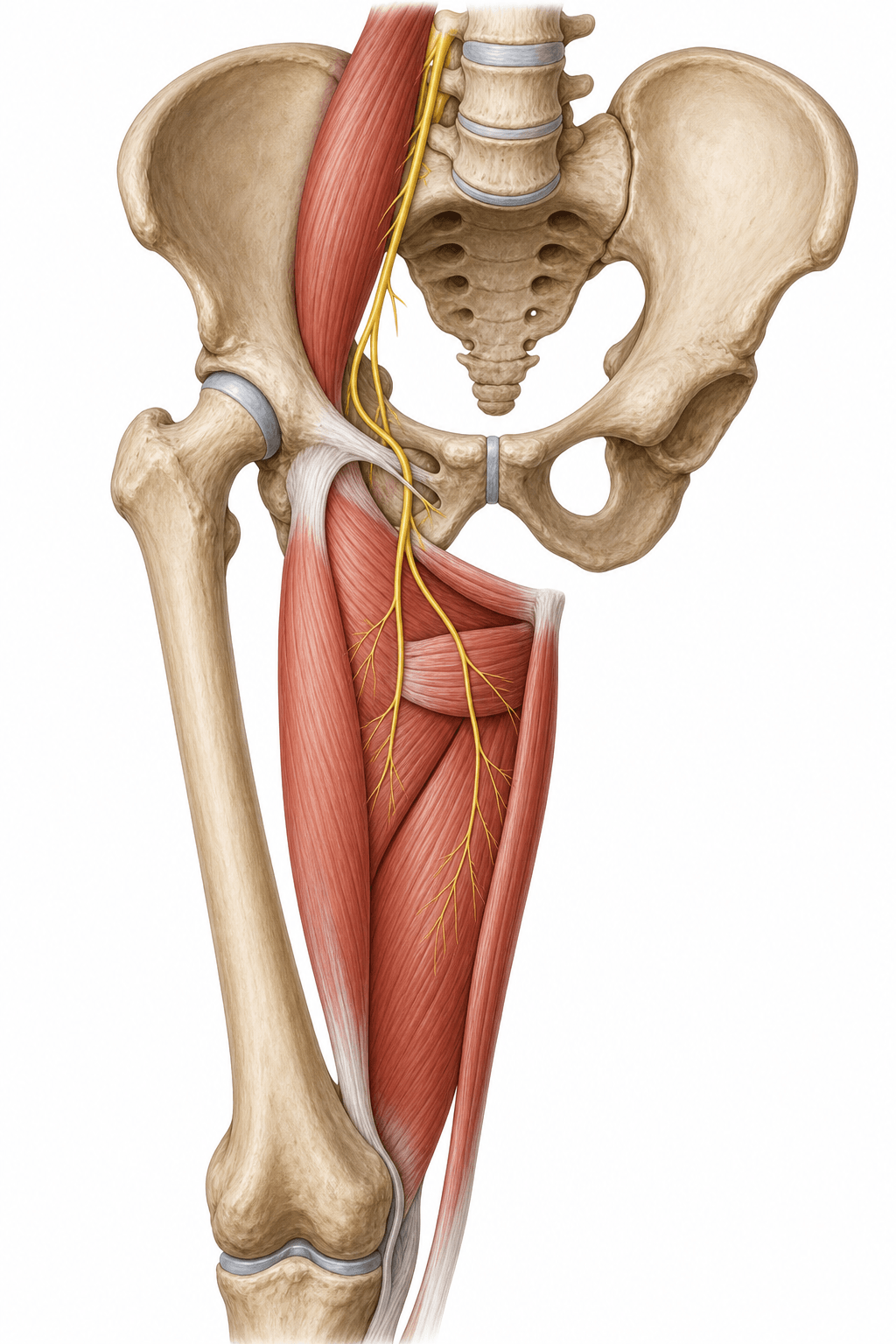
- The obturator nerve arises from the ANTERIOR divisions of the anterior rami of L2, L3 and L4 (contrast: the femoral nerve is from the POSTERIOR divisions).
- It emerges from the MEDIAL border of psoas major, runs along the lateral pelvic wall, and enters the thigh through the OBTURATOR CANAL.
- It supplies the thigh ADDUCTOR muscles (adductor longus/brevis/magnus adductor part, gracilis, obturator externus).
- It carries sensation from a small area of the MEDIAL THIGH.
- It also gives an articular branch to the hip and (via the posterior branch) the knee - the anatomical basis of referred hip-to-knee pain.
- The Howship-Romberg sign (medial thigh/knee pain on hip extension/adduction/internal rotation) suggests obturator nerve compression, classically by an obturator hernia.
- “Anterior vs posterior divisions: obturator = anterior division; femoral = posterior division (both L2-L4).
- “It supplies BOTH the hip and knee with articular branches - hence obturator pathology can refer pain to the medial knee.
- “Proximal obturator nerve is the highest-risk segment during pelvic lymph node dissection.
Medial thigh/knee pain and paraesthesia provoked by hip extension, adduction or internal rotation, relieved by hip flexion. It reflects obturator nerve compression - classically by an obturator hernia (typically a thin, elderly woman with bowel obstruction).
It is an easily-missed cause of bowel obstruction and groin/medial-knee pain. Recognising the sign points to the obturator canal and prompts the right imaging - a high-yield exam association.
Origin & Course
Origin
- The obturator nerve arises from the anterior divisions of the anterior rami of L2, L3 and L4.
- This contrasts with the femoral nerve (same roots, but posterior divisions).
- It forms within psoas major and emerges from its medial border (the femoral nerve emerges from the lateral border - a classic comparison).
Motor & Sensory Supply
The obturator nerve is the motor nerve of the thigh adductor compartment: adductor longus, adductor brevis, gracilis, obturator externus, and the adductor part of adductor magnus (the hamstring part of adductor magnus is supplied by the sciatic/tibial division).
- Action: adduction of the thigh (and gracilis assists knee flexion/medial rotation).
- Sensory: a variable patch of medial thigh skin via the cutaneous branch of the anterior division (the accessory obturator nerve, when present, is a minor variant).
- Articular: branches to both the hip and the knee, explaining why hip pathology can refer to the medial knee.
Clinical Correlations
Causes
- Pelvic surgery: pelvic lymph node dissection (e.g. during radical prostatectomy), gynaecological surgery, and acetabular/quadrilateral-plate fracture fixation - the proximal nerve is the highest-risk segment.
- Obturator hernia: compression in the obturator canal producing the Howship-Romberg sign.
- Hip surgery / arthroscopy and pelvic masses.
- Adductor spasticity is treated with obturator nerve blocks/neurectomy in cerebral palsy.
OAFPObturator vs Femoral Nerve
Hook:Obturator = Anterior divisions/Adductors; Femoral = Posterior divisions/quadriceps.
Evidence Base
Obturator Nerve Injury Risk Map in Pelvic Surgery
- Series of 3558 laparoscopic/robotic radical prostatectomies; obturator nerve injury in 0.1% during pelvic lymph node dissection
- Across the literature the PROXIMAL part of the obturator nerve accounts for ~77.8% of reported injuries
- Recognising injury intra-operatively allowed successful simultaneous repair (clip removal or microsuture)
- No long-term adductor or neurologic deficit after timely repair
Obturator-to-Femoral Nerve Transfer (Feasibility)
- Cadaveric feasibility study transferring obturator motor branches (to gracilis/adductor longus) to the motor portion of the femoral nerve
- Tension-free direct neurorrhaphy was possible in all specimens (mean overlap 26 mm; matched nerve diameters)
- Access in the thigh was straightforward compared with pelvic transfer
- Proposed as a reproducible reconstruction for proximal femoral nerve injury
Viva Scenarios
Practise clinical reasoning and management decisions out loud
“A thin, elderly woman presents with bowel obstruction and intermittent medial thigh and knee pain that worsens when she extends and internally rotates the hip. What sign is this and what is the likely cause?”
Guidelines, Registries & Global Practice
Global Practice Picture
Obturator nerve anatomy is foundational for pelvic and acetabular surgery and for regional anaesthesia. Internationally consistent teaching points are: protect the proximal nerve during pelvic lymph node dissection and quadrilateral-plate fixation; recognise the Howship-Romberg sign of obturator hernia; and use the obturator block for adductor spasticity and to prevent the adductor jerk in bladder surgery.
Side-by-Side Synthesis
- Obturator nerve
- L2-L4 anterior divisions
- Femoral nerve (for contrast)
- L2-L4 posterior divisions
- Obturator nerve
- Medial border
- Femoral nerve (for contrast)
- Lateral border
- Obturator nerve
- Obturator canal
- Femoral nerve (for contrast)
- Under inguinal ligament
- Obturator nerve
- Thigh adductors
- Femoral nerve (for contrast)
- Quadriceps
- Obturator nerve
- Medial thigh
- Femoral nerve (for contrast)
- Anterior thigh + saphenous
- Obturator nerve
- Obturator hernia (Howship-Romberg), PLND
- Femoral nerve (for contrast)
- Anterior THA retractor, iliacus haematoma
Anatomy
- L2-L4 anterior divisions
- Medial border of psoas
- Through the obturator canal
- Anterior + posterior branches
Supply
- Motor: thigh adductors + obturator externus
- Sensory: medial thigh
- Articular branches to hip AND knee
- Adductor magnus = dual (obturator + sciatic)
Clinical
- Howship-Romberg = obturator hernia
- Proximal nerve at risk in pelvic LN dissection
- Deficit: weak adduction, medial thigh numbness
- Obturator block for adductor spasticity