Occipitocervical Instability & Dissociation
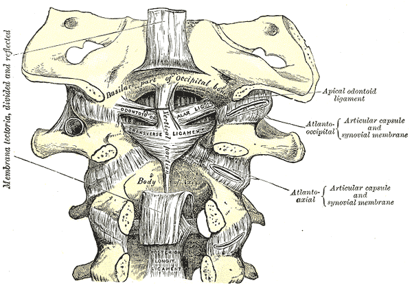
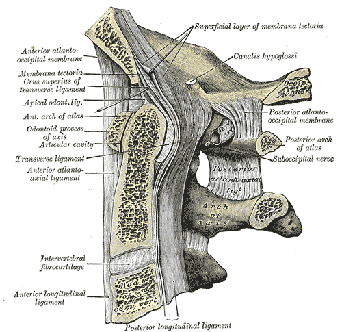
- The craniocervical junction (occiput-C1-C2) has SHALLOW bony articulations, so its stability is overwhelmingly LIGAMENTOUS: the TRANSVERSE ligament (cruciate complex) holds the odontoid against the anterior atlas, the ALAR ligaments restrain rotation and distraction between the dens and occipital condyles, and the TECTORIAL MEMBRANE and apical/atlanto-occipital ligaments span the dens/atlas to the occiput.
- TRAUMATIC atlanto-occipital dissociation (AOD) is a rare, HIGHLY UNSTABLE, HIGHLY LETHAL injury from high-energy hyperextension/distraction (e.g. motor-vehicle and pedestrian trauma); many die at the scene, and CHILDREN are predisposed by their anatomy. Survivors may have brainstem/upper-cervical-cord injury and lower cranial-nerve deficits.
- ATRAUMATIC craniocervical instability occurs in RHEUMATOID ARTHRITIS (pannus and ligament erosion causing atlanto-axial instability and cranial settling/basilar invagination), DOWN SYNDROME (ligamentous laxity, os odontoideum), skeletal dysplasias, and congenital os odontoideum - at-risk groups to screen (e.g. before anaesthesia/sport).
- DIAGNOSIS is by CT of the craniocervical junction using objective measurements - the C1-CONDYLE INTERVAL (CCI; the most sensitive for AOD), the HARRIS lines (basion-dental interval BDI and basion-axial interval BAI, normally each under ~12 mm), and the Powers ratio - plus MRI to assess the LIGAMENTS, cord and oedema.
- A critical safety point: in a DISTRACTION-type AOD, CERVICAL TRACTION is CONTRAINDICATED (it can distract the junction and worsen neurological injury) - immobilise rigidly and avoid traction.
- Definitive treatment of unstable craniocervical injury/instability is POSTERIOR OCCIPITOCERVICAL STABILISATION AND FUSION (commonly C0-C2); the construct and levels depend on the pathology (e.g. C1-C2 fusion for isolated atlanto-axial instability).
- “CCJ stability is LIGAMENTOUS - transverse (odontoid to atlas), alar (rotation/distraction), tectorial membrane; the bone alone is not enough.
- “AOD is highly lethal and easily missed - use the CCI and Harris lines (BDI/BAI <~12 mm) on the trauma CT, and DO NOT apply traction to a distraction injury.
- “Think atraumatic CCJ instability in RHEUMATOID ARTHRITIS and DOWN SYNDROME; treat unstable injuries with occipitocervical fusion (C0-C2).
A highly lethal, easily-missed distraction injury of the occiput-C1 junction from high-energy trauma; children are predisposed. Survivors risk brainstem/cord injury. Do NOT apply traction - it can worsen a distraction injury.
In rheumatoid arthritis (pannus, cranial settling) and Down syndrome (laxity, os odontoideum), instability develops insidiously - screen these groups (e.g. before anaesthesia/intubation and contact sport) for atlanto-axial/craniocervical instability and myelopathy.
Anatomy - a Ligament-Dependent Junction
The craniocervical junction (CCJ) - the occiput, atlas (C1) and axis (C2) - provides nearly half of cervical flexion-extension and rotation, yet its bony articulations are shallow and incongruent. Its stability therefore depends on ligaments:
- the transverse ligament (part of the cruciate ligament) holds the odontoid (dens) against the anterior arch of the atlas - the key restraint to anterior atlanto-axial translation;
- the alar ligaments run from the dens to the occipital condyles, restraining rotation and distraction;
- the tectorial membrane (the cephalad continuation of the posterior longitudinal ligament) and the apical and atlanto-occipital membranes/ligaments span the dens/atlas to the occiput. Disruption of these - by trauma or disease - produces craniocervical instability.
Causes
Atlanto-occipital dissociation is a rare, highly unstable ligamentous injury of the occiput-C1 junction from high-energy hyperextension/distraction - typically motor-vehicle and pedestrian trauma. It carries very high morbidity and mortality (often fatal at the scene from brainstem/upper cord injury), and children and young adults are disproportionately affected because of their relatively horizontal occipital condyles and large head. Survivors may present with cranial-nerve palsies, brainstem/cord signs or, with milder unilateral injuries, neck pain - CCJ ligamentous injury is best thought of as a spectrum, a subset of which can be managed non-operatively.
Diagnosis & Measurements
AOD is frequently missed, so look for it actively on the craniocervical CT using objective measurements:
- C1-condyle interval (CCI) - the gap between the occipital condyle and the C1 lateral mass; an increased CCI is the most sensitive sign of AOD (in adults and children).
- Harris lines - the basion-dental interval (BDI) (basion to the tip of the dens) and the basion-axial interval (BAI) (basion to the posterior axial line); each should normally be under about 12 mm.
- Powers ratio (basion-posterior arch / opisthion-anterior arch) - greater than ~1 suggests anterior dislocation (less reliable for distraction/posterior injuries). MRI assesses the ligaments, cord signal, oedema and epidural haematoma, confirming the soft-tissue injury. Always evaluate the whole cervical spine and the patient's neurology.
| 0 | 1 | 2 |
|---|---|---|
| C1-condyle interval (CCI) | Occipital condyle to C1 lateral mass gap | Most sensitive for AOD (adults & children) |
| Basion-dental interval (BDI) | Basion to tip of dens (Harris) | Normally under ~12 mm |
| Basion-axial interval (BAI) | Basion to posterior axial line (Harris) | Normally under ~12 mm |
| Powers ratio | Basion-posterior arch / opisthion-anterior arch | Greater than ~1 = anterior AOD (misses distraction/posterior) |
Management
In a distraction-type atlanto-occipital dissociation, cervical traction is contraindicated - it can further distract the already-disrupted junction and worsen neurological injury or cause death. Immobilise the head and neck rigidly (collar/sandbags; halo with great caution), avoid traction, and proceed to definitive stabilisation. This is a classic exam safety point.
Unstable craniocervical injury or instability is treated with posterior OCCIPITOCERVICAL STABILISATION AND FUSION - commonly occiput to C2 (C0-C2) - using an occipital plate and C1/C2 (and occipital) screw-rod constructs with bone graft. The levels and construct are tailored to the pathology: isolated atlanto-axial instability (e.g. transverse-ligament rupture, os odontoideum) may be treated by C1-C2 fusion (e.g. Goel-Harms/Magerl), whereas occipito-atlantal disruption requires inclusion of the occiput. In rheumatoid disease, decompression and fusion address instability/cranial settling and myelopathy. Milder, stable unilateral ligamentous injuries may be managed non-operatively with immobilisation and follow-up. Recognising the injury early, before deterioration, is the key to survival and outcome.
Evidence & Key Studies
Atlanto-occipital dissociation
- AOD is a rare, highly unstable craniocervical injury with very high morbidity and mortality, usually from high-energy hyperextension trauma; children and young adults are frequently affected.
- The diagnostic method of choice is CT assessment of the C1-condyle interval (CCI) together with cervical MRI.
- Standard treatment of stable patients with unstable AOD injuries is posterior occipitocervical stabilisation and fusion (C0-C2).
Unilateral atlanto-occipital injury: a case series and detailed radiographic description
- Craniocervical ligamentous injury behaves as a spectrum rather than a dichotomous diagnosis; all patients had a widened condyle-C1 interval (>2 mm).
- A subset of milder unilateral atlanto-occipital injuries was managed non-operatively without delayed neurologic injury or death.
- Supports careful radiographic characterisation (condyle-C1 interval) and individualised operative-versus-non-operative decisions.
According to PubMed, the lethality, CCI-based diagnosis and occipitocervical-fusion treatment of AOD come from the cited Vachata series, and the spectrum concept / condyle-C1 interval from the cited Lepard series. The stabilising-ligament anatomy, the Harris lines (BDI/BAI) and Powers ratio, and the no-traction caution are standard, well-established craniocervical teaching. (See also our Jefferson Fracture and Rheumatoid Cervical Spine material.)
Clinical Decision Scenarios
Practise clinical reasoning and management decisions out loud
“A young patient after a high-speed motor-vehicle collision has neck pain and a subtle neurological deficit. Why must you consider atlanto-occipital dissociation, what stabilises the craniocervical junction, and how would you diagnose it?”
“How would you manage a confirmed unstable atlanto-occipital dissociation, what must you avoid, and in which non-traumatic patients do you worry about craniocervical instability?”
Mnemonics & Memory Aids
CCJ
Hook:CCJ: check the ligaments, CT-measure it, and (just) no traction - fuse occiput to C2.
DROP
Hook:DROP the traction in AOD: distraction, rheumatoid/Down at risk, occipitocervical fusion, Powers/CCI/Harris.
Anatomy
- CCJ stability is LIGAMENTOUS (shallow bony joints)
- Transverse ligament (odontoid-atlas), alar (rotation/distraction), tectorial membrane
- Occiput-C1-C2 provide ~half of cervical flexion/rotation
Causes
- Traumatic: atlanto-occipital dissociation (high-energy, highly lethal, children predisposed)
- Rheumatoid arthritis: atlanto-axial instability + cranial settling/basilar invagination
- Down syndrome (laxity/os odontoideum), congenital os odontoideum, dysplasias
Diagnosis
- CT: C1-condyle interval (most sensitive), Harris BDI/BAI (<~12 mm), Powers ratio
- MRI for ligaments/cord/oedema
- AOD frequently missed - look for it actively
Management
- Distraction AOD: NO traction - rigid immobilisation
- Unstable: posterior occipitocervical fusion (C0-C2); C1-C2 for isolated atlanto-axial
- Milder stable unilateral injuries: non-operative with follow-up; screen RA/Down before anaesthesia