Rigid stabilisation of the craniocervical junction — occiput to C2 or beyond
- Indications: rheumatoid cranial settling/basilar invagination, atlanto-occipital dislocation, os odontoideum with irreducible compression, condyle destruction by tumour, occipitalised atlas with instability, failed C1-C2 fusion, Down syndrome instability
- Occipital bone is thickest in the midline keel and at the external occipital protuberance; it thins rapidly laterally — midline bicortical screws give the strongest purchase
- Venous sinus anatomy (torcular Herophili at the EOP, transverse sinuses running laterally) dictates safe screw zones below and lateral to these landmarks
- Cervical anchors follow the Goel-Harms philosophy: C1 lateral mass and C2 pedicle screws; C2 pars or translaminar screws are bailouts for a high-riding vertebral artery
- The single most examinable technical step: set the occipito-cervical angle in neutral (match the preoperative standing lateral) BEFORE final tightening — over-flexion causes postoperative dysphagia and airway compromise. Know the number: in Miyata's series EVERY patient whose O-C2 angle fell more than 10 degrees below the preoperative value developed dyspnoea or dysphagia, and NO patient whose angle increased did. Aim for a non-negative change, not an absolute angle.
- Vertebral artery injury protocol: complete the screw for tamponade, do NOT instrument the contralateral side, obtain postoperative angiography
- “Awake fibreoptic intubation plus pre-positioning neuromonitoring baseline for unstable craniocervical junctions
- “Basilar invagination is usually decompressed indirectly by reduction (traction or Goel joint distraction) rather than transoral odontoidectomy
- “Fusion in slight O-C2 flexion or excessive extension both cause problems: flexion causes dysphagia; marked extension causes downgaze difficulty and subaxial kyphosis compensation
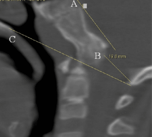
Fusing the O-C2 segment in flexion narrows the oropharyngeal airway space and causes dysphagia and dyspnoea — a signature complication. Measure the O-C2 angle (McGregor line to inferior endplate of C2) intraoperatively against the preoperative neutral standing lateral before final tightening.
The torcular Herophili lies deep to the external occipital protuberance; transverse sinuses run laterally along the superior nuchal line. Bicortical midline screws must be planned on CT. If a sinus is breached: leave or place the screw to tamponade, never remove and re-drill; manage CSF leak with bone wax, screw insertion, and lumbar drainage if persistent.
Preoperative CT angiography for a high-riding VA (narrow C2 isthmus) and ponticulus posticus at C1. If the VA is injured during C2 screw placement: complete that screw (tamponade), abandon contralateral instrumentation, obtain postoperative angiography.
Rheumatoid cranial settling and atlanto-occipital dislocation are cord-at-risk states. Awake fibreoptic intubation, neuromonitoring baselines before and after positioning, Mayfield fixation with fluoroscopic confirmation of alignment before draping.
Indications and Goals
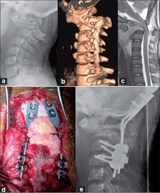
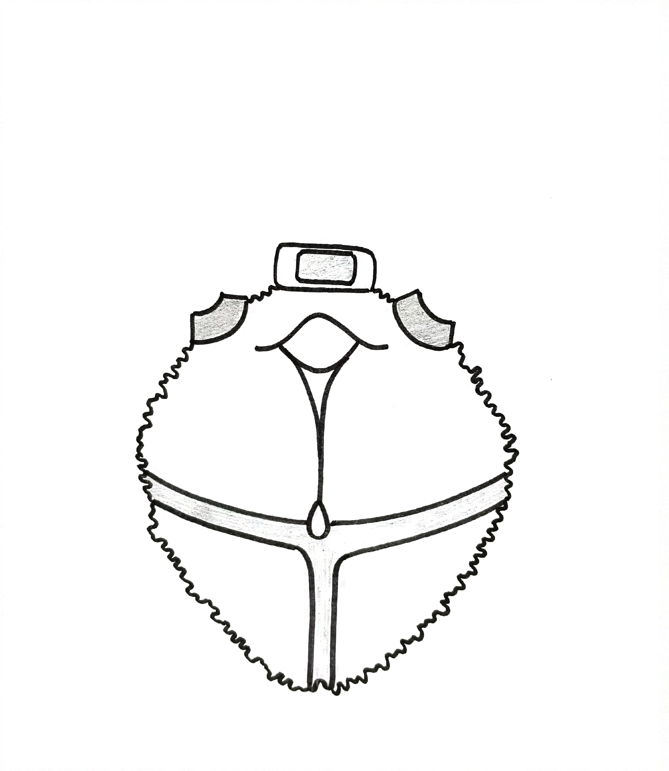
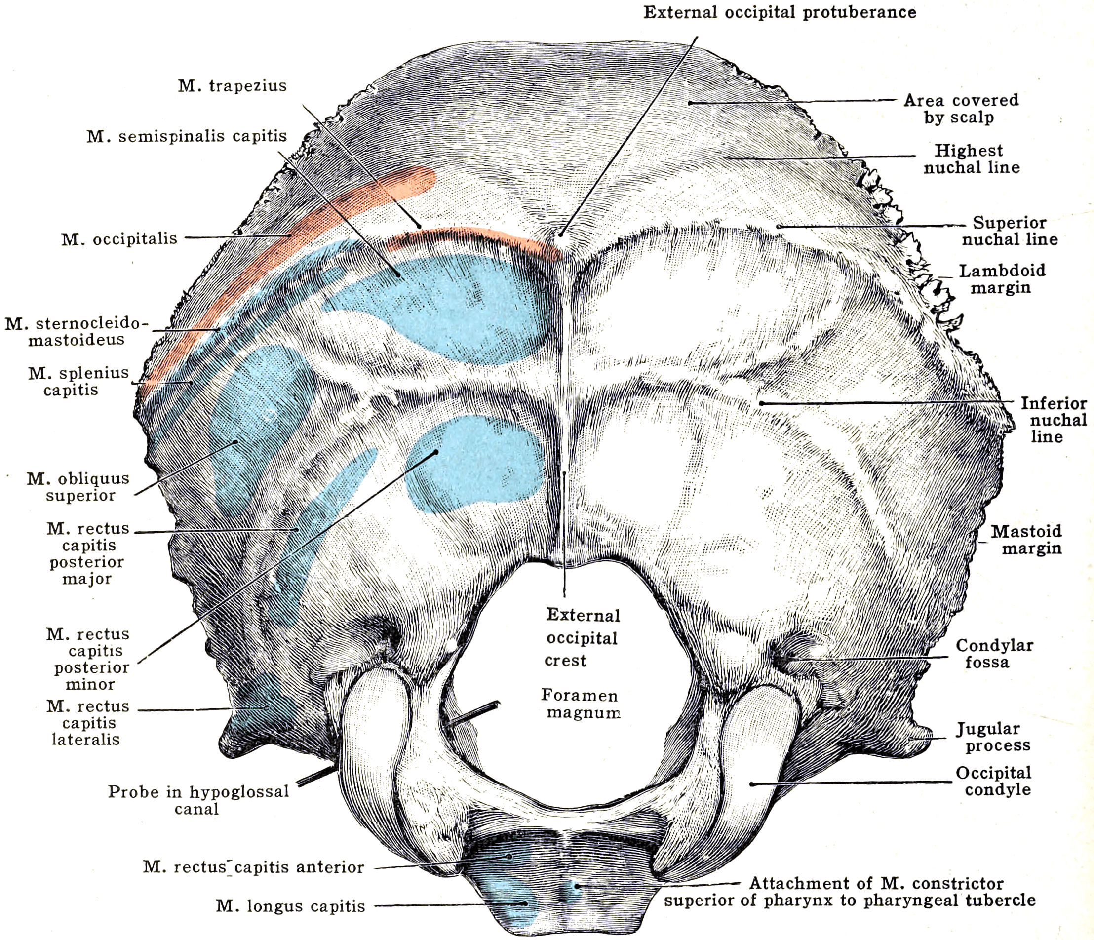
Occipitocervical fusion is indicated when the occipito-atlantal joint is unstable, destroyed, or must be crossed to obtain fixation.
- Examples
- Rheumatoid basilar invagination / cranial settling, atlanto-occipital erosion
- Notes
- Commonest historical indication; declining with biologic therapy
- Examples
- Atlanto-occipital dislocation, occipital condyle fracture with craniocervical instability (Anderson-Montesano type III / unstable Tuli)
- Notes
- AOD survivors need urgent O-C fusion; halo alone inadequate
- Examples
- Os odontoideum with irreducible compression, occipital condyle hypoplasia, atlas assimilation (occipitalisation) with instability, Klippel-Feil variants
- Notes
- Assimilated C1 removes the option of C1 lateral mass fixation
- Examples
- Down syndrome atlantoaxial and occipitoatlantal instability, skeletal dysplasias, Morquio
- Notes
- Small anatomy, poor bone, higher complication rates
- Examples
- Tumour destruction of occipital condyles or C1-C2 (metastasis, chordoma, myeloma)
- Notes
- Often combined with tumour resection; may need cement augmentation
- Examples
- Failed atlantoaxial fusion, iatrogenic condyle resection (far lateral approaches greater than 50 percent condyle removed)
- Notes
- Extend construct cranially for salvage
Preoperative Planning
- Occipital bone thickness mapping: thickest at the external occipital protuberance and midline nuchal keel — up to 12 to 15 mm — thinning rapidly laterally to 3 to 6 mm. Plan screw lengths and entry points on sagittal and axial CT.
- C1 anatomy: lateral mass height and width, ponticulus posticus (arcuate foramen) over the VA groove — present in roughly 15 percent, contraindicating a broad C1 entry over the arch.
- C2 anatomy: pedicle/isthmus dimensions. Mandel's cadaveric study of 205 C2 vertebrae is where the number comes from, and the criterion is less than 5 mm in EITHER height OR width — not 5 mm for one and 4 mm for the other — because that is the point at which a 3.5 mm screw becomes technically difficult. A high-riding vertebral artery on that side is a reason to abandon a transarticular or full-length pars screw and drop back to a deliberately short pars screw or a translaminar screw, which avoids the artery altogether. Know the frequency, because it is not rare: isthmus height was under 5 mm in 11.7 per cent of specimens and width under 5 mm in 2.4 per cent, and on CT Paramore found 18 per cent of 94 patients had a high-riding transverse foramen prohibiting transarticular screws on at least one side, with a further 5 per cent feasible-but-risky — so roughly one patient in five is unsuitable on at least one side. Two anatomical asymmetries worth carrying: the right isthmus is generally the smaller, and low isthmus height was three times commoner in women (18 of 24 specimens).
- Why you will see three different prevalences for "high-riding vertebral artery" — they are three different definitions, not a disagreement. Quoting a figure without its criterion is how this gets muddled. Mandel measures the bony isthmus in height and width with a single 5 mm floor and finds roughly 10 per cent at risk. Paramore reconstructs the CT along the intended screw path and judges suitability, giving 18 to 23 per cent unsuitable on at least one side. The widely used Neo criteria — isthmus thickness under 5 mm or internal height under 2 mm — are the most inclusive and identify about one patient in three (see
/topics/vertebral-artery-injuryfor that series). Higher numbers reflect a broader definition, not a more dangerous population. Pick one definition, state it when you quote a percentage, and measure it yourself on the sagittal reconstruction. - CT angiography where VA anomaly suspected: dominant VA, anomalous medial loop, fenestration.
Evolution of Constructs
Corticocancellous onlay from occiput to laminae with wire fixation. No rigid control — required prolonged halo immobilisation; pseudarthrosis rates high; no ability to hold reduction.
Contoured rods wired sublaminarly and through occipital burr holes. Better than onlay but still semi-rigid; sublaminar wire passage risks in a stenotic canal; halo often still used.
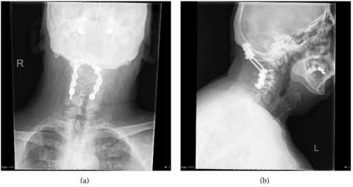
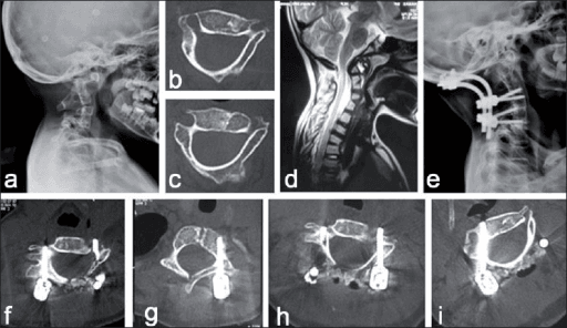
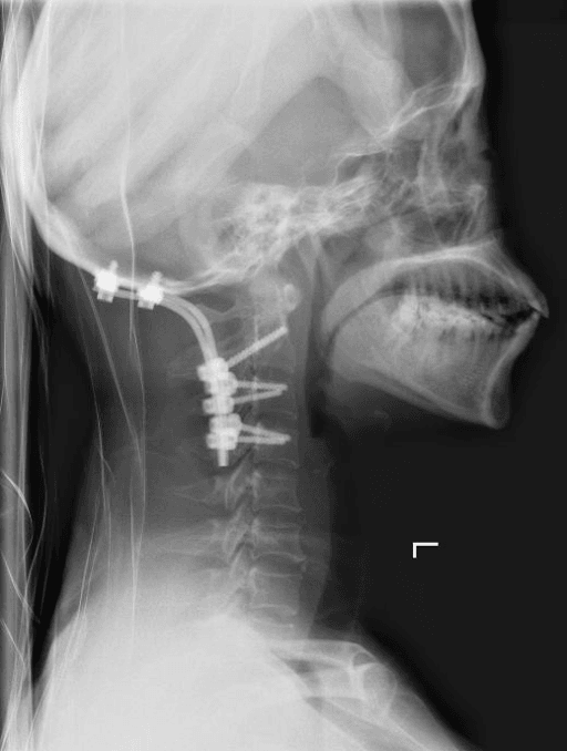
Midline keel plates with bicortical screws linked by rods to C1 lateral mass and C2 pedicle screws (Goel-Harms concept extended cranially). Rigid, allows reduction manoeuvres against the construct, halo unnecessary, fusion rates above 95 percent.
Fixation Options
- Entry point
- Junction of posterior arch and lateral mass, with the C2 root retracted and protected
- Trajectory
- Medially angled 10 to 15 degrees, cephalad toward the anterior tubercle
- Contraindication / risk
- Venous plexus bleeding; C2 root irritation causing occipital neuralgia
- Bailout role
- Not available if the atlas is assimilated — skip to C2 and occiput
- Entry point
- Lateral pars, palpating the medial pedicle wall
- Trajectory
- 20 to 30 degrees medial and cephalad
- Contraindication / risk
- Contraindicated with a high-riding vertebral artery on that side
- Bailout role
- Workhorse and strongest anchor — the reference against which bailouts are judged
- Entry point
- Same starting zone as the pedicle screw
- Trajectory
- Same trajectory family but shorter and steeper, stopping short of the transverse foramen
- Contraindication / risk
- Shorter purchase; still hazardous if the artery is very dominant
- Bailout role
- First bailout for a high-riding vertebral artery
- Entry point
- Base of the contralateral spinous process
- Trajectory
- Crossed screws down the contralateral lamina
- Contraindication / risk
- Dorsal canal breach; weaker pull-out; lamina must be intact and grafting surface is compromised
- Bailout role
- Vertebral-artery-free bailout when both pedicle and pars are unsafe
- Entry point
- Lateral mass of C3 to C5
- Trajectory
- Magerl or An trajectories
- Contraindication / risk
- Added levels increase stiffness, adjacent segment load and dysphagia risk
- Bailout role
- Extends the construct when C1/C2 bone is poor — rheumatoid osteopenia, tumour, failed C2 fixation
Stop at C2 when anchors are good — every subaxial level added increases stiffness, adjacent segment load, and dysphagia risk without fusion benefit. Extend to C3-C5 lateral masses for rheumatoid osteopenia, tumour, or failed C2 fixation.
Operative Technique (PIPADRAW)
PIPADRAW: occipitocervical fusion operative sequence
Position: prone, Mayfield pins, reverse Trendelenburg (reduces venous bleeding), head in neutral or the planned reduced position; confirm alignment on lateral fluoroscopy before draping. Eyes free, abdomen free.
Imaging/equipment: fluoroscopy (biplanar ideal) or navigation; neuromonitoring running; occipital plate system with polyaxial cervical screws; high-speed burr; cell salvage available.
Preparation: midline mark inion to lower cervical spine; antibiotics; tranexamic acid per protocol.
Approach: midline posterior incision from the EOP to below the caudal instrumented level; strictly midline avascular raphe (ligamentum nuchae) dissection; subperiosteal exposure of the occiput below the superior nuchal line, C1 posterior arch (no more than 12 to 15 mm lateral to midline on the cranial surface — VA groove), and C2 lamina/pars.
Dissection at-risk zones: VA in the C1 sulcus arteriosus and lateral to C2 pars; C2 venous plexus around the C1-C2 joint; greater occipital nerve.
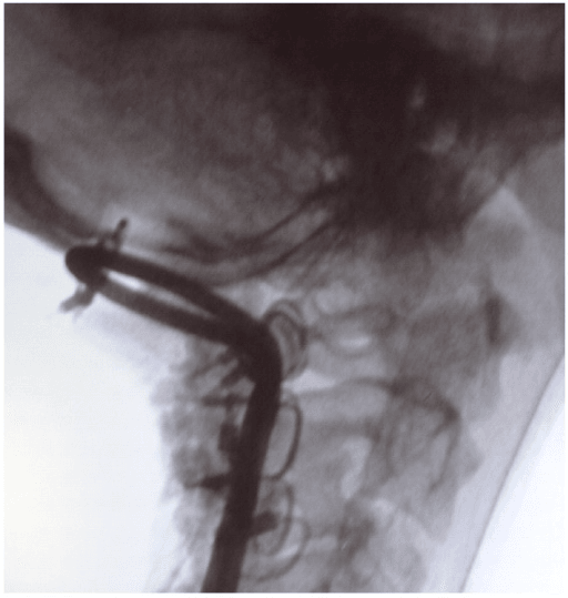
Reduction: for reducible basilar invagination — preoperative or intraoperative skull traction, or Goel-type C1-C2 joint distraction with intra-articular spacers/cages, translating the odontoid caudally and ventrally away from the brainstem; hold the reduction with the rods.
Alignment — the critical step: before final tightening, obtain a true lateral and confirm the O-C2 angle and posterior occipitocervical angle match the preoperative neutral standing film; confirm no fixed rotation or coronal tilt (check that the mandible, EOP, and spinous processes align).
The number to hold in your head while you do it. Miyata measured the change in O-C2 angle (postoperative minus preoperative) against the cross-sectional area of the oropharynx in 29 fusions. The relationship is linear, and the ends of it are unambiguous: every patient whose angle fell more than 10 degrees lost more than 40 per cent of oropharyngeal area and developed dyspnoea, dysphagia or both — and no patient whose angle increased developed either. So the target is a non-negative change from that patient's own preoperative film, not any particular absolute angle. Note also that all five affected patients in that series had short constructs: a short fusion is no protection, because the angle is set at the occiput regardless of how far down the construct runs.
Fixation: occipital plate with bicortical midline keel screws; C1 lateral mass and C2 pedicle (or bailout) screws; contour and seat rods; compression/distraction as needed; final tighten.
Grafting: decorticate occiput and C1/C2 laminae with a burr; structural corticocancellous iliac crest or rib strut onlay from occiput to laminae, supplemented with local autograft/cancellous chips; secure graft under the construct or with cables.
Closure: meticulous layered closure over prominent hardware — approximate deep muscle to the nuchal fascia, avoid dead space; drain per preference; skin without tension (fragile rheumatoid skin — consider nylon sutures).
Aftercare: rigid collar 6 to 12 weeks per construct security; upright lateral radiograph before discharge; formal swallowing assessment before oral intake if any dysphagia; CT at 6 to 12 months if fusion in doubt.
- Is the O-C2 angle at or slightly more extended than the preoperative neutral? Any flexion risks dysphagia and airway compromise. 2. Is there any rotation or tilt? A fixed rotated head is a devastating, avoidable outcome — check midline landmarks on AP fluoroscopy and clinically under the drapes.
Complications: Prevention and Management
- Where
- C2 pars/pedicle screw trajectory; lateral C1 arch exposure
- Mechanism
- Drilling or screw breach of a high-riding, medialised or dominant vessel
- Avoid by
- Preoperative CT angiography; avoid C2 pedicle screw with high-riding VA (use translaminar or short pars); keep C1 arch dissection within 1.5 cm of midline subperiosteally
- If injured
- Complete the screw to tamponade; do NOT instrument the contralateral side; postoperative angiography; endovascular management of pseudoaneurysm or arteriovenous fistula
- Where
- Occipital squama, near and above the external occipital protuberance
- Mechanism
- Bicortical drilling or overlong screws breaching inner table into the sinus
- Avoid by
- Place screws at least 2 cm below the EOP in the thick midline keel; incremental drilling with a depth stop; bicortical purchase only where thickness is mapped on CT
- If injured
- Insert the screw to tamponade; bone wax; head-up positioning; avoid air embolism; lumbar drain for persistent CSF leak
- Where
- Foramen magnum, C1-C2 canal, occipital inner table
- Mechanism
- Sublaminar passage, subperiosteal dissection of the posterior arch, or malpositioned screw; also traction or malreduction
- Avoid by
- Neuromonitoring; avoid sublaminar instrumentation at the cervicomedullary junction; fluoroscopic or navigated screw placement; reduce and hold before final tightening
- If injured
- Stop, decompress and reposition hardware; repair or patch dura with sealant; maintain mean arterial pressure; postoperative imaging and steroid decision case by case
- Where
- Anterior to occipital condyle, above the C0-C1 joint
- Mechanism
- Overlong or anteriorly directed condylar or occipital screw
- Avoid by
- Limit condyle screw length on preoperative CT; keep trajectory posteromedial; use midline keel occipital fixation where possible
- If injured
- Recognise tongue deviation early; back out or shorten the screw; speech and swallow assessment
- Where
- O-C2 (McGregor to C2) angle at final tightening
- Mechanism
- Fusion in flexion narrows the oropharyngeal airway and inlet — dysphagia and dyspnoea
- Avoid by
- Set the O-C2 angle to the preoperative neutral value before locking; check lateral fluoroscopy with the plate provisionally seated; never fuse in kyphosis
- If injured
- Swallowing team, modified diet, nasogastric feeding; revision of alignment if severe and structural
- Where
- Occiput-to-C2 graft bed and screw-bone interfaces
- Mechanism
- Inadequate decortication, poor graft, rigid-construct failure, smoking or metabolic bone disease
- Avoid by
- Rigid segmental fixation, thorough decortication, structural autograft, optimise vitamin D and nutrition, smoking cessation, halt biologics perioperatively in rheumatoid disease
- If injured
- Revision with augmented fixation, fresh autograft, extend the construct
- Where
- Midline occipitocervical closure; subaxial spine below the construct
- Mechanism
- Thin irradiated or rheumatoid skin over prominent hardware; kyphotic or overlong fusion loading the subaxial spine
- Avoid by
- Tension-free layered closure, low-profile implants, shortest construct that achieves stability, do not fuse in kyphosis
- If injured
- Early debridement with implant retention if fusion incomplete, plastics input for flap cover; surveillance and extension of fusion for symptomatic subaxial instability or myelopathy
- Where
- O-C1 and C1-C2 segments
- Mechanism
- Obligatory sacrifice of nodding and rotation with construct inclusion
- Avoid by
- Counsel that roughly 50 percent of head rotation is lost when C1-C2 is included, plus loss of O-C1 nodding
- If injured
- Preoperative counselling on driving, blind spots and occupational implications; no operative rescue
- Prevention
- CT angiography, avoid pedicle screw with high-riding VA, limit lateral C1 arch exposure
- Management
- Complete the screw to tamponade; do NOT instrument the contralateral side; postoperative angiography; endovascular management of pseudoaneurysm/fistula
- Prevention
- Screws at least 2 cm below EOP, incremental drilling with stop, bicortical only where mapped safe
- Management
- Insert screw (tamponade), bone wax, head-up; lumbar drain for persistent CSF leak
- Prevention
- Set O-C2 angle to preoperative neutral before final tightening; avoid flexed fusion
- Management
- Swallowing team, modified diet, nasogastric feeding; revision of alignment if severe and structural
- Prevention
- Rigid fixation, thorough decortication, structural autograft, optimise metabolic factors, smoking cessation
- Management
- Revision with augmented fixation, fresh autograft, extend construct
- Prevention
- Do not fuse in kyphosis; shortest construct that achieves stability
- Management
- Surveillance; extend fusion for symptomatic instability/myelopathy
- Prevention
- Tension-free layered closure, biologic cessation in rheumatoid disease, caution in irradiated tissue
- Management
- Early debridement, retain implants if fusion incomplete, plastics input for flap cover
- Prevention
- Counsel: roughly 50 percent of head rotation lost with C1-C2 inclusion plus loss of O-C1 nodding
- Management
- Preoperative counselling — driving, occupational implications
Guidelines, Registries & Global Practice
- Global epidemiology of indications varies markedly: rheumatoid craniocervical disease has declined in high-resource settings with biologic therapy but remains a major indication where access to disease-modifying treatment is limited; congenital craniovertebral junction anomalies (atlas assimilation, os odontoideum, basilar invagination) form a large proportion of practice in South Asian centres, where the Goel joint-distraction philosophy was developed; trauma (atlanto-occipital dislocation) dominates in high-energy road trauma populations.
- Society guidance: AO Spine craniovertebral junction knowledge forum publications and AANS/CNS cervical spine guidelines address atlanto-occipital dislocation (early rigid internal fixation, avoid traction) and odontoid pathology; there is no single dedicated O-C fusion guideline — practice is technique- and evidence-driven.
- Registry evidence: craniocervical procedures are low-volume and not captured by arthroplasty registries; evidence rests on institutional series and multicentre spine study groups, which consistently report fusion rates above 95 percent with modern screw-rod constructs.
- Resource-setting variation: where navigation and CT angiography are unavailable, surgeons rely more on translaminar C2 fixation and generous fluoroscopy; halo immobilisation with wiring constructs remains a legitimate option where modern implants are unaffordable, accepting longer immobilisation and lower union rates.
Controversies & Areas of Uncertainty
- Occipital plate versus condyle screws: condyle screws lower profile and preserve squamous occiput but carry hypoglossal canal and VA risk; keel plates remain standard.
- Posterior-only reduction versus anterior decompression for basilar invagination: the Goel distraction school treats most cases posteriorly; some centres still favour endonasal/transoral odontoidectomy for truly irreducible ventral compression — the threshold for "irreducible" is contested.
- Caudal extent of fusion: stop at C2 versus routine extension to C3/C4 in poor bone — longer constructs are stiffer but increase adjacent segment load and dysphagia.
- Allograft, BMP, and graft strategy: structural autograft remains gold standard; off-label BMP use at the craniocervical junction is debated because of swelling risk near the airway.
- Prophylactic O-C fusion in asymptomatic Down syndrome instability: most advocate observation with activity modification unless neurological signs or marked instability — thresholds vary between guidelines.
Memory Aids
CAPVertebral Artery Injury Response
Hook:CAP the bleeding artery: Complete, Abort the other side, Picture it after.
KEELOccipital Screw Safety
Hook:Screw the KEEL, respect the sinuses.
MCQ Practice Points
A: The midline nuchal keel at and just below the external occipital protuberance — up to 12 to 15 mm — thinning rapidly laterally. Bicortical midline screws provide the strongest fixation.
A: Fusing with a reduced (flexed) O-C2 angle, which narrows the oropharyngeal airway space — causes dysphagia and dyspnoea. Match the preoperative neutral standing lateral before final tightening.
A: Complete the screw to tamponade, do not instrument the contralateral side, and obtain postoperative angiography.
A: Approximately 50 percent of head rotation (C1-C2 provides about half of cervical axial rotation), plus loss of O-C1 flexion-extension — an essential preoperative counselling point.
A: A high-riding vertebral artery — narrow C2 isthmus (height less than 5 mm or internal width less than 4 mm). Use a short pars screw or translaminar screw instead on that side.
A: The torcular Herophili (confluence of sinuses), with the transverse sinuses running laterally along the superior nuchal line — occipital screws are placed caudal to these landmarks.
Exam Viva Scenarios
Practise clinical reasoning and management decisions out loud
“A 62-year-old woman with long-standing rheumatoid arthritis presents with progressive gait disturbance and hand clumsiness. MRI shows basilar invagination with the odontoid indenting the medulla and cord signal change. Outline your management.”
“During right C2 pedicle screw insertion for an occipitocervical construct you encounter torrential arterial bleeding from the screw track. What do you do now and for the rest of the operation?”
“Three days after an O-C3 fusion, a patient cannot manage saliva and reports breathlessness when supine. Lateral radiograph shows the head fused in a flexed position relative to the preoperative films. Explain the mechanism and your management.”
“A 24-year-old polytrauma patient survives a high-speed collision. CT shows an increased condyle-C1 interval bilaterally consistent with atlanto-occipital dislocation; he is intubated with intact motor responses. How do you manage the craniocervical junction?”
Indications
- Rheumatoid basilar invagination / cranial settling
- Atlanto-occipital dislocation (trauma) — no traction, early fusion
- Congenital: os odontoideum with irreducible compression, atlas assimilation, condyle hypoplasia
- Tumour destruction of condyles or C1-C2; failed atlantoaxial fusion; Down syndrome instability
Planning
- CT: occipital keel thickness (12 to 15 mm midline maximum), C2 isthmus for high-riding VA, ponticulus posticus
- MRI: compression, cord signal, pannus; dynamic films for reducibility
- Preoperative neutral standing lateral defines the target O-C2 angle
- Awake fibreoptic intubation; neuromonitoring before and after positioning
Technique
- Midline keel plate, bicortical screws below the superior nuchal line — torcular/transverse sinuses at risk
- C1 lateral mass plus C2 pedicle (Goel-Harms); pars or translaminar bailout; subaxial extension for poor bone
- Goel C1-C2 joint distraction reduces basilar invagination posteriorly
- Set O-C2 angle to preoperative neutral and exclude rotation/tilt BEFORE final tightening
- Decorticate; structural iliac crest/rib onlay occiput to laminae plus local autograft
Complications
- VA injury: complete the screw, no contralateral instrumentation, postoperative angiography
- Sinus breach/CSF leak: screw tamponade, bone wax, lumbar drain if persistent
- Flexed fusion causes dysphagia and dyspnoea — the signature exam complication
- Pseudarthrosis under 5 percent with modern constructs; wound breakdown in rheumatoid/irradiated tissue
- Counsel: roughly 50 percent rotation lost with C1-C2 inclusion; fusion rates above 95 percent
Evidence Base
O-C2 Angle as a Predictor of Dyspnea and/or Dysphagia After Occipitocervical Fusion
- 29 consecutive occipitocervical or occipitocervicothoracic fusions, with O-C2 angle on plain radiographs and axial CT cross-sectional area of the oropharynx just cranial to the epiglottis measured before and after surgery
- The CHANGE in angle is what matters: dOC2A = postoperative minus preoperative O-C2 angle, and it correlated LINEARLY with the percentage change in oropharyngeal cross-sectional area
- EVERY patient with dOC2A below MINUS 10 DEGREES had an oropharyngeal area reduction worse than minus 40 per cent, and EVERY one of them developed dyspnoea and/or dysphagia
- NO patient with a POSITIVE dOC2A developed either complication
- 5 of 29 were affected (4 dysphagia, 1 both) - and all had undergone SHORT occipitocervical fusions, so a short construct is no protection
- The angle is measurable intraoperatively, which is the point: it is an index you can act on before final tightening