The most rigid external cervical immobilisation — a shrinking but still essential tool
- Anterior pins go 1 cm above the lateral third of the supraorbital rim, below the skull equator, anterior to the temporalis — medial placement risks the supraorbital and supratrochlear nerves and the frontal sinus; lateral placement risks the thin squamous temporal bone and painful temporalis penetration.
- The patient closes their eyes while anterior pins are tightened — tightening on open eyes anchors forehead skin and causes lagophthalmos (inability to close the lids).
- Adult torque is 8 inch-pounds, tightening diagonally opposed pairs simultaneously; retorque ONCE at 24 to 48 hours if loose, then never routinely again.
- Halo in the frail elderly carries a well-documented respiratory, aspiration and mortality burden - Tashjian's 78 patients over 65 with an odontoid fracture had 42 per cent in-hospital mortality in a halo versus 20 per cent without, and 66 per cent versus 36 per cent major complications, with no difference in injury severity or baseline medical condition. Modern practice favours a rigid collar or primary surgical fixation instead.
- Imaging is mandatory after application and after ANY adjustment of the halo or vest, because reduction can be lost silently.
- Snaking - paradoxical intersegmental motion between two fixed ends - persists despite apparently rigid external fixation, and it is NOT confined to the subaxial spine: Lind's prospective series found the halo restricted motion most below C2 and LEAST above C2. The halo suits odontoid, C1 ring and hangman fractures because those fractures unite in an orthosis, not because it is mechanically better at those levels.
- “Ring is sized to leave 1 to 2 cm clearance all round the skull; low-profile open-back rings ease MRI-compatible and supine care.
- “Posterior pins sit diagonally opposite the anterior pins, roughly at the 4 and 8 o'clock positions, posterolateral above the ear.
- “Suspected inner table penetration: stop, remove the pin, insert a fresh pin at an adjacent site, nurse head-up, image, and watch for CSF leak, meningitis and seizure.
- “Type II odontoid nonunion risk rises with age, displacement greater than 5 mm, angulation, posterior displacement and delay in treatment — factors that push toward surgery over halo.
- “Weaning: interval imaging in the halo, then remove and obtain upright and flexion-extension views out of orthosis to confirm healing before discharging immobilisation.
Multiple series link halo use in patients over about 75 years with pneumonia, aspiration, dysphagia and excess mortality. For geriatric type II odontoid fractures the modern choice is a rigid collar or surgical stabilisation — not a halo.
Too medial — supraorbital/supratrochlear nerve injury, frontal sinus breach. Too lateral — squamous temporal bone (thin, penetration risk) and temporalis (pain on chewing, loosening). Too high — above the skull equator the ring migrates upward.
Anterior pins tightened with eyes closed to avoid lagophthalmos. Tighten diagonally opposed pairs simultaneously to keep the ring centred and the head neutral.
Reduction can be lost when the vest is loosened for skin care, when a pin is exchanged, or as swelling settles. Radiographs after application and after any adjustment are non-negotiable.
Biomechanics — What a Halo Actually Does
- Motion control
- Minimal — proprioceptive reminder only
- Anchorage
- Soft tissues of neck; no bony purchase
- Key weakness
- Does not restrict any plane meaningfully
- Typical indication
- Comfort in muscular strain or whiplash; never for instability
- Motion control
- Partial — limits flexion and extension more than rotation or lateral bend
- Anchorage
- Chin, occiput and upper sternum
- Key weakness
- Skin pressure at chin and occiput; poor rotational control
- Typical indication
- Stable fractures, post-operative support, clearance protocols
- Motion control
- Better subaxial and rotational control than a collar
- Anchorage
- Chin and occiput plus thoracic extension
- Key weakness
- Still no skeletal fixation; upper cervical control remains limited
- Typical indication
- Mid and lower cervical stable injuries needing more than a collar
- Motion control
- The most rigid non-operative option — but the quoted 'roughly 75 percent of gross motion' comes from 7 patients (Koch and Nickel 1978), and Lind's 31-patient prospective series found 70 percent of normal motion still available
- Anchorage
- Skeletal: four skull pins into the calvarium, uprights to a thoracic vest, load to trunk
- Key weakness
- Snaking (segmental flexion at one level with compensatory extension at another) persists despite ring rigidity, and Lind found restriction LEAST above C2; performance falls further with a loose vest in the obese, cachectic or barrel-chested patient
- Typical indication
- Occiput to C2 injuries: odontoid, C1 ring, hangman variants — chosen because those fractures unite in an orthosis, not because the halo is more rigid there
The halo is a three-point-plus skeletal construct: four skull pins fix the ring to the calvarium, uprights connect ring to a thoracic vest, and the vest transmits load to the trunk. Motion control is therefore only as good as the vest-to-torso interface.
- Practical implication
- The most rigid non-operative option, but not internal fixation — and the figure comes from seven patients compared supine versus upright (Koch and Nickel 1978)
- Practical implication
- Lind 1988 found 70 percent of normal motion still available across 31 patients sampling ordinary activities; treat 75 percent control as a best case, not an expectation
- Practical implication
- Lind found restriction greatest below C2 and least above C2. The halo suits odontoid, C1 ring and hangman fractures because those fractures unite in an orthosis — not because the halo is mechanically better up there
- Practical implication
- Segmental flexion at one level with compensatory extension at another occurs between any two fixed ends; it is the reason an unstable subaxial injury needs internal fixation
- Practical implication
- A loose vest in an obese, cachectic or barrel-chested patient converts a halo into an expensive collar
- Practical implication
- Failure mode is loosening at the bone-pin interface and vest slippage
The construct fixes the two ends — skull and thorax — and leaves the intervening motion segments free to translate against each other. That is the mechanism of snaking, and it applies to the whole column between the pins and the vest, including the occipitocervical junction: Lind's measurements found least restriction above C2, not most.
So do not defend the halo on rigidity. Defend it on healing. An odontoid, C1 ring or hangman fracture unites in external immobilisation; an unstable subaxial flexion-distraction or facet injury does not unite in anything short of internal fixation, however well the neck is splinted. The fracture pattern chooses the treatment, not the orthosis's motion figures.
Indications and Contraindications
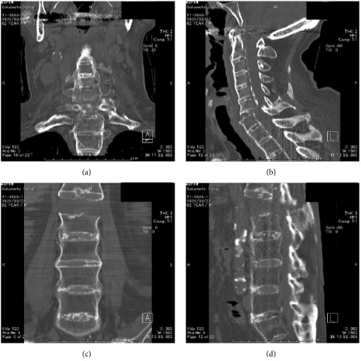
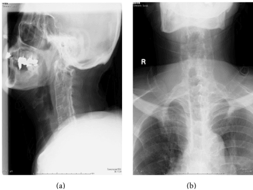
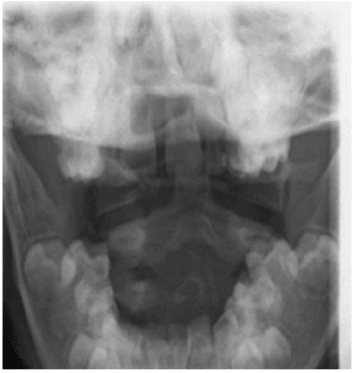
- 1Step 1 — Localise the injury
Halo controls the occiput to C2 complex; it does not reliably control subaxial (C3 to C7) segments, where vest-to-skull motion produces snaking.
Subaxial unstable injury: exclude halo and proceed to internal fixation.
- 2Step 2 — Screen physiology before pattern
Age over roughly 75 years, frailty, restricted chest excursion, severe COPD, pectus or rib deformity, unstable thoracic or sternal injury, cognitive impairment, severe scalp disease, obesity or cachexia preventing vest fit.
Positive screen: collar or primary surgical stabilisation, not a halo.
- 3Step 3 — Check pin-site feasibility
Skull fracture at intended sites, craniotomy flap, shunt hardware, thin or pathological bone.
No safe cranial anchorage: collar or surgery.
- 4Step 4 — C1 ring (Jefferson) fracture
Assess the transverse atlantal ligament by MRI (Dickman type) and lateral mass overhang on open-mouth view, total overhang less than 7 mm by the corrected Spence rule suggesting an intact ligament.
Ligament competence decides orthosis versus C1 to C2 fusion.
- 5Step 5 — Odontoid fracture
Type III routinely unites in external immobilisation. Type II nonunion risk rises with older age, displacement greater than 5 mm, angulation greater than 10 degrees, posterior displacement, comminution and delayed presentation.
Geriatric type II is the commonest place the halo is now wrongly chosen.
- 6Step 6 — Hangman (traumatic spondylolisthesis of C2)
Levine-Edwards classification governs treatment.
Type IIa and type III require surgical stabilisation.
- 7Step 7 — Paediatric and adjunct roles
Children tolerate the halo well, heal quickly and may avoid fusing a growing spine. Halo also supports revision occipitocervical fusion, poor bone quality, tumour resection or infection.
These remain the most durable modern indications.
- 8Step 8 — Default when halo fails the screen
The alternatives are a rigid collar for stable or low-demand patterns, or internal fixation for unstable ones.
Halo is now a targeted intervention, not the automatic conservative option.
Halo immobilisation in patients over roughly 75 years is now regarded as a high-risk intervention rather than the conservative option. Restricted chest excursion, forced neck extension impairing swallow, falls in a top-heavy device and immobility combine to produce pneumonia, aspiration and death. If a geriatric odontoid fracture cannot be managed in a collar, the safer answer is usually surgical stabilisation, not a halo.
Equipment, Sizing and Pre-Application Checklist
Full CT of the cervical spine; MRI where ligamentous integrity (transverse ligament, discoligamentous complex) determines halo versus surgery. Document neurology before touching the patient.
Open-back or closed ring sized to leave 1 to 2 cm clearance circumferentially from the skull, sitting below the skull equator (widest diameter) so it cannot migrate cephalad. MRI-compatible graphite/titanium rings where imaging is anticipated.
Measure chest circumference. The vest should sit on the shoulders and iliac crests, not compress the abdomen; sheepskin lining; check no pressure over sternum, scapular spine or iliac crest. Have a spare and a paediatric size available.
Four people minimum: one for in-line stabilisation of the head, two applying pins, one for the vest. Consent covering pin infection, loosening, nerve injury, dural penetration, scarring, pressure sores, dysphagia and respiratory complications.
Shave posterior sites if needed, antiseptic prep, local anaesthetic infiltrated to periosteum at all four sites. Sedation/analgesia as needed. Sterile disposable pins each time — never reuse.
Pin Placement Anatomy — The Examinable Core
Safe zone: 1 cm superior to the orbital rim, below the skull equator, and anterior to the temporalis muscle. On the mediolateral axis, be precise about which zone you mean: the supraorbital and supratrochlear nerves exit at the medial third, so the anatomically permissible territory is the lateral two-thirds - but the target you aim for is the lateral third, above the outer canthus. Aiming laterally within the permissible zone buys margin from the nerves medially while still staying anterior to the temporalis and off the thin squamous temporal bone laterally.
- Structure at risk
- Supraorbital and supratrochlear nerves (exiting at the medial third, roughly the mid-pupillary line)
- Consequence of breach
- Forehead numbness, painful neuroma
- Structure at risk
- Frontal sinus
- Consequence of breach
- Pin instability, sinus penetration, infection
- Structure at risk
- Squamous temporal bone (thinnest calvarial bone)
- Consequence of breach
- Inner table penetration, dural breach
- Structure at risk
- Temporalis muscle and fascia
- Consequence of breach
- Pain on chewing, early loosening
- Structure at risk
- Above the skull equator
- Consequence of breach
- Ring migrates cephalad, loss of purchase
- Structure at risk
- Orbital rim / globe
- Consequence of breach
- Orbital penetration — catastrophic
Practical marking: ask the patient to look straight ahead; the safe target lies above the outer canthus/lateral eyebrow, not above the pupil.
Instruct the patient to close the eyes gently while anterior pins are tightened. Forehead skin is tented by the advancing pin; if fixed with the eyes open (frontalis contracted and brows elevated), skin tethering leaves the patient unable to close the eyelids — lagophthalmos with exposure keratopathy.
SAFE-BROWAnterior Pin Safe Zone
Hook:Aim for the outer brow, one finger-breadth up — never above the pupil.
Application Technique (PIPADRAW)
Halo vest: application to weaning sequence
Position — Supine on a trolley with the head supported off the end on a board or held by an assistant maintaining manual in-line stabilisation; the occiput must be accessible for the posterior half of the vest. Alternatively sitting for cooperative, neurologically intact patients.
Imaging/equipment — Fluoroscopy or a portable lateral radiograph available to confirm alignment during and after application. Ring, uprights, vest, torque screwdriver (calibrated), sterile pins, local anaesthetic, antiseptic.
Preparation — Mark the four pin sites with the ring held in position (below the equator, 1 to 2 cm clearance, level, and clear of the ears). Infiltrate local anaesthetic down to periosteum.
Approach — Percutaneous; no incision. Position the ring using temporary positioning pins/blocks so it does not need to be repositioned once pins engage.
Every halo vest patient must have the anterior vest release tool taped to the vest and staff must know how to remove the anterior shell for CPR while an assistant maintains manual in-line stabilisation. Cardiac arrest in a halo is otherwise unmanageable.
Ongoing Management and Pin-Site Care
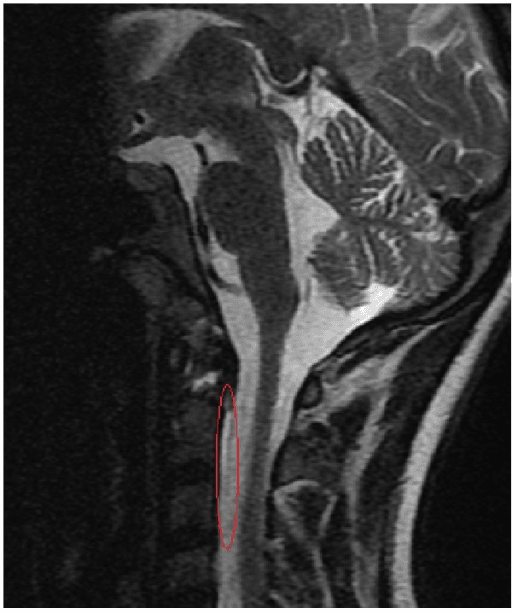
- Frequency
- Daily to twice daily
- Detail
- Half-strength hydrogen peroxide, chlorhexidine or saline per local protocol; keep crusts from occluding the site
- Frequency
- Once at 24 to 48 hours only
- Detail
- Retighten to specification if loose. Do NOT repeatedly retorque thereafter — repeated torquing erodes the outer table and creates a loose pin tract
- Frequency
- Daily
- Detail
- Log-roll with a second person; inspect sternum, scapulae, iliac crests, occiput
- Frequency
- Per protocol, one shell at a time
- Detail
- Never both shells off simultaneously; maintain alignment
- Frequency
- Daily inpatient, each clinic visit
- Detail
- Any new deficit means urgent imaging
- Frequency
- After application, after any adjustment, and at intervals (commonly 2 weeks, 6 weeks, and at completion)
- Detail
- Erect lateral views
- Frequency
- Especially in older or comorbid patients
- Detail
- Early speech and language therapy input if dysphagia
- Frequency
- Continuous
- Detail
- Halo raises the centre of gravity and restricts visual field
Pins settle in the first day or two as periosteum and outer table compress. One retorque at 24 to 48 hours captures this. Beyond that, a pin that will not hold torque is a failed pin — remove it and place a new pin at an adjacent safe site.
Complications — Recognition and Management
- Approximate frequency
- 36 percent of patients (Garfin, 179 patients) — the commonest problem. Counted by pin it is 25 percent of 716 pins
- Management
- Single retorque within 24 to 48 hours; thereafter exchange to a fresh adjacent site. About two-thirds of loose or infected pins ultimately need change or removal
- Approximate frequency
- 20 percent of patients, or 9 percent of pins (Garfin). In children 22 of 37 (Dormans), concentrated anteriorly
- Management
- Minor: local care plus oral antibiotics. Loose, purulent or deep: remove pin, culture, new pin at adjacent site, systemic antibiotics; consider imaging for abscess/osteomyelitis
- Approximate frequency
- 1 percent (Garfin); 1 of 37 children (Dormans)
- Management
- Remove the pin, nurse head elevated, urgent CT, neurosurgical referral; watch for CSF leak, pneumocephalus, meningitis, seizure, abscess; may need operative repair
- Approximate frequency
- Nerve injury 2 percent (Garfin); from medial pin placement
- Management
- Reposition pin laterally; numbness often improves but may persist; neuropathic pain treated medically
- Approximate frequency
- Not separately quantified in the reference series — reported in a meaningful minority
- Management
- Re-image, reposition ring below the equator, re-reduce and re-lock; consider surgery if unstable
- Approximate frequency
- 2 percent overall (Garfin), but disproportionately in older patients — part of the 66 percent major complication rate Tashjian found in the elderly
- Management
- Reduce extension, sit upright for meals, speech and language assessment, consider abandoning halo
- Approximate frequency
- 11 percent (Garfin); notably none attributable to the vest in Dormans's children
- Management
- Refit, pad, treat wound; occiput, sternum, scapulae and iliac crests are the hot spots
- Approximate frequency
- Disproportionately in the elderly; 42 percent in-hospital mortality with a halo versus 20 percent without in Tashjian's over-65 odontoid series
- Management
- Chest physiotherapy, early mobilisation, low threshold to convert to collar or surgery
- Approximate frequency
- Related to immobility; not quantified in the halo series
- Management
- Prophylaxis per local protocol and mobilisation
- Approximate frequency
- Cosmetically disfiguring in 9 percent (Garfin); severe pin discomfort in 18 percent
- Management
- Counsel preoperatively; anterior scars are visible
- Approximate frequency
- Notably higher in type II odontoid with risk factors
- Management
- Assess with CT; convert to surgical fixation (odontoid screw or posterior C1-C2 fusion)
PINSHalo Complications
Hook:Every halo review asks: are the PINS tight, is the spine reduced, and is the patient eating and breathing?
Weaning and Confirming Healing
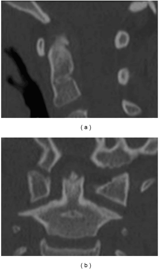
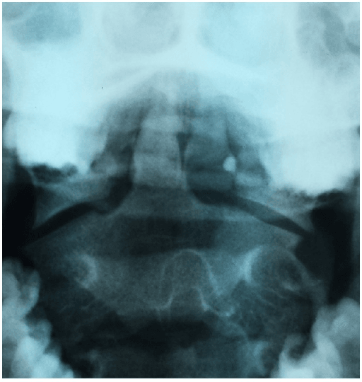
Post-application and erect radiographs. Pin torque check once at 24 to 48 hours. Establish pin care and swallow/respiratory safety. Review at 1 to 2 weeks with radiographs.
Radiographs at intervals to confirm maintained alignment. Any displacement, intolerable pin problems or new neurology triggers reconsideration of surgery.
Typical treatment duration is 8 to 12 weeks (longer in some type II odontoid, shorter in children). CT is the most reliable assessment of bony bridging, particularly at the dens.
Remove pins, clean and dress sites (rarely need closure). Transition to a rigid collar for a short period. Erect radiographs out of halo.
Supervised, patient-controlled flexion-extension radiographs out of the orthosis. Absence of abnormal translation or angulation confirms stability and permits discharge from immobilisation. Abnormal motion or persistent nonunion means surgical stabilisation.
Some odontoid fractures achieve a stable fibrous union rather than bony union. If dynamic views show no abnormal motion and the patient is asymptomatic, a stable fibrous union in a low-demand patient may be accepted; instability on dynamic views is an indication for fusion.
Halo Traction Variants
- Use
- Severe rigid kyphoscoliosis, cervicothoracic deformity, basilar invagination, nutritional optimisation before major reconstruction
- Key points
- Ring attached to an overhead pulley in wheelchair, walker and bed; weight escalated gradually (commonly starting a few kilograms and increasing to a proportion of body weight) over weeks with daily cranial nerve and neurological examination
- Use
- Rare; severe rigid deformity
- Key points
- Higher neurological and pin complication burden; largely historical for halo-pelvic
- Use
- Positioning and gradual correction during deformity surgery
- Key points
- Allows controlled distraction with continuous neuromonitoring
- Use
- Awake, cooperative patient with serial neurological checks and imaging
- Key points
- Gardner-Wells tongs are usually preferred for pure traction; halo ring is the choice when the ring will subsequently be converted to a vest
During halo-gravity traction, assess cranial nerves (especially VI — abducens palsy causing diplopia is an early warning), limb power and sensation daily, and reduce weight immediately if any deficit appears. Over-distraction can cause brainstem or cord injury, particularly with an unrecognised occipitocervical dissociation — traction is contraindicated where craniocervical distraction injury is present.
Guidelines, Registries and Global Practice
Global epidemiology. Cervical spine injury incidence follows two peaks worldwide: young adults after road traffic collisions and high-energy trauma, and older adults after low-energy falls. The ageing populations of Europe, North America, Japan and Australasia have made the geriatric type II odontoid fracture the single most frequent cervical fracture in many trauma systems — precisely the group in which halo use has fallen away.
Society guidance where it genuinely addresses this topic
- Relevant position
- Support external immobilisation (halo or collar) for many type II and III odontoid and selected C1 fractures, while highlighting increased nonunion in older patients and displaced type II injuries
- Relevant position
- Injury classification (upper cervical and subaxial systems) drives the decision between external immobilisation and internal fixation; emphasises ligamentous assessment before choosing a halo
- Relevant position
- Emphasise timely imaging, spinal clearance protocols and specialist spinal referral; external immobilisation is an interim or definitive measure only within a documented plan
- Relevant position
- Recommends CT as first-line imaging in significant blunt cervical trauma in adults and cautions on collar-related harm from prolonged immobilisation — the same reasoning underpins minimising prolonged halo use
- Relevant position
- Consistently frame halo as a niche technique with a specific occipitocervical indication set, best preserved in paediatric practice and deformity traction
Registry and database evidence. There is no implant registry for orthoses. The relevant evidence base is national trauma registries and administrative databases, which have repeatedly documented rising surgical fixation and falling halo use for geriatric odontoid fractures over the past two decades, together with the mortality signal associated with halo use in that group.
Practice variation by resource setting.
- High-resource settings — halo largely reserved for paediatric trauma, halo-gravity traction, adjunct to complex reconstruction and selected young patients declining surgery.
- Middle-income and rural settings — halo retains a wider definitive role where instrumentation, intraoperative imaging or neuromonitoring are limited; competence in application remains a core skill.
- Humanitarian and conflict settings — the halo is a comparatively cheap, reusable frame that can definitively treat many upper cervical injuries without an operating theatre; the limiting factors are pin-care supplies and follow-up imaging.
- Consequently, halo application is a globally examinable skill even where it is no longer a common local treatment.
Controversies and Areas of Uncertainty
- Type II odontoid in the middle-aged patient (50 to 70 years). Union rates in a halo fall progressively with age, but surgical risk rises. Where exactly the crossover lies is unresolved; displacement, fracture obliquity, bone quality and patient priorities drive individual decisions.
- Is fibrous union acceptable? For low-demand patients with a stable fibrous union on dynamic views, many surgeons accept it. Others argue that any motion at C1-C2 risks late myelopathy and warrants fusion.
- Collar versus halo in stable C1 fractures. With an intact transverse ligament, a rigid collar produces comparable outcomes to halo in many series with far fewer complications — halo use here is increasingly hard to justify.
- Optimal pin-site care solution. Chlorhexidine, saline, dilute hydrogen peroxide and dry dressings all have advocates; robust comparative evidence is lacking and practice is institutional.
- Retorquing philosophy. The single 24 to 48 hour retorque is widely taught, but some units never retorque, arguing that any pin that loosens should simply be exchanged.
- Paediatric pin number and torque. Six to eight low-torque pins is standard teaching, but exact torque values are extrapolated from small series and cadaveric skull thickness data rather than trials.
- Halo-gravity traction protocols. Weight escalation rates, maximum weight as a proportion of body weight, and duration before surgery vary widely between deformity centres.
MCQ Practice Points
A: One centimetre superior to the orbital rim, over the lateral third of the rim (lateral two-thirds of the supraorbital ridge), below the skull equator and anterior to the temporalis muscle. Medial placement risks the supraorbital and supratrochlear nerves and the frontal sinus; lateral placement risks the thin squamous temporal bone and temporalis.
A: Tightening with the eyes open and brows elevated tethers the forehead skin in an elevated position, leaving the patient unable to close the lids afterwards — lagophthalmos with corneal exposure.
A: Adults 8 inch-pounds; children 2 to 4 inch-pounds with 6 to 8 pins. Torque is checked once at 24 to 48 hours. Beyond that, a pin that will not hold torque should be removed and replaced at an adjacent site rather than repeatedly retorqued.
A: Say the textbook answer, then show you know what is behind it. The quoted figure is approximately 75 percent of gross cervical motion — from Koch and Nickel, seven patients, comparing supine with upright. Lind's prospective series of 31 patients, sampling a range of exercises, found 70 percent of normal motion still available, and found the halo restricted motion most below C2 and least above C2 — the opposite of the usual "best at occiput to C2" claim. Paradoxical intersegmental snaking is real but is not confined to the subaxial spine. The reason a halo suits odontoid, C1 ring and hangman fractures is that those fractures unite in external immobilisation, not that the halo is mechanically better at those levels.
A: Pin loosening. In Garfin's 179-patient reference series it affected 36 percent of patients, with pin-site infection in 20 percent. Quote the denominator you mean: counted by pin rather than by patient the same series gives 25 percent loosening and 9 percent infection, and about two-thirds of loose or infected pins needed changing or removal. In children (Dormans, 37 patients) complications reached 68 percent, infection commonest and concentrated at the anterior pins.
A: Reduced vital capacity from the vest, forced neck extension causing dysphagia and aspiration, falls in a top-heavy device and immobility together produce a well-documented excess of pneumonia and mortality, with poorer union rates than in the young. A rigid collar or surgical stabilisation is preferred.
A: Interval radiographs in the halo, CT to assess bony union, then removal, a short period in a rigid collar, and supervised flexion-extension radiographs out of the orthosis demonstrating no abnormal translation or angulation.
Exam Viva Scenarios
Practise clinical reasoning and management decisions out loud
“A 24-year-old motorcyclist has an undisplaced type II odontoid fracture, neurologically intact, with no other injuries. He declines surgery after a full discussion. Talk me through applying a halo vest, including pin placement.”
“An 84-year-old woman on anticoagulation falls at home and has a type II odontoid fracture with 4 mm of posterior displacement. She is frail, lives alone and has COPD. The referring team asks you to apply a halo. What is your response?”
“Three days after halo application, a patient reports a headache and the nurse notes clear fluid weeping from a right anterior pin site. What do you do?”
“A 5-year-old sustains a C2 synchondrosis fracture in a road traffic collision, reduced by positioning. How does halo application differ in children?”
Core numbers
- Motion control quoted as 75 percent (Koch and Nickel, 7 patients) - but Lind's 31 patients found 70 percent of normal motion remaining, and restriction LEAST above C2
- Ring clearance 1 to 2 cm; sits below the skull equator
- Anterior pin: 1 cm above the lateral third of the orbital rim
- Adult torque 8 inch-pounds; paediatric 2 to 4 inch-pounds with 6 to 8 pins
- Torque re-check once at 24 to 48 hours only
- Typical treatment 8 to 12 weeks
Pin anatomy
- Anterior: above outer brow, not above the pupil, anterior to temporalis, below equator
- Medial hazards: supraorbital and supratrochlear nerves, frontal sinus
- Lateral hazards: squamous temporal bone (thinnest), temporalis muscle
- Posterior pins diagonally opposite, roughly 4 and 8 o'clock, above and behind the ear
- Pins perpendicular to the skull; tighten diagonally opposed pairs simultaneously
Indications
- C1 ring fracture with intact transverse ligament
- Type III and selected young type II odontoid fractures
- Levine-Edwards type I and reduced type II hangman fractures
- Paediatric cervical trauma — well tolerated, avoids fusing a growing spine
- Adjunct after complex cervical reconstruction
- Halo-gravity traction for rigid deformity or basilar invagination
Contraindications
- Cranial fracture or defect at intended pin sites
- Respiratory compromise or severe chest wall deformity
- Unstable thoracic or rib injury preventing vest loading
- Frailty and advanced age — respiratory complications and mortality
- Severe scalp disease, non-compliance, cognitive impairment
- Type IIa hangman fracture — do not apply traction
Complications
- Pin loosening about one third; pin-site infection 10 to 20 percent
- Dural penetration: remove pin, head up, CT, neurosurgical referral
- Supraorbital nerve injury from medial pin placement
- Ring migration and loss of reduction — image after every adjustment
- Dysphagia and aspiration from over-extension
- Pressure sores, venous thromboembolism, pneumonia, nonunion
Weaning
- Interval radiographs in halo; CT to assess bony union
- Remove, transition to rigid collar, erect radiographs
- Flexion-extension views out of orthosis to confirm stability
- Instability or symptomatic nonunion means surgical fixation
- Stable fibrous union may be acceptable in low-demand patients
Safety essentials
- Vest release tool taped to the vest for emergency CPR access
- Written pin-care instructions and 24-hour contact
- Never remove both vest shells at once
- Neurological examination after application and at every review
- Never reuse pins; never repeatedly retorque a failing pin
Evidence
Halo-Vest Immobilization Increases Early Morbidity and Mortality in Elderly Odontoid Fractures
- 78 patients over 65 years with a type II or III odontoid fracture, from a single trauma registry between 1997 and 2004; mean age 80.7 years
- 38 patients (49 per cent) were treated with a halo vest and 40 (51 per cent) without - 27 in a cervical orthosis and 13 operatively
- There was NO difference in injury severity or baseline medical condition between the halo and non-halo groups
- In-hospital mortality was 42 per cent with a halo versus 20 per cent without (p = 0.03)
- Major complications occurred in 66 per cent of halo patients versus 36 per cent of non-halo patients (p = 0.003)
Fractures of the Odontoid Process of the Axis
- The classification that still governs odontoid fracture management worldwide: type I avulsion of the tip, type II fracture at the base of the dens, type III fracture extending into the cancellous body of C2
- Type II is the fracture that behaves badly with external immobilisation; type III, being through cancellous bone with a broad surface area, generally unites
Injuries Involving the Transverse Atlantal Ligament - Classification and Treatment Guidelines Based Upon Experience with 39 Injuries
- 39 patients with transverse atlantal ligament injury studied with plain films, thin-section CT and MRI
- Type I (n = 16) is disruption of the SUBSTANCE of the ligament; type II (n = 23) is a fracture or avulsion of the tubercle where the ligament inserts on the C1 lateral mass
- Type I injuries were incapable of healing satisfactorily without internal fixation and should have early surgery
- Type II injuries were treated initially in a RIGID CERVICAL ORTHOSIS with a 74 per cent success rate; 26 per cent failed immobilisation and needed delayed fixation
- A type II injury renders the ligament physiologically incompetent even though its substance is not torn
Bursting Atlantal Fracture Associated with Rupture of the Transverse Ligament
- The origin of the 'rule of Spence' - combined overhang of the C1 lateral masses on the open-mouth view is used as an indirect sign that the transverse ligament has failed
- The observation was made on cadaveric loading together with clinical cases