Type II High Risk | Age Determines Treatment | Collar vs Fusion
- Type II is most common (60%) and most problematic (40% nonunion with collar)
- Type III heals well with collar - fracture into cancellous C2 body
- Elderly Type II - consider primary posterior C1-C2 fusion (Harms/Magerl)
- Anterior odontoid screw preserves rotation for acute displaced Type II (young)
- Transverse ligament integrity determines stability
- “Type II is at watershed blood supply zone - high nonunion risk
- “Age over 65, displacement over 5mm, angulation over 10° = surgical factors
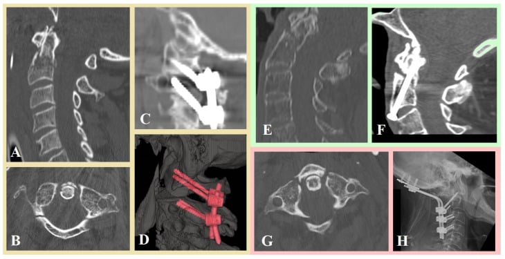
- “Harms technique (C1 lateral mass + C2 pedicle screws) is gold standard posterior
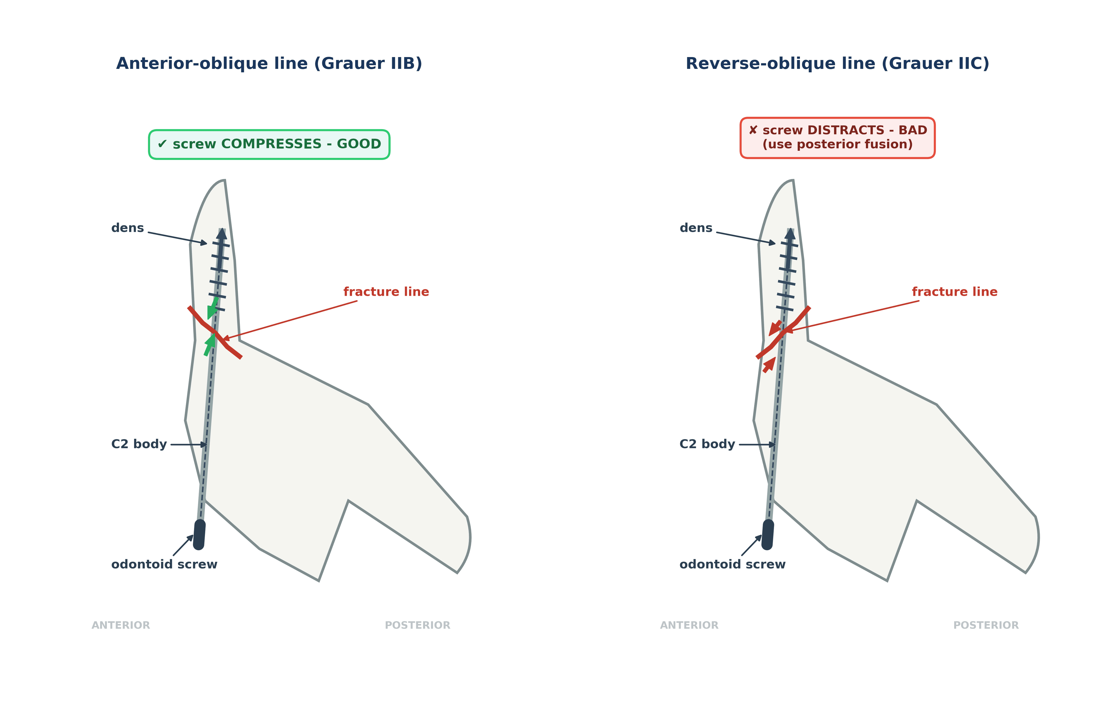
- “Anterior screw contraindicated by the REVERSE OBLIQUE line (Grauer IIC, posterosuperior to anteroinferior), barrel chest, fixed kyphosis and severe osteoporosis - note Apfelbaum calls this same unfavourable line ANTERIOR oblique
Overview and Epidemiology
Odontoid fractures have a bimodal age distribution. Young adults break the dens in high-energy trauma; the elderly break it in a low-energy fall onto osteoporotic bone, and in the elderly the odontoid is the most common cervical fracture pattern. Every treatment decision in that older group comes back to the same balance: the risk of the operation against the morbidity of a nonunion.
Mechanism. In the young the energy comes from a motor vehicle accident, diving or sport; in the elderly, from a fall. The direction of the force predicts the direction of displacement: hyperflexion displaces the dens anteriorly, hyperextension displaces it posteriorly, and a rotational component may be present.
Associations. Look beyond the dens:
- Head injury is common with a high-energy mechanism
- Other cervical spine fractures in 10-20%
- Vertebral artery injury is rare, but assess for it
- Thoracolumbar fractures: check the whole spine
Anatomy and Biomechanics
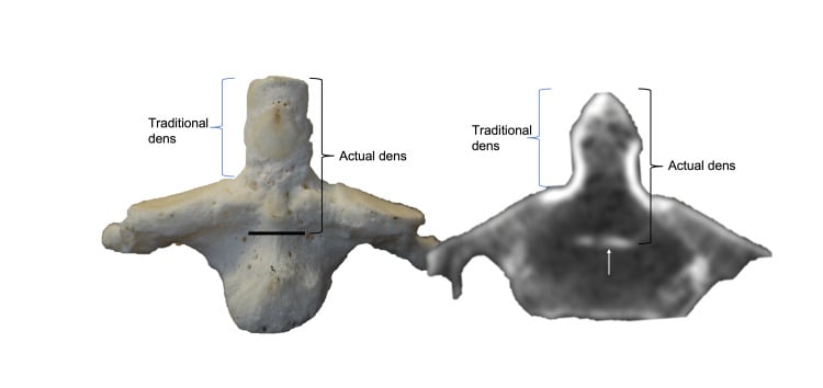
The axis. C2 is unlike any other vertebra. The odontoid process (dens) projects superiorly into the ring of C1, articulates with the anterior arch of the atlas in front, and is held there from behind by the transverse ligament. 50% of cervical rotation occurs at C1-C2.
The blood supply. The tip of the dens is fed by the apical arcade and the posterior descending arteries; the base is fed by ascending arteries from the C2 body; the body itself has the vertebral body vessels. The base of the dens, the level of a Type II fracture, is the watershed between the two supplies, so a fracture there lies in the bone with the poorest supply. That is the anatomical reason Type II fractures fail to unite so often, and why a Type I fracture through the tip and a Type III fracture through the cancellous body both heal well.
The transverse ligament (TAL) is the primary stabiliser of C1-C2. It holds the dens against the anterior arch of C1, and when it ruptures the result is atlantoaxial instability. Its integrity is read indirectly from the atlantodental interval on the lateral radiograph or CT, and directly on MRI; the numbers are in Investigations.
The alar ligaments run from the tip of the dens to the occipital condyles and limit axial rotation. A Type I fracture is an avulsion at their origin, which is why a Type I fracture is a prompt to check for occipitocervical instability.
The rule of thirds. At the level of C1 the spinal canal is divided into three: one third odontoid, one third spinal cord, one third free space filled with CSF. That free third is a physiological reserve, and it is why neurological injury is relatively rare in odontoid fractures despite what can be significant displacement: there is room for the cord to escape.
Classification Systems
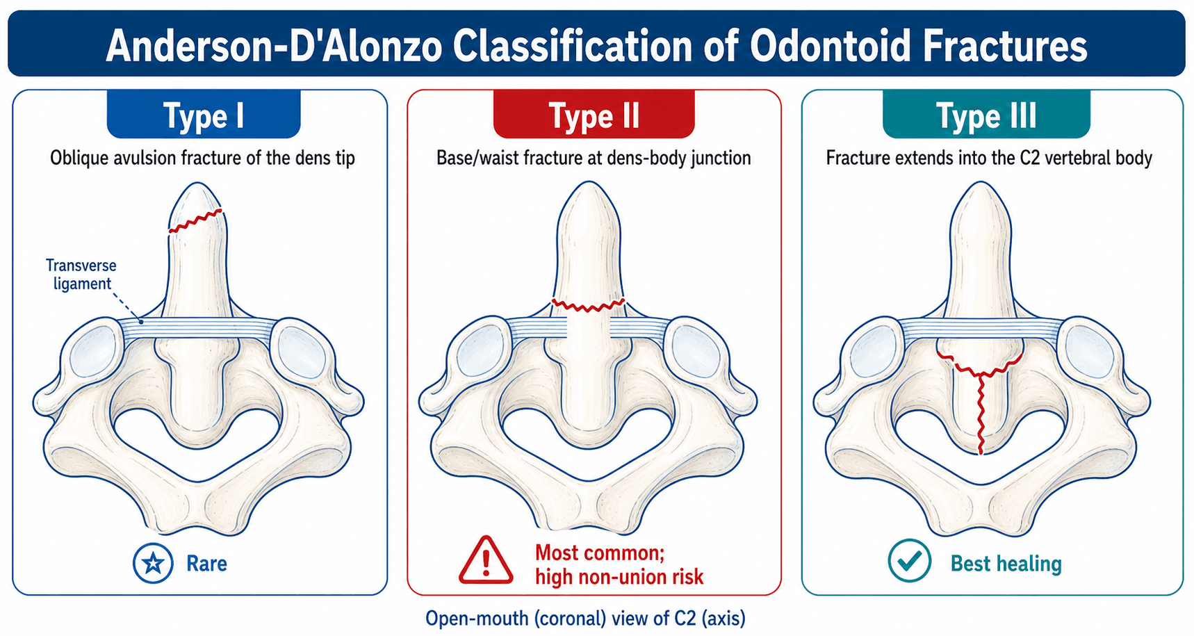
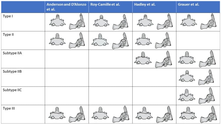
Anderson and D'Alonzo (1974) is the standard classification and it works by location: tip, base or body. Location predicts blood supply and blood supply predicts union, which is why the type frames the treatment. Grauer's modification (2005) subclassifies Type II by the orientation of the fracture line, which is what decides whether an anterior screw can work.
- Location
- Tip avulsion
- Frequency
- 5%
- Stability
- Stable
- Treatment
- Collar 6-8 weeks
- Location
- Base of dens
- Frequency
- 60%
- Stability
- Unstable
- Treatment
- Collar vs surgery
- Location
- Type II + comminution
- Frequency
- Rare
- Stability
- Very unstable
- Treatment
- Posterior fusion
- Location
- Into C2 body
- Frequency
- 35%
- Stability
- Often stable
- Treatment
- Collar 10-12 weeks
The comminuted Type IIA is Hadley's addition to the system, as the classification panel below shows. A comminuted IIA is very unstable and goes to posterior fusion, not to a screw.
Clinical Assessment
History. The mechanism sorts itself by age: a motor vehicle accident in the young, a fall in the elderly. Ask about neck pain, which is posterior or suboccipital; about head injury, which is common after a high-energy mechanism; and about previous neck problems, particularly degenerative disease. Neurological symptoms are rare, because the cord has space.
Examination. Keep the immobilisation on until the spine is cleared. Palpate for tenderness over the C2 spinous process, examine the cord and the roots fully, and complete the trauma survey for other injuries. The vascular examination is rarely abnormal, but it is part of the assessment.
Neurology. Neurological injury is uncommon, under 10%, despite what can be significant displacement: the canal at C1-C2 is large (the rule of thirds) and gradual displacement allows the cord to adapt. When a deficit is present, suspect cord injury; urgent decompression may be needed.
Red flags. Features that require immediate attention:
- Neurological deficit (rare but urgent)
- Rapidly progressive symptoms
- Respiratory compromise (high cord injury)
- Vascular symptoms (vertebral artery)
- Polytrauma with haemodynamic instability
Investigations
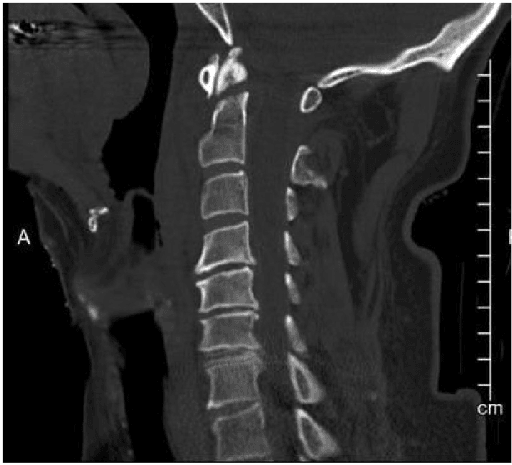
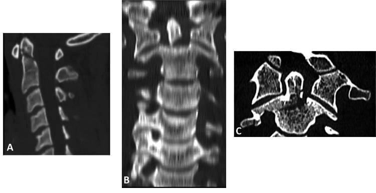
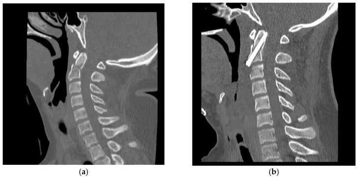
CT is the gold standard for diagnosis and the source of the bony detail for classification and surgical planning. It shows the fracture type, the displacement, the angulation and any comminution, and the sagittal reconstructions are essential because they show the orientation of the fracture line, on which the Grauer subtype and the case for an anterior screw depend. Always get the CT first: MRI alone may miss a subtle fracture.
MRI is for the transverse ligament and the rest of the soft tissue. It is required when the ADI is increased or instability is suspected, it shows cord oedema when there is a neurological concern, and it is the next test when the CT findings do not explain the symptoms.
Flexion-extension radiographs assess atlantoaxial instability, and only for a fracture that has healed or whose stability has been confirmed. Never in the acute setting with an unstable fracture.
Measurements. The numbers read off the lateral film or the CT:
- Normal
- Under 3mm (adult)
- Abnormal
- Over 3mm
- Significance
- TAL rupture
- Normal
- 0
- Abnormal
- Over 5mm
- Significance
- Surgical indication
- Normal
- 0
- Abnormal
- Over 10°
- Significance
- Surgical indication
- Normal
- Over 13mm
- Abnormal
- Under 13mm
- Significance
- Cord compression risk
The ADI is the distance from the anterior arch of C1 to the dens. Under 3mm is normal in an adult and under 5mm in a child; over 3mm in an adult suggests the transverse ligament has ruptured.
Differential Diagnosis
The radiographic appearance of the dens can be deceptive. Several entities mimic or coexist with an acute odontoid fracture and must be actively distinguished, because the management differs substantially.
- Distinguishing Feature
- Sharp lucent line at dens base, soft-tissue swelling
- Clue on Imaging
- Cortical disruption, prevertebral haematoma, marrow oedema on STIR
- Pitfall
- Best diagnostic standard
- Distinguishing Feature
- Smooth corticated ossicle, hypertrophied anterior C1 arch
- Clue on Imaging
- Rounded margins, no oedema, often wide gap
- Pitfall
- Mistaken for acute nonunion - it is chronic
- Distinguishing Feature
- Small ossicle above a normal dens body
- Clue on Imaging
- Above the level of the transverse ligament, stable
- Pitfall
- Usually incidental, not unstable
- Distinguishing Feature
- Apparent lucency from overlapping arches or teeth
- Clue on Imaging
- Disappears on a true open-mouth or CT view
- Pitfall
- False-positive fracture call on plain film
- Distinguishing Feature
- Torticollis, asymmetric dens-lateral mass spacing
- Clue on Imaging
- Dynamic CT shows fixed rotation
- Pitfall
- Coexists with trauma in children
- Distinguishing Feature
- Lytic destruction, age-atypical pain
- Clue on Imaging
- Bone destruction without clear trauma, marrow replacement on MRI
- Pitfall
- Low-energy fracture can be the first sign of malignancy
Os odontoideum is worth knowing as a condition, not just a line in the table. It is a rounded, well-corticated ossicle separated from a hypoplastic dens by a wide gap above the level of the transverse ligament, with a characteristically hypertrophied anterior arch of C1. Its origin is debated, congenital (failed fusion of the dens ossification centres) against post-traumatic (an unrecognised childhood dens fracture that healed as a rounded nonunion), and it is associated with Down syndrome, Klippel-Feil, Morquio and the skeletal dysplasias.
Two morphologies. Orthotopic, with the ossicle in the normal dens position and moving with the C1 arch; and dystopic, with the ossicle fused to the clivus or occiput, which is the more unstable of the two.
The clinical problem is atlantoaxial instability. Because the dens-equivalent is not anchored to C2, the transverse ligament has lost its post, so flexion-extension views and the space available for the cord (the posterior atlantodental interval) matter more than the ADI. An asymptomatic, stable, incidental os odontoideum may be observed with activity precautions; instability, neurological symptoms or signs, or a reduced space available for the cord mandate posterior C1-C2 fusion.
An os odontoideum has smooth corticated margins and no marrow oedema, which is chronicity; an acute fracture has sharp irregular edges and prevertebral soft-tissue swelling. Treating a chronic, well-compensated os odontoideum as an acute fracture, or the reverse, is a common examination error, and collaring a chronic unstable lesion that actually needs fusion is the expensive version of it. Dynamic flexion-extension imaging, when safe, clarifies whether it is stable.
Management Algorithm
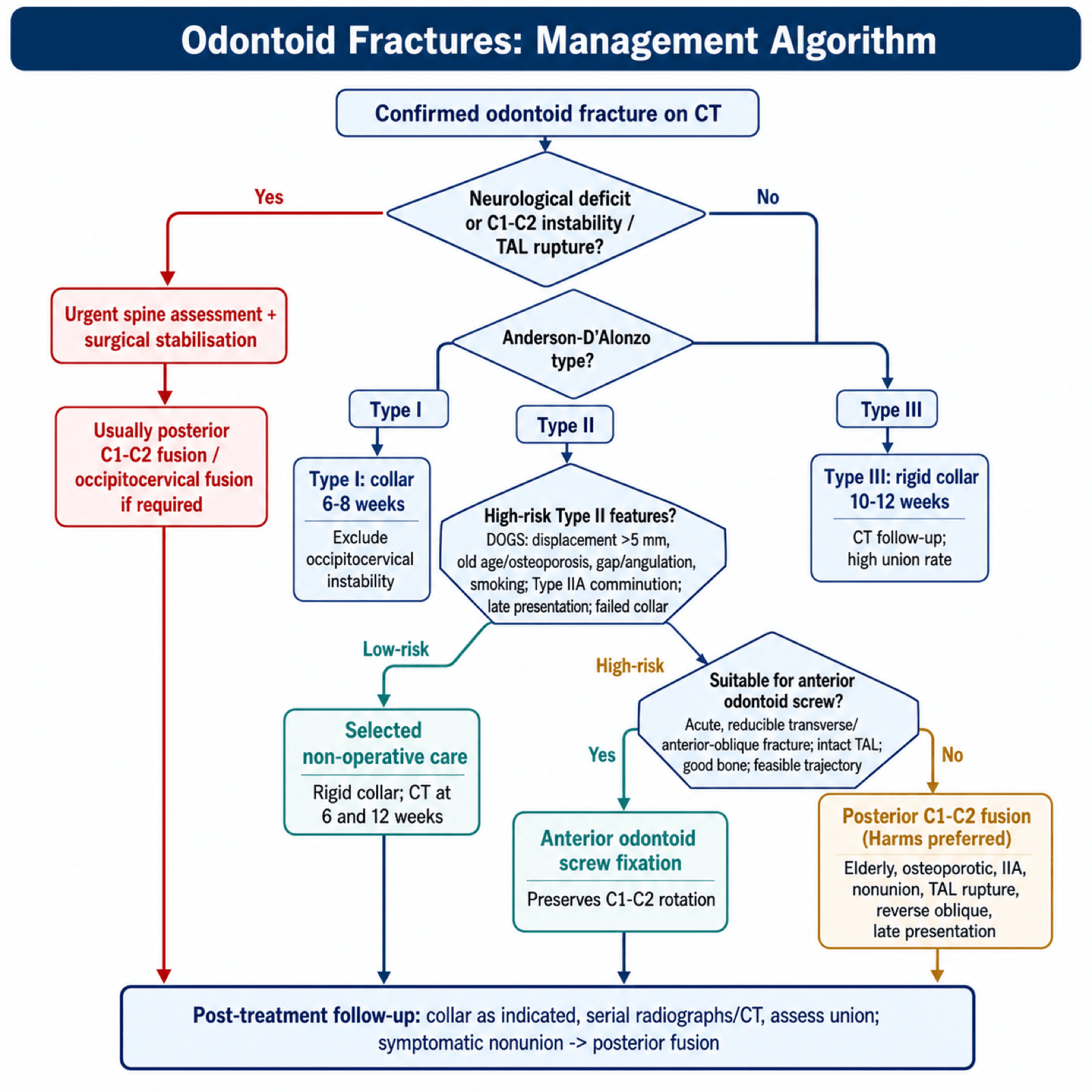
The decision turns on the fracture type, the patient's age and the stability of the atlantoaxial joint. Type I and Type III fractures are treated in a collar and usually unite. Type II is the controversy, because the nonunion rate with a collar is 30-40% and higher again in the elderly, so a Type II is risk-stratified: the DOGS features, comminution (IIA), late presentation and a failed collar all push it towards surgery. Whatever the type, a neurological deficit, C1-C2 instability or a transverse ligament rupture goes to urgent assessment and surgical stabilisation.
The age threshold. The DOGS factors and the summary use 65. The halo-failure series (Lennarson) and the AANS guidance both use 50, and the evidence base records that Lennarson's 50 was the dichotomy chosen rather than a tested cut-point, and that biological risk is continuous.
Type I. Rigid collar for 6-8 weeks, with union in 95% or more. Because this is an alar ligament avulsion, check for occipitocervical instability before calling it benign.
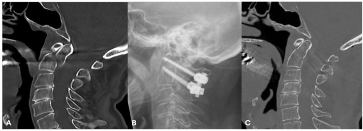
Type II, selected patients. A rigid collar (Philadelphia, Miami J) or a halo, best suited to the young patient with minimal displacement (under 5mm) and a transverse line. Union is 50-60% and lower in the elderly (the union table under Outcomes gives the figures by age), so follow closely with CT at 6 and 12 weeks.
Type III. Usually non-operative unless the displacement is significant: a rigid collar for 10-12 weeks, longer because the fracture is larger, with union in 85% or more. A halo is rarely needed.
- Collar
- Moderate
- Halo
- Best
- Collar
- Better
- Halo
- Poor
- Collar
- Fewer
- Halo
- Pin infections, loosening
- Collar
- Better
- Halo
- High complication rate
- Collar
- 50-60%
- Halo
- 60-70%
The figures are worse than the commonly repeated "40% complications", and they are two separate numbers. In Tashjian's series (J Trauma 2006;60(1):199-203, PMID 16456456) of 78 patients over 65 with a type II or III odontoid fracture (mean age 80.7), comparing those managed with a halo vest against those managed without:
- In-hospital MORTALITY 42% with a halo versus 20% without (p = 0.03)
- MAJOR COMPLICATIONS 66% with a halo versus 36% without (p = 0.003)
There was no difference in injury severity or baseline medical condition between the groups. The mechanisms are respiratory compromise and aspiration, pin loosening and infection, falls with the added head mass and altered balance, and poor tolerance generally.
Three caveats to state alongside them. Allocation was not randomised, so some confounding by indication is inevitable - frailty, dementia, respiratory reserve and swallowing were not measured, and those are exactly what a clinician weighs when choosing a collar. The endpoint is in-hospital death, not union, so this is not evidence that a collar heals the fracture better - only that the halo costs more lives while it is on. And the comparator is mixed: the 40 "without halo" patients were 27 in a collar and 13 operated, while 4 of the 38 halo patients wore the halo after surgery - so this is halo versus collar-or-surgery, not the head-to-head against a collar that the recommendation implies. The direction of the finding is not in doubt; its precision is.
Practical position: in the patient over 75 with a type II fracture, choose a rigid collar or surgical stabilisation; the halo is now widely regarded as a relative contraindication in this group. Full detail in halo vest application and management.
Surgical Technique
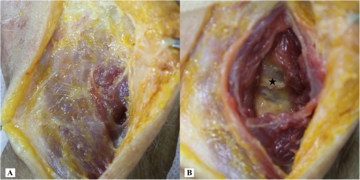
Proper positioning is essential for safe screw placement and to prevent intraoperative complications.
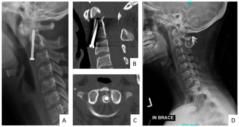
Anterior odontoid screw
- Supine on a radiolucent table
- Head extended (chin tuck to expose the neck)
- Shoulder roll
- Arms at the sides, or taped down
- C-arm access for AP and lateral views
Posterior C1-C2 fusion
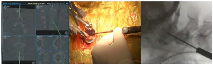
- Prone on a Jackson or Mayfield frame
- Head neutral or in slight flexion
- Mayfield pins or Gardner-Wells tongs
- Arms tucked
- Fluoroscopy or navigation
Complications
- Incidence
- 30-40% (collar)
- Prevention
- Patient selection, surgery for high-risk
- Management
- Posterior fusion
- Incidence
- Variable
- Prevention
- Maintain reduction
- Management
- Osteotomy if symptomatic
- Incidence
- 0.5-2%
- Prevention
- Preoperative CT planning
- Management
- Angiography, observation
- Incidence
- 5-10%
- Prevention
- Fluoroscopy/navigation
- Management
- Revision if symptomatic
- Incidence
- 10-20%
- Prevention
- Gentle retraction
- Management
- Usually temporary
- Incidence
- 2-5%
- Prevention
- Bone graft, rigid fixation
- Management
- Revision fusion
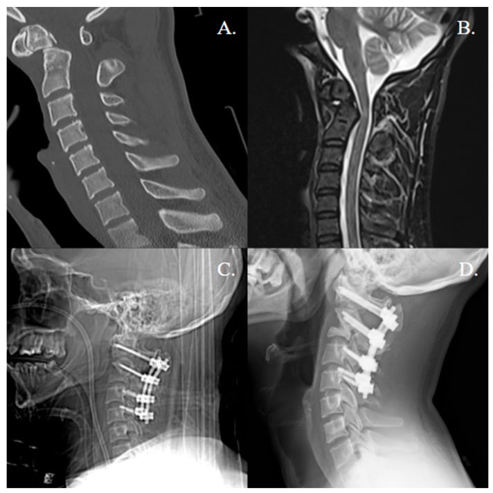
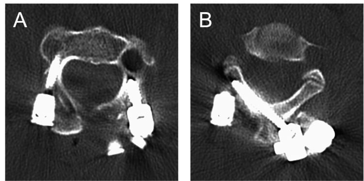
Type II nonunion. A symptomatic nonunion is confirmed on CT, more than 6 months out with no bridging bone, and then assessed: flexion-extension views for instability, and MRI for the transverse ligament. Posterior C1-C2 fusion by the Harms technique is the gold standard. An anterior screw rarely works, because the fracture site is fibrous tissue, and if the C1 lateral mass is compromised, consider extending the fusion to the occiput.
Postoperative Care
The first six weeks. Every surgical patient goes into a collar, with wound care, DVT prophylaxis and analgesia in the first fortnight. Radiographs at 2 and 6 weeks; gentle range of movement when comfortable, and physiotherapy for the surrounding muscles.
The 6-week CT decides the rest. If fusion or healing is progressing, the collar continues to the duration in the table; if it is not, the plan is reassessed. When the CT shows union or fusion, wean the collar, confirm stability with flexion-extension radiographs, and progress physiotherapy to range of movement and strengthening.
- Collar Duration
- 6-8 weeks
- Follow-up
- CT at 6 weeks
- Collar Duration
- 6-8 weeks
- Follow-up
- CT at 6 weeks for fusion
- Collar Duration
- 8-12 weeks
- Follow-up
- May need Halo if unstable
- Collar Duration
- 10-12 weeks (Type II/III)
- Follow-up
- CT at 6 and 12 weeks
Outcomes and Prognosis
Union. The figures by type and treatment, which are the ones the treatment decision is built on:
- Collar
- 95%+
- Halo
- N/A
- Surgery
- N/A
- Collar
- 60%
- Halo
- 70%
- Surgery
- 90-95%
- Collar
- 40-50%
- Halo
- 50% (high complications)
- Surgery
- 90-95%
- Collar
- 85%+
- Halo
- 90%+
- Surgery
- 95%+
Mortality. An elderly Type II fracture treated in a collar carries a 15-25% one-year mortality, and nonunion contributes to it; function is often limited with a fibrous union. Surgery brings its own perioperative risk, but early mobilisation reduces complications, some studies report lower mortality in the elderly, and long-term function is better if fusion is achieved.
The elderly Type II dilemma. Nonunion with a collar is frequent, the halo carries the Tashjian mortality and complication figures given under Management, and surgery carries perioperative risk. Recent evidence favours primary posterior fusion in the fit elderly patient for better union and earlier mobilisation. Be precise about survival: the AOSpine GOF cohort found a 30-day survival advantage with surgery, but its long-term mortality signal was a trend with a confidence interval crossing 1, and the evidence base below is explicit that it should not be quoted as a survival benefit.
Guidelines, Registries & Global Practice
- Most common cervical fracture in patients over 70 worldwide
- Incidence rising with ageing populations across high-income countries
- Type II predominates (about 60% of dens fractures)
- Low-energy falls in the elderly now outnumber high-energy injuries
- Up to 80% of geriatric cases are osteoporotic-bone fractures
- Major trauma / spinal unit referral for all odontoid fractures
- CT first, MRI for ligament and cord assessment
- Elderly falls pathways should actively screen the C-spine
- Shared decision-making essential in frail elderly patients
Side-by-Side Society Guidance
- Emphasis
- Evidence-based management of dens fractures
- Type II Elderly Stance
- Surgery recommended for displacement over 5mm or age over 50; halo or surgery both options
- Emphasis
- Upper cervical injury classification, operative algorithms
- Type II Elderly Stance
- Favours operative stabilisation in unstable type II, individualised in frail elderly
- Emphasis
- Spinal injury assessment and major trauma pathways
- Type II Elderly Stance
- Multidisciplinary decision; nonoperative acceptable if surgery high-risk
- Emphasis
- Geriatric trauma, fragility-fracture care
- Type II Elderly Stance
- Early mobilisation prioritised; stable nonunion an accepted goal if unfit
Registry and Resource-Setting Notes
- No dedicated dens-fracture implant registry exists; outcome data derive from prospective cohorts such as the AOSpine North America Geriatric Odontoid Fracture (GOF) study rather than arthroplasty-style registries.
- High-resource settings: ready CT/MRI access, navigation or fluoroscopy for screw placement, and early posterior C1-C2 fixation in fit elderly.
- Limited-resource settings: reliance on plain radiographs and rigid collars, restricted MRI for ligament assessment, and a lower threshold for nonoperative management - making accurate stability assessment and falls prevention even more important.
Controversies and Areas of Uncertainty
Odontoid fracture management is one of the most genuinely contested areas in spinal trauma. A strong viva candidate can articulate where the evidence is soft and argue both sides.
The AOSpine GOF data suggest a 30-day survival advantage and higher fusion with surgery, but selection bias is unavoidable - fitter patients are offered surgery. No randomised trial has settled the question, and the competing systematic review found stable nonunion is an acceptable goal with under 5% needing revision.
In frail elderly patients, stable fibrous nonunion may give equivalent function and pain to bony union. Chasing radiographic union with high-risk surgery or prolonged halo may add morbidity without functional benefit.
Anterior screw preserves rotation but fails in osteoporotic bone, reverse-oblique lines and chronic fractures. Posterior C1-C2 fusion is more reliable but sacrifices rotation. The trade-off (motion vs union reliability) is unresolved for the borderline young-elderly patient.
Halo offers marginally better immobilisation but with high complication rates in the elderly. Many units have largely abandoned the halo in older patients, yet high-quality comparative data remain limited.
State the principle first ("treatment is individualised because the evidence is observational, not randomised"), then give the balance (survival/fusion advantage of surgery vs selection bias and the acceptability of stable nonunion), and finish with a patient-specific plan. Examiners reward candidates who acknowledge uncertainty rather than dogmatically insisting on one answer.
MCQ Practice Points
Q: Which odontoid fracture type has the highest nonunion rate?
A: Type II - fracture through the waist/base of the dens. This is at the watershed blood supply zone, leading to 30-40% nonunion with collar treatment. Type I and III have much higher union rates.
Q: What percentage of cervical rotation occurs at C1-C2?
A: 50% - This is why anterior odontoid screw fixation (which preserves the joint) is preferred over posterior fusion (which sacrifices rotation) when feasible.
Q: What is a contraindication to anterior odontoid screw fixation?
A: Reverse oblique (posterior-superior to anterior-inferior) fracture line - The screw would distract rather than compress the fracture. Other contraindications: barrel chest, TAL rupture, elderly with osteoporosis, chronic nonunion.
Q: What ADI measurement suggests transverse ligament injury in an adult?
A: Greater than 3mm - Normal ADI in adults is under 3mm. In children, up to 5mm may be normal. Increased ADI indicates TAL incompetence and atlantoaxial instability.
Q: Why is Halo vest avoided in elderly patients with odontoid fractures?
A: High complication rate (40%+) - Including pin loosening, pin infection, respiratory complications, falls, and death. In elderly with Type II, primary posterior C1-C2 fusion often preferred.
Q: What is the fusion rate difference between single and double anterior odontoid screws?
A: No significant difference - Meta-analysis shows similar fusion rates (~90%). Single screw is technically easier and sufficient.
Exam Viva Scenarios
Practise clinical reasoning and management decisions out loud
“78-year-old male fell at home. CT shows Type II odontoid fracture with 6mm posterior displacement. Neurologically intact. Past history of COPD and diabetes. How do you manage?”
“25-year-old male, MVA, Type II odontoid fracture, 4mm anterior displacement, transverse fracture line on CT. No TAL injury on MRI. Would you consider anterior screw fixation?”
“65-year-old female was treated with collar for Type II odontoid fracture 4 months ago. CT shows persistent fracture line with no bridging bone. She has ongoing neck pain. How do you assess and manage?”
Classification
- Type I: Tip (rare, stable, collar)
- Type II: Waist (60%, 40% nonunion, controversial)
- Type IIA: Comminuted Type II (posterior fusion)
- Type III: Into C2 body (85% union with collar)
Key Numbers
- ADI over 3mm = TAL injury (adult)
- Displacement over 5mm = surgical factor
- Angulation over 10° = surgical factor
- Age over 65 = increased nonunion risk
- 50% cervical rotation at C1-C2
Anterior Screw Contraindications (BARREL)
- B: Barrel chest
- A: Angulation posteriorly
- R: Reverse oblique fracture
- R: Ruptured TAL
- E: Elderly/osteoporotic
- L: Late presentation
Treatment by Type
- Type I: Collar 6-8 weeks
- Type II (young): Collar vs anterior screw
- Type II (elderly): Posterior fusion (Harms)
- Type IIA: Posterior fusion (no screw)
- Type III: Collar 10-12 weeks
Surgical Options
- Anterior screw: Preserves rotation, for ideal Type II
- Harms (C1-C2): Gold standard posterior, sacrifices rotation
- Magerl: Transarticular, higher VA risk
- Wiring: If screws not possible
Evidence Base
AOSpine GOF Mortality Study: Surgery vs Non-Op in the Elderly
- Retrospective multicentre cohort of 322 patients aged 65 or older with type II odontoid fracture (165 operative, 157 nonoperative). Nonoperative treatment carried higher 30-day mortality (adjusted HR 3.00, 95% CI 1.51-5.94) with a trend toward higher long-term mortality (HR 1.35, 95% CI 0.97-1.89). Overall 30-day mortality 14%, maximal follow-up mortality 44%.
AOSpine GOF Treatment Predictors
- Prospective multicentre cohort of 159 patients aged 65 or older with radiographically confirmed type II odontoid fracture. Independent predictors of treatment failure were older age (OR 1.08 per year), initial nonsurgical treatment (OR 3.09), male sex (OR 4.33) and baseline neurological comorbidity (OR 4.13). 12-month mortality 18.2%.
Nonunion Outcomes With Nonoperative Treatment
- Subgroup of the AOSpine GOF study (58 nonoperatively treated elderly type II fractures). 30% developed primary or secondary nonunion and 22% required delayed surgery, yet NDI and SF-36 outcomes did not differ by union status at 12 months.
Anterior Odontoid Screw Fixation (Landmark Series)
- 147 consecutive patients undergoing direct anterior odontoid screw fixation. Fusion rate 88% for recent fractures (6 months or less) but only 25% for remote fractures (18 months or more). Fusion was independent of patient age, sex and number of screws (one vs two); horizontal and posterior-oblique fracture lines fused better than anterior-oblique.
Age as a Risk Factor for Halo Failure
- Case-control study of 33 isolated type II dens fractures treated by halo immobilisation. Age over 50 years was a highly significant risk factor for nonunion (P = 0.002), with a 21-fold higher risk of halo failure. Sex, displacement amount and direction were not significant.
Halo-Vest Immobilization Increases Early Morbidity and Mortality in Elderly Odontoid Fractures
- 78 patients over 65 with a type II or III odontoid fracture from one trauma registry, 1997-2004; mean age 80.7 years (50 type II, 17 type III, 11 combined)
- 38 (49%) were managed with a halo vest and 40 (51%) without - the latter being 27 in a rigid collar and 13 operated
- IN-HOSPITAL MORTALITY 42% with a halo versus 20% without (p = 0.03); 24 patients (31%) died during the admission overall
- MAJOR COMPLICATIONS 66% with a halo versus 36% without (p = 0.003)
- There was NO difference in injury severity or baseline medical condition between the halo and non-halo groups
- The authors' conclusion is unambiguous: outcomes after halo-vest treatment appear inferior to those achieved with a collar or with operative fixation
Transverse Atlantal Ligament Injury Classification
- Analysis of 39 transverse atlantal ligament injuries defined the Dickman classification: Type I (midsubstance disruption, n=16) cannot heal without internal fixation and needs early surgery; Type II (bony avulsion from the C1 tubercle, n=23) is initially treated in a rigid orthosis with a 74% success rate.
Harms Technique: Posterior C1-C2 Polyaxial Screw-Rod Fixation
- Original description of C1 lateral mass plus C2 pars/pedicle polyaxial screws connected by rods in 37 patients. Solid fusion was achieved in all patients with no vascular or neurological injury, and the technique allows intraoperative reduction without sacrificing the joint surfaces if temporary.