Craniocervical Junction Disruption | High-Energy Trauma | Often Fatal
- Atlanto-occipital dislocation (AOD) is a high-energy craniocervical junction injury with high mortality
- Traynelis classification guides direction of displacement and stability assessment
- Powers ratio, condyle-C1 interval (CCI), and basion-dens interval (BDI) are key radiographic measurements
- Immediate rigid immobilisation in neutral position; traction is contraindicated
- Definitive treatment is occipitocervical fusion after reduction and stabilisation
- “AOD often missed on initial lateral cervical X-ray; CT is essential
- “Type II distraction injuries carry highest risk of neurological catastrophe
- “Paediatric patients have higher relative incidence due to ligamentous laxity
- “Never apply traction before confirming AOD type on imaging
Overview and Epidemiology
Atlanto-occipital dislocation (AOD) is one of the most lethal spinal injuries. Overall mortality is 70-85 percent, and only 15-30 percent of patients reach hospital alive. Those who do need immediate recognition, because inappropriate traction or a delayed diagnosis converts a potentially survivable injury into a fatal one.
Mechanism. High-energy trauma. A high-energy motor vehicle collision is the most common adult mechanism and often comes with associated injuries; a fall from height delivers a significant axial load or hyperextension force; a pedestrian impact is a high-speed collision with direct craniocervical force. Children have a higher relative incidence, and in a child a lower-energy mechanism can produce AOD; the anatomical reasons are set out below.
What the survivor faces. Brainstem and upper cervical cord injury are common. Survivors may have cranial nerve palsies, quadriparesis or respiratory failure, and associated injuries include head injury, thoracic trauma and long-bone fractures. Survivors require a lifelong occipitocervical fusion and rehabilitation.
Anatomy and Pathophysiology
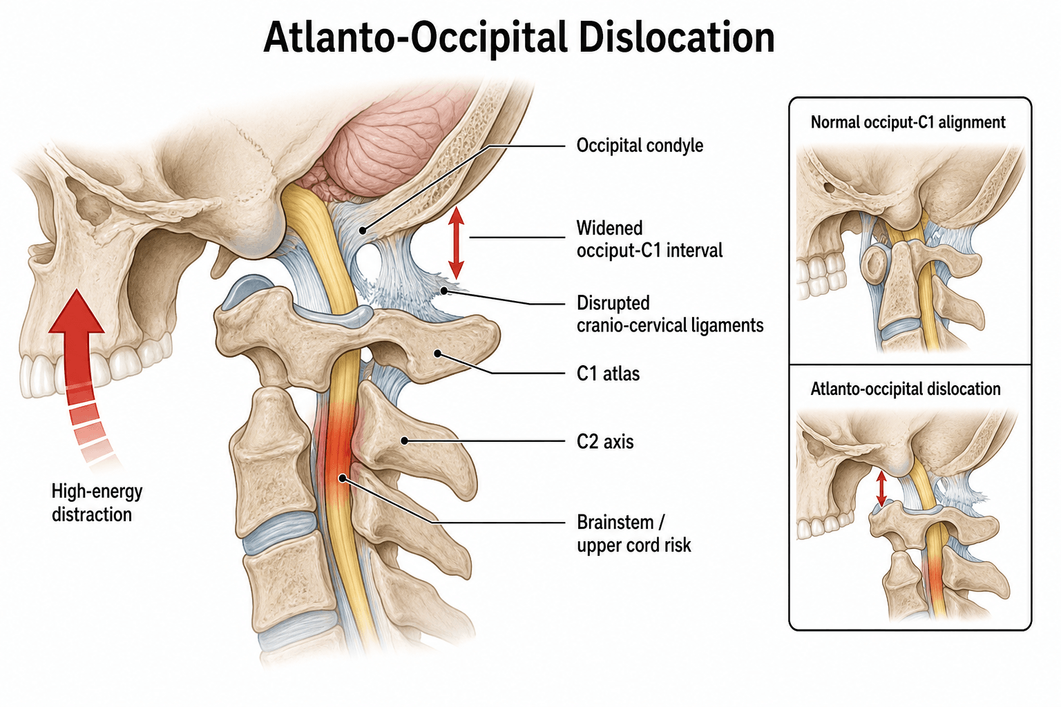
A ligamentous injury. The occipital condyles articulate with the superior facets of C1, and the bony craniocervical junction is intrinsically unstable: it relies almost entirely on ligaments. AOD is therefore fundamentally a soft-tissue injury. The atlanto-occipital joint depends on the paired alar ligaments, the apical odontoid ligament, the tectorial membrane and the joint capsules; once these restraints are disrupted the occiput can translate or distract relative to C1, and the vertebral arteries and brainstem are immediately at risk.
- Function
- Limit rotation and lateral bending
- When Disrupted
- Allow anterior or posterior translation
- Function
- Secondary stabiliser of dens to occiput
- When Disrupted
- Contributes to longitudinal instability
- Function
- Posterior restraint to translation
- When Disrupted
- Permits anterior dislocation when torn
- Function
- Primary joint stability
- When Disrupted
- Complete disruption in all Traynelis types
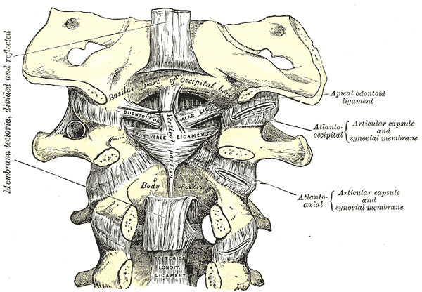
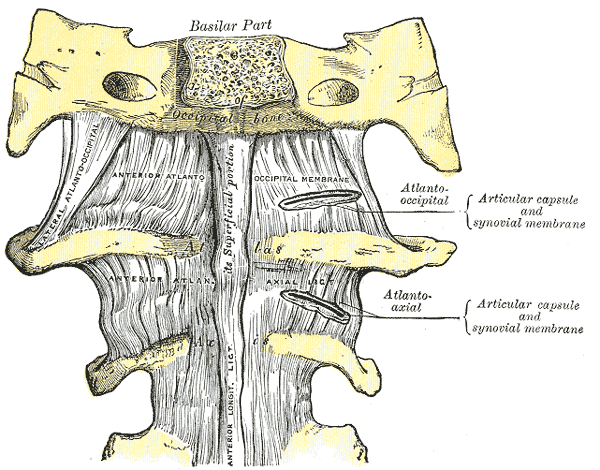
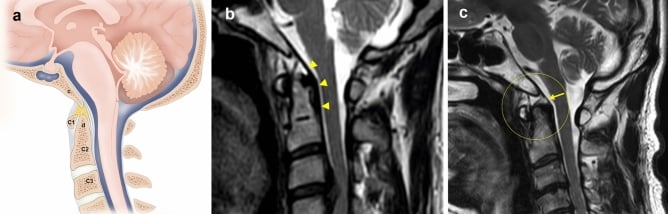
Why Type II is the most dangerous. Longitudinal distraction disrupts all the ligamentous tethers in sequence, from anterior to posterior, which is why it produces the greatest instability. The occiput separates from C1, and the brainstem and vertebral arteries are stretched.
Children. Ligamentous laxity and a larger head-to-body ratio make children more susceptible, and the fulcrum of injury is higher (C2-C3 in infants). AOD must be excluded in any child with an unexplained neurological deficit after trauma, even with a seemingly minor mechanism.
Classification
The Traynelis classification describes the direction in which the occiput has moved on C1. It is the most widely used system because the direction directly informs the reduction manoeuvre and the decision to avoid traction.
- Direction
- Anterior displacement of occiput on C1
- Stability
- Unstable
- Key Imaging Finding
- Occipital condyles anterior to C1 facets on lateral view
- Direction
- Longitudinal distraction
- Stability
- Most unstable
- Key Imaging Finding
- Increased BDI and CCI, vertical separation
- Direction
- Posterior displacement of occiput on C1
- Stability
- Unstable
- Key Imaging Finding
- Occipital condyles posterior to C1 facets
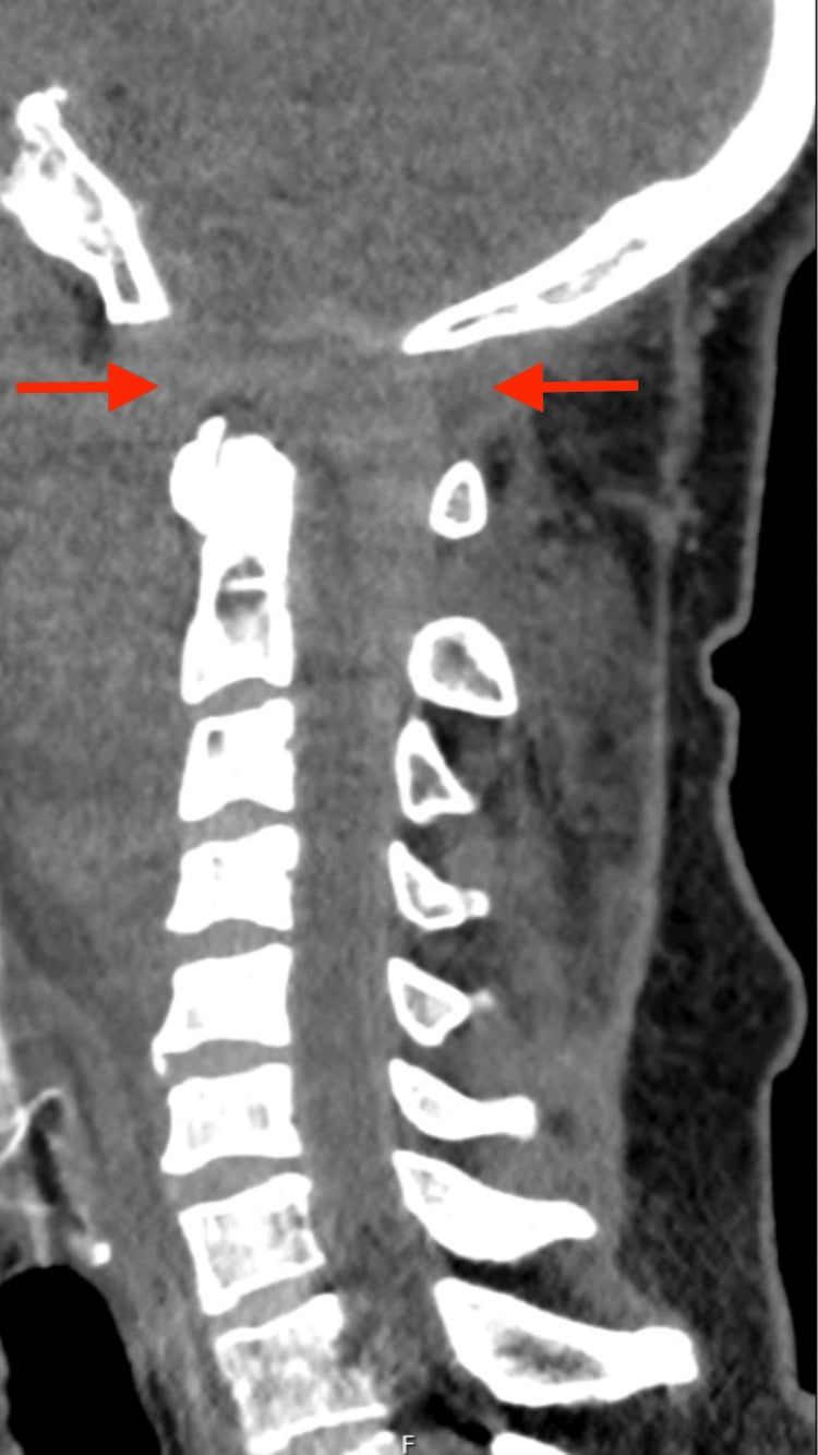
Related Injury: Occipital Condyle Fractures (Anderson-Montesano)
Occipital condyle fractures sit on the same craniocervical-junction spectrum as atlanto-occipital dislocation and appear in its differential. The Anderson-Montesano classification grades morphology and the Tuli classification grades stability; between them they determine whether the fracture is stable or unstable.
- Pattern
- Comminuted / impaction fracture from axial loading
- Stability and treatment
- Stable (alar ligaments and tectorial membrane intact) - rigid collar
- Pattern
- Condyle fracture as part of a basilar skull fracture (linear extension)
- Stability and treatment
- Usually stable - collar
- Pattern
- Avulsion of the condyle at the alar ligament attachment
- Stability and treatment
- Potentially UNSTABLE (the alar ligament is a CCJ stabiliser) - halo or occipitocervical fusion if displaced, bilateral, or with ligamentous injury
The Type III is the one to worry about. It is an alar-ligament avulsion, and because the alar ligament is a craniocervical-junction stabiliser, a displaced or bilateral Type III, or one with associated ligamentous injury, is unstable and may need halo immobilisation or occipitocervical fusion. Types I and II are usually stable and treated in a collar. Like AOD, occipital condyle fractures are frequently missed on plain films and need CT.
The Tuli classification grades instead by displacement and ligamentous integrity: Type 1 non-displaced; Type 2A displaced but stable; Type 2B displaced and unstable. It maps more directly onto management.
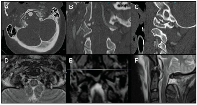
Clinical Assessment
History. The mechanism is a high-energy motor vehicle collision, a fall or a pedestrian strike. A patient who was unconscious at the scene raises suspicion dramatically, and associated head, chest and abdominal trauma are common. The awake patient reports severe neck pain, occipital headache and cranial nerve symptoms.
Examination. Secure the airway first: a high cervical injury risks respiratory arrest. Examine the cranial nerves, especially IX-XII, and record motor and sensory levels. Look at the neck for swelling, step-off and crepitus, but do not manipulate it until it has been imaged, and look for associated scalp lacerations, facial fractures and long-bone injuries.
The unconscious patient. An unconscious polytrauma patient cannot report neck pain, so AOD must be excluded by CT in every high-energy mechanism patient who arrives intubated or with a Glasgow Coma Scale under 8. A normal lateral cervical radiograph does not rule it out.
- Key Discriminating Feature
- Abnormal Powers ratio, CCI, or BDI
- Investigation
- Urgent CT with measurements
- Key Discriminating Feature
- Fixed rotation, normal CCI/BDI
- Investigation
- Dynamic CT or open-mouth view
- Key Discriminating Feature
- Unilateral condyle displacement, stable joint
- Investigation
- CT classification (Anderson-Montesano)
- Key Discriminating Feature
- Lateral mass spread on open-mouth view
- Investigation
- CT for transverse ligament integrity
The other craniocervical injuries competing for this diagnosis are developed in full elsewhere: atlantoaxial rotatory subluxation, Jefferson fracture, and, for the chronic and congenital instability that reaches clinic rather than the resuscitation room, craniocervical junction instability.
Investigations
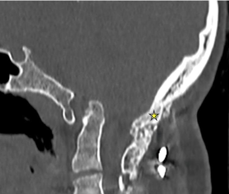
CT first. Non-contrast CT of the cervical spine is the first-line investigation in every high-energy trauma patient, especially the unconscious and those with neck pain: thin slices (1 mm or less) from occiput to T1 with sagittal and coronal reformats. CT with multiplanar reconstructions is mandatory because plain lateral cervical radiographs are unreliable, missing up to 50 percent of cases; the Powers ratio on a film requires precise identification of the basion and opisthion, which are often obscured. Any measurement exceeding its threshold requires immediate spine team notification, so document the exact values in the radiology report and communicate abnormal values immediately to the trauma and spine teams.
The order of measurement. Calculate the condyle-C1 interval bilaterally first, then the Powers ratio and the basion-dens interval; the basion-axial interval is worth recording but is poorly reproducible on reformatted CT. The condyle-C1 interval carries most of the diagnostic weight because it is the only one of these that measures the joint that is actually injured. The classic craniocervical lines are far less sensitive than their familiarity suggests.
- Normal Value
- Mean 1.28 mm in children; no normal joint above 2.5 mm
- Abnormal Threshold
- 2.5 mm or more is outside normal; 4 mm or more is the tested diagnostic criterion
- View Required
- Reformatted coronal and sagittal CT
- Normal Value
- 0.8-1.0
- Abnormal Threshold
- Greater than 1.0 - but only 37.5% sensitive
- View Required
- Lateral cervical X-ray or sagittal CT
- Normal Value
- Under 8.5 mm on CT; under 12 mm on a lateral film
- Abnormal Threshold
- Over 12 mm on a lateral film; 9-12 mm on CT is already outside normal
- View Required
- Sagittal CT or lateral X-ray
- Normal Value
- Under 12 mm on a lateral film
- Abnormal Threshold
- Greater than 12 mm - only 31% sensitive, and hard to reproduce on CT
- View Required
- Lateral X-ray; poorly reproducible on reformatted CT
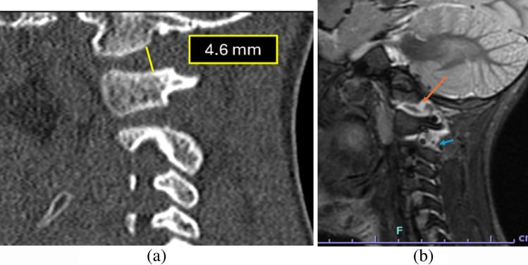
The condyle-C1 interval. Measured on the reformatted coronal and sagittal images between the occipital condyle and the superior articular facet of C1. In Pang's 89 children the mean was 1.28 mm, no normal joint exceeded 2.5 mm, the two sides were markedly symmetrical and the value did not change with age, so abnormal widening or asymmetry signifies AOD. The normal values and the 100 percent sensitivity come from paediatric series; the use of the CCI in adults is a reasonable extrapolation rather than a separately validated threshold.
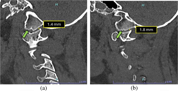
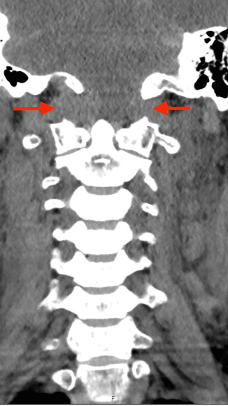
The Powers ratio. The distance from the basion to the posterior arch of C1, divided by the distance from the opisthion to the anterior arch of C1 (BC over OA), on a lateral radiograph or sagittal CT. A ratio greater than 1.0 indicates anterior displacement of the occiput on C1, the Type I pattern. Its weakness is mechanical: the ratio compares two distances that both lengthen in pure distraction, so it can stay under 1.0 while the head is lifted off the spine in a Type II injury, and it behaves erratically in the posterior Type III pattern.
The Harris rule of 12s. The basion measurements answer the question the Powers ratio cannot. The basion-dens interval (BDI), from the basion to the tip of the dens, should be under 12 mm; the basion-axial interval (BAI), from the basion to the posterior axial line (the rostral extension of the posterior cortex of the C2 body), should also be under 12 mm. Either value over 12 mm is abnormal.
The Wackenheim clivus-canal line. A line drawn along the posterior surface of the clivus and extended caudally should fall tangential to the posterior aspect of the tip of the dens. In AOD the occiput-to-dens relationship is disrupted, so the line no longer touches the dens normally; in basilar invagination it is the dens that crosses the line.
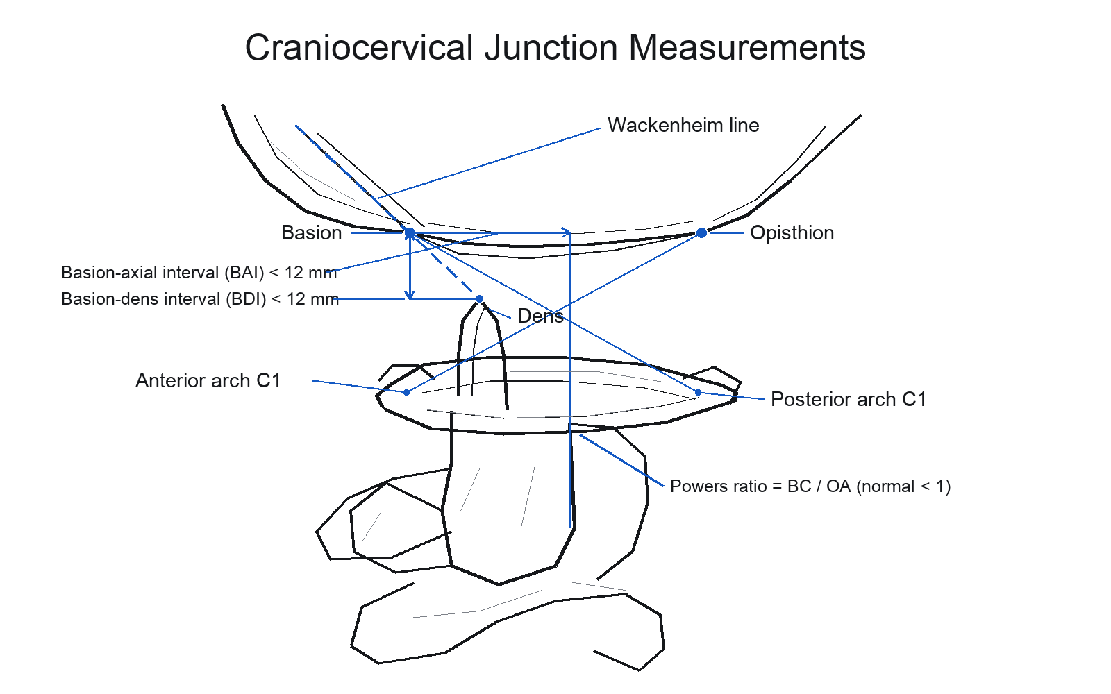
Plain-film numbers do not transfer unchanged to CT. Both 12 mm cut-offs were derived on plain radiographs. On multidetector CT, 95% of uninjured adults have a BDI under 8.5 mm, not 12 mm, so a CT basion-dens interval of 9 to 12 mm passes the classic rule while sitting outside the normal range; the error runs towards under-calling the injury, on exactly the study a trauma patient actually gets. The same series found the BAI difficult to reproduce on CT at all, and narrowed two other familiar craniocervical values: the atlantodental interval to under 2 mm rather than 3 mm, and the atlanto-occipital interval to 0.5-1.4 mm (PMID 17893223; 200 consecutive adult MDCT studies). The Powers ratio was the one measurement that did not shift between the two modalities.
Tested head to head against a CCI of 4 mm or more, the classic criteria are strikingly insensitive: Wholey's dens-basion interval 50%, Powers ratio 37.5%, Harris basion-axial interval 31%, Sun's interspinous ratio 25%, which is a false-negative rate of 50 to 75%. Harris also carried a 50% false-positive rate against the Powers ratio's 10%, so it is not the safer test in either direction. The CCI was positive in all 16 patients with AOD and in none of 10 patients with other upper cervical injuries (PMID 18091277; retrospective diagnostic-accuracy study, 16 AOD and 10 non-AOD controls, predominantly paediatric - a small, single-centre series, but the only direct comparison available).
Use the lines as positive evidence when they are abnormal, and never as permission to clear a craniocervical junction when they are normal. The reformatted CCI, measured bilaterally, is what decides.
The normal-CCI trap. A single normal measurement cannot overrule a globally abnormal craniocervical injury pattern, particularly when an associated C1-C2 dissociation changes the geometry. Read every CT alignment measure together with the ligament pattern rather than treating any one criterion as definitive.
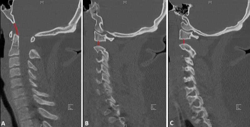
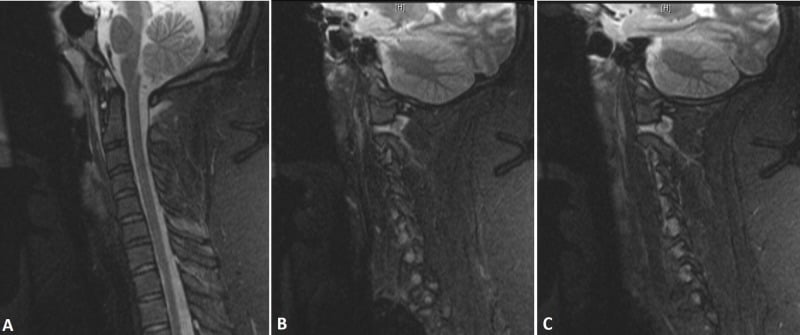
CT angiography. If the CT is positive, CTA assesses vertebral artery integrity before any reduction or surgical planning. A vertebral artery occlusion or dissection alters the timing of surgery and the anticoagulation decisions.
MRI. An adjunct for the neurological deficit unexplained by CT, and for assessing ligamentous and cord injury: tectorial membrane disruption, cord oedema and brainstem signal change. The soft-tissue signs are more sensitive than any of the classic craniocervical lines, with tectorial membrane damage at 71%, extra-axial blood at C1-C2 at 75% and perimedullary subarachnoid blood at 63%, but each carries a false-positive rate of 25 to 40%, so they raise or lower suspicion rather than settle it (PMID 18091277). MRI must never delay collar immobilisation, the CT or spine-team notification; the reformatted CCI remains the measurement that makes or refutes the diagnosis.
Management
The goal. Prevent further distraction or translation while obtaining definitive imaging, then secure the junction. Rigid neutral immobilisation from the scene onward is the single most important intervention.
At the scene. A rigid cervical collar applied in the neutral position, log-roll precautions with manual in-line stabilisation, airway management without neck extension if possible, and rapid transport to a trauma centre with spine capability.
In the emergency department. Maintain neutral alignment and do not remove the collar until the CT has been reviewed. CT of the cervical spine within 30 minutes of arrival; calculate all the measurements (Powers ratio, CCI, BDI, BAI) and notify the spine team immediately if any threshold is exceeded.
After CT, by type.
- Type I and III: gentle closed reduction under fluoroscopy if displaced, then halo or collar
- Type II: strict neutral position, no traction attempted
- All types: occipitocervical fusion within 24-72 hours once medically optimised
Apply a rigid cervical collar or halo ring in the neutral position only, log-roll with in-line stabilisation, and obtain urgent CT from occiput to T1 with sagittal and coronal reconstructions. Traction is absolutely contraindicated until imaging has excluded a distraction-type injury, and in Type II it can be fatal: applying weight can convert a partial ligamentous disruption into complete separation, increase the longitudinal gap, stretch the brainstem and cause immediate cardiorespiratory arrest and death. If the CT demonstrates vertical separation, maintain the neutral collar or halo without any distractive force and make no attempt at closed reduction using traction; reduction, if required, is performed surgically after exposure.
Complications
- Incidence
- The majority die at the scene, from brainstem transection or vascular disruption
- Risk Factors
- High-energy mechanism, delayed diagnosis
- Management
- Prevention via early recognition and immobilisation
- Incidence
- 10-20 percent of survivors
- Risk Factors
- Type II distraction, aggressive manipulation
- Management
- CTA confirmation, anticoagulation if dissection
- Incidence
- 5-15 percent
- Risk Factors
- Smoking, osteoporosis, inadequate fixation
- Management
- Revision with extended construct or bone stimulator
- Incidence
- Common in survivors (IX-XII)
- Risk Factors
- Brainstem stretch at time of injury
- Management
- Supportive, may improve over months
- Incidence
- High in upper cervical cord involvement
- Risk Factors
- Phrenic nerve or brainstem injury
- Management
- Prolonged ventilation, tracheostomy
Outcomes and Prognosis
By type. No source separates survivorship by Traynelis type. In a Type I or III injury diagnosed early, the cranial nerve and cord injury is variable and long-term function is good with a successful fusion and rehabilitation. In Type II the mechanism stretches the brainstem directly, so the rate of brainstem injury is high and long-term function is poor.
Children. Relative survival is higher: in Pang's 16 AOD patients, 11 (69%) made an excellent neurological recovery. Fusion is still required, with growth to consider.
The best prognosis follows early recognition, a Type I or III injury, successful closed reduction, rigid occipitocervical fusion within 72 hours and the absence of vertebral artery injury. The poor prognosis follows a Type II distraction pattern, unconsciousness at the scene, an associated severe head injury and a diagnosis delayed beyond 24 hours.
Two numbers to distrust. Type-specific survival figures in circulation ("20-30% for Type I/III, under 10% for Type II") have no source that separates survivorship by Traynelis type; quote overall survival to hospital and the mechanism instead. And early fusion is credited with "improving survival" only by selection: it is performed on patients who have already survived to theatre, and no series compares it against non-fusion in matched patients. What early stabilisation changes is the neurological outcome and the security of the junction in a survivor, not the probability of surviving. Saying it improves survival is a common and unearned extrapolation.
Guidelines, Registries & Global Practice
- Rare but lethal: Represents less than 1 percent of cervical spine injuries but carries the highest mortality
- Adult peak: High-speed motor vehicle collisions in unrestrained occupants
- Paediatric peak: Falls and pedestrian impacts; higher relative incidence than adults
- Survival bias: Most data come from patients who reach trauma centres alive
- High-resource: Immediate CT with multiplanar reconstructions, CTA, and 24-hour spine coverage for fusion
- Limited-resource: Plain films plus clinical assessment; transfer to centre with CT and surgical capability
- Universal principle: Rigid neutral immobilisation from the scene is the single most important intervention worldwide
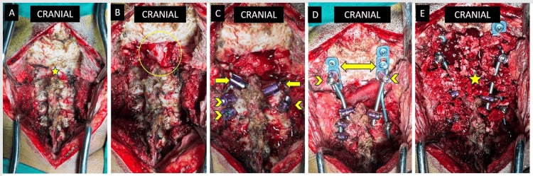
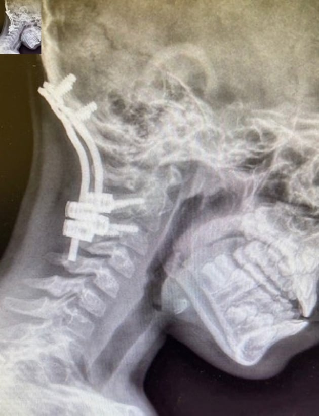
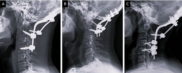
- Surgery: Occipital plating with segmental screw fixation is standard wherever neurosurgical or orthopaedic spine expertise exists
- Diagnosis Emphasis
- CT with CCI, Powers ratio, BDI/BAI on every high-energy trauma
- Initial Management
- Rigid collar or halo in neutral; no traction until type known
- Surgical Timing
- Urgent fusion within 24-72 hours once stable
- Diagnosis Emphasis
- Clinical suspicion plus CT measurements; document all values
- Initial Management
- Log-roll and collar; transfer to major trauma centre if AOD suspected
- Surgical Timing
- Same-day or next-day fusion when patient optimised
- Diagnosis Emphasis
- High index of suspicion in unconscious polytrauma patients
- Initial Management
- ATLS protocol with spine precautions; CT before any manipulation
- Surgical Timing
- Early surgical stabilisation; recognise it secures the junction rather than proving a survival benefit
There is no dedicated international registry for atlanto-occipital dislocation because of its rarity and high pre-hospital mortality. Evidence is drawn from case series and expert consensus. The consistent message across all guidelines is: maintain neutral rigid immobilisation, obtain CT with measurements immediately, avoid traction in distraction injuries, and proceed to occipitocervical fusion as soon as the patient is medically stable.
Record in every high-energy trauma patient:
- Traynelis type once CT is obtained
- Exact values of Powers ratio, CCI (bilateral), BDI, and BAI
- Decision to avoid traction and rationale
- Time of spine team notification and surgical plan A missed or mismanaged atlanto-occipital dislocation is a recurring source of preventable death and litigation worldwide. Always measure and document the craniocervical parameters on CT.
Controversies & Areas of Uncertainty
Some surgeons attempt gentle closed reduction under fluoroscopy for Type I and III injuries before fusion, while others proceed directly to open reduction and fixation. No randomised data exist; practice is driven by surgeon experience and the degree of initial displacement.
Halo immobilisation provides superior rigidity but carries pin-site and pulmonary complications. Segmental screw-and-rod constructs are so stable that some centres use only a rigid collar for 8-12 weeks. Choice remains institution-dependent.
Standard constructs stop at C2. Extension to C3 is used when C2 bone quality is poor or additional stability is required. No comparative data define the threshold for extension.
CTA is routine, but the decision to anticoagulate a dissected vertebral artery before fusion versus after remains variable. Multidisciplinary discussion with neurosurgery and vascular neurology is recommended.
MCQ Practice Points
Q: What is the most unstable Traynelis type of atlanto-occipital dislocation? A: Type II (longitudinal distraction). Type II produces sequential disruption of all ligamentous tethers and creates the greatest risk of brainstem stretch. Traction is contraindicated because it can complete the distraction and cause immediate cardiorespiratory arrest.
Q: What is the abnormal threshold for the condyle-C1 interval (CCI) on coronal CT? A: 2.5 mm or more. The CCI is measured on the coronal reconstruction between the occipital condyle and the superior articular facet of C1. Values of 2.5 mm or more indicate atlanto-occipital instability and mandate urgent spine consultation. CCI is the most sensitive single measurement; the normal values and the 100 percent sensitivity come from paediatric series, and its use in adults is a reasonable extrapolation rather than a separately validated threshold.
Q: Why is traction contraindicated in certain atlanto-occipital dislocations? A: Traction risks completing a partial distraction injury (Type II). In longitudinal distraction injuries, the ligaments are already torn in sequence; any additional distractive force can separate the occiput completely from C1, stretching the brainstem and vertebral arteries and causing immediate death. Neutral rigid immobilisation is used instead until surgical fusion.
Q: What is the Powers ratio and what value indicates anterior atlanto-occipital dislocation? A: Powers ratio is basion-posterior C1 arch distance divided by opisthion-anterior C1 arch distance. A ratio greater than 1.0 indicates anterior displacement of the occiput on C1 (Traynelis Type I). Although still taught, modern practice relies on CT measurements (CCI, BDI) because plain-film landmarks are often obscured.
Q: What is the definitive treatment for a survivor of atlanto-occipital dislocation? A: Occipitocervical fusion using an occipital plate with C1 lateral mass and C2 pedicle screws. After anatomic reduction (Types I and III), rigid internal fixation provides immediate stability. Post-operative halo or collar immobilisation for 8-12 weeks is standard. Wiring techniques have been replaced by modern screw-rod constructs with higher fusion rates.
Q: Why must atlanto-occipital dislocation be excluded even after apparently minor trauma in children? A: Children have ligamentous laxity and a larger head-to-body ratio that allows the craniocervical junction to be injured at lower energy than would be required in adults. A normal plain cervical series does not exclude AOD; CT with CCI measurement is mandatory in any child with neck pain or neurological symptoms after trauma.
Exam Viva Scenarios
Practise clinical reasoning and management decisions out loud
“A 32-year-old unrestrained driver is brought in after a high-speed head-on collision. He is unconscious with a Glasgow Coma Scale of 6. He has been placed in a rigid collar. Lateral cervical X-ray shows an increased basion-dens interval. What is your immediate management and suspected diagnosis?”
“A 4-year-old child is brought to the emergency department after a fall from a first-floor balcony. The child is conscious but complains of severe occipital headache and refuses to move the neck. The mechanism seems relatively low energy. Open-mouth and lateral cervical views appear normal. What is your next step?”
Key Anatomy
- Atlanto-occipital joint relies on alar ligaments, apical ligament, tectorial membrane, and capsules
- Occipital condyles articulate with C1 superior facets; disruption allows translation or distraction
- Brainstem and vertebral arteries are immediately at risk with any displacement
- Type II distraction produces the greatest instability and highest mortality
Traynelis Classification
- Type I: Anterior displacement of occiput on C1 (most common survivable pattern)
- Type II: Longitudinal distraction (most unstable; avoid traction)
- Type III: Posterior displacement of occiput on C1
- Classification directly informs reduction strategy and traction safety
Radiographic Diagnosis
- Powers ratio greater than 1.0 indicates anterior dislocation (lateral view or sagittal CT)
- CCI of 2.5 mm or more on coronal CT is the most sensitive single measurement
- BDI or BAI over 12 mm is abnormal - but that is the plain-film value; on CT a BDI over 8.5 mm is already outside normal
- CT with multiplanar reconstructions is mandatory; plain films miss up to 50 percent
Immediate Management
- Rigid cervical collar or halo in neutral position from the scene
- Log-roll precautions; secure airway without neck extension if possible
- Urgent CT occiput to T1 with measurements; CTA for vertebral arteries
- Never apply traction before confirming Traynelis type on CT
Definitive Treatment
- Occipitocervical fusion (occipital plate + C1 lateral mass + C2 pedicle screws)
- Achieve anatomic reduction first (Types I and III) under fluoroscopy
- Post-operative halo or rigid collar for 8-12 weeks
- Early fusion (within 72 hours) is standard for survivors - but quote it as securing the junction, not as a proven survival benefit
Evidence Base and Key Trials
Traumatic atlanto-occipital dislocation. Case report
- ONE new case plus a review of the 17 previously reported patients surviving AOD more than 48 hours - not a case series
- Proposed the three-part direction-based classification (I anterior, II longitudinal, III posterior) still used worldwide
- Most survivors were children, and many had excellent neurological outcomes
- Fusion was favoured for long-term stability
- On CT the paper is more cautious than this card first said - it concluded CT may CONFIRM the diagnosis 'when necessary', not that it is required
The pathological anatomy of fatal atlanto-occipital dislocations
- Described the ligamentous and osseous disruption patterns in fatal AOD cases
- Confirmed that alar ligaments and tectorial membrane failure allow fatal displacement