Cartilage Lesions | MACI | OAT | Restoration
- Medial femoral condyle is most common location
- ICRS grading classifies cartilage damage
- Microfracture for small (less than 2cm²) contained lesions
- MACI/ACI for larger lesions (greater than 2-4cm²)
- OAT (mosaicplasty) for small-moderate lesions
- “Address alignment, meniscal, and ligament issues concurrently
- “Microfracture produces fibrocartilage (Type I collagen)
- “ACI/MACI produces hyaline-like cartilage (Type II)
- “OAT transfers hyaline cartilage with bone from non-weight-bearing area
Overview and Classification
An osteochondral defect of the knee is damage to the articular cartilage and, potentially, to the subchondral bone beneath it. Treatment aims at pain relief, better function and the prevention of osteoarthritis.
Where. The medial femoral condyle (MFC) is the most common weight-bearing location for symptomatic lesions. Patellofemoral lesions, of the patella or trochlea, are next most frequent and behave differently: they tolerate microfracture poorly, and ACI/MACI has historically had less reliable results there.
ICRS grading. The ICRS grade classifies the cartilage damage by depth, and grades III and IV are generally the ones treated.
- Grade I - superficial softening, fibrillation
- Grade II - partial-thickness lesion, less than 50% depth
- Grade III - deep lesion, greater than 50% depth or down to subchondral bone
- Grade IV - full thickness with exposed subchondral bone
Pathophysiology
Why cartilage does not heal. Articular (hyaline) cartilage is avascular, aneural and alymphatic, and adult chondrocytes have very limited mitotic capacity. A full-thickness chondral injury that does not reach the subchondral vascular bed therefore mounts essentially no intrinsic repair and persists as a defect.
Depth decides the response.
- A partial-thickness (chondral) injury has no access to marrow, so no clot forms and there is no healing response; the defect remains and its rim degenerates over time.
- A full-thickness osteochondral injury breaches the subchondral plate, and a marrow clot carrying mesenchymal stem cells fills the defect. It heals with mechanically inferior fibrocartilage (Type I collagen) rather than true hyaline cartilage (Type II), which is the biological basis of microfracture and of its long-term deterioration.
Why a defect progresses. Once cartilage is lost, higher peak contact stress accelerates degeneration of the adjacent cartilage, and abnormal load transfer to the subchondral bone produces marrow oedema and cysts. That is the substrate for progression to focal, then generalised, osteoarthritis. Anything that raises focal contact stress (malalignment, meniscal deficiency, instability) magnifies the cascade, which is why concurrent pathology must be corrected.
Patient Selection and Concurrent Pathology
The candidate. Cartilage restoration is for a focal lesion in a knee that is otherwise sound:
- Young (typically under 40-50)
- Active and motivated
- Single-compartment disease
- Focal lesion, not diffuse osteoarthritis
- Normal or correctable alignment
- Stable knee
Treat the knee, not only the defect. Alignment, meniscal and ligament problems are addressed concurrently with the cartilage.
- Alignment. Varus or valgus malalignment must be corrected by osteotomy (HTO, DFO); uncorrected malalignment is a leading cause of failure.
- Meniscus. Meniscal deficiency increases contact stress. Consider meniscal allograft transplantation (MAT) after a previous meniscectomy.
- Ligaments. ACL deficiency causes abnormal kinematics. Reconstruct the ligament if the knee is unstable.
Lesion Containment and Shoulder Quality
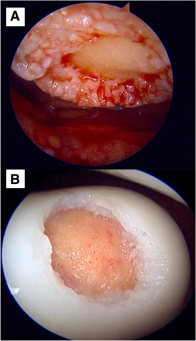
Containment and shoulder quality determine whether a marrow clot or a cell-based graft can stay stable in the defect. Assess both at arthroscopy, together with size, before committing to a technique, because the result may change the planned operation.
Containment. A contained lesion is surrounded on all sides by an intact peripheral rim (wall) of healthy cartilage. That wall holds a marrow clot (microfracture, AMIC) or an ACI/MACI graft inside the defect, and those techniques need it to succeed.
Uncontained lesions have a deficient rim, typically at the very edge of a condyle, in a bipolar ("kissing") lesion, or where the shoulder has broken down. A clot or cell graft cannot be retained, so marrow stimulation and cell therapy do poorly. These lesions favour structural replacement that needs no containing wall, OAT or osteochondral allograft, which bring their own bone and cartilage.
Shoulder quality. The shoulder is the transition between the defect and the surrounding cartilage. A good shoulder is healthy, full-thickness cartilage firmly anchored to subchondral bone, which lets the surgeon debride the lesion to stable vertical walls and create a well-contained defect.
A poor shoulder (soft, fibrillated, delaminating or undermined cartilage) makes the contained appearance illusory: the walls will not hold a repair. That predicts failure of marrow stimulation regardless of lesion size and again favours OAT or OCA, so a small lesion with a poor shoulder is not a good microfracture candidate.
Clinical Presentation
History. Expect a young or active patient with prior trauma, a twisting injury or an insidious onset. Ask about previous knee surgery, especially meniscectomy or ACL reconstruction, and about any previous cartilage procedure.
Symptoms. Pain is activity-related and often localised to the joint line or the front of the knee, with an effusion after activity. Mechanical symptoms (catching, locking, giving way) suggest an unstable fragment or a loose body.
Examination. Look for focal condylar or patellofemoral tenderness, an effusion and possibly crepitus. Always assess limb alignment (varus or valgus, a gait thrust), meniscal signs and ligament stability, because these determine whether any cartilage procedure can succeed.
Red flags against restoration. Diffuse or bipolar pain, older age with established osteoarthritis, and inflammatory features all count against a restoration procedure.
Differential diagnosis. A focal cartilage or subchondral signal change on MRI is not always a treatable osteochondral defect. Distinguish the following before committing to a restoration procedure.
- Typical Patient / Clue
- Young, post-traumatic or insidious; mechanical pain
- Key Imaging Feature
- Sharp-bordered cartilage defect ± exposed bone, ICRS III-IV
- Distinguishing Point
- Discrete, well-shouldered lesion in otherwise healthy joint - the restoration candidate
- Typical Patient / Clue
- Adolescent/young adult; classic lateral aspect of medial femoral condyle
- Key Imaging Feature
- Subchondral fragment with crescentic lucent interface
- Distinguishing Point
- Bone-side process; stability (T2 rim, cysts) drives treatment, not size alone
- Typical Patient / Clue
- Older patient (over 55), acute medial pain, often female
- Key Imaging Feature
- Subchondral insufficiency fracture line, oedema on weight-bearing MFC
- Distinguishing Point
- Not a graft candidate; often related to insufficiency fracture/meniscal root tear
- Typical Patient / Clue
- Older, diffuse/bipolar wear
- Key Imaging Feature
- Joint-space narrowing, osteophytes, bipolar cartilage loss
- Distinguishing Point
- Diffuse not focal - cartilage restoration contraindicated; consider osteotomy/arthroplasty
- Typical Patient / Clue
- Acute trauma
- Key Imaging Feature
- Reticular subchondral oedema, intact cartilage
- Distinguishing Point
- Self-limiting; treat the injury, not the signal
- Typical Patient / Clue
- Middle-aged, popping injury or insidious
- Key Imaging Feature
- Root signal, meniscal extrusion, secondary MFC oedema
- Distinguishing Point
- Drives rapid chondral overload; repair the root rather than chase the cartilage
Investigations
Radiographs. Weight-bearing films assess the joint space, exclude established osteoarthritis and look for OCD or loose bodies:
- AP
- Lateral
- Skyline
- Rosenberg (45° flexion PA)
Alignment films. Long-leg standing alignment films are mandatory before any restoration procedure, to quantify the mechanical axis.
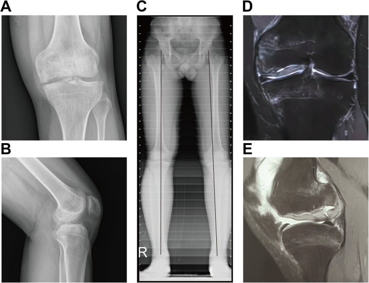
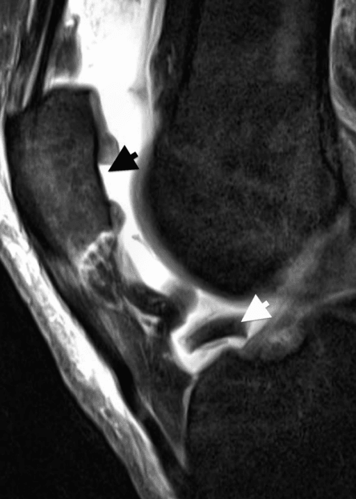
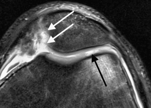
MRI is the key modality. It maps the lesion's location, size and depth (ICRS grade), the state of the subchondral bone (oedema, cysts) and any concurrent meniscal or ligament pathology. On follow-up, cartilage-sensitive sequences and quantitative mapping (for example T2 or dGEMRIC) assess the repair tissue.
Arthroscopy. Diagnostic arthroscopy defines the true ICRS grade, lesion size, containment and shoulder quality, and the lesion can be measured directly with a probe. It is often also the staging step, the chondrocyte harvest, for ACI/MACI.
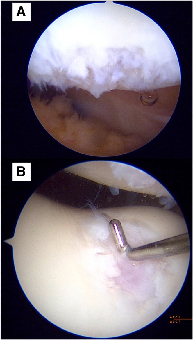
Lesion size measured intra-operatively at arthroscopy is frequently larger than the MRI estimate — never finalise a size-based treatment plan from imaging alone, and consent the patient for the next technique up.
Treatment Options
Choosing by size. Treatment selection is size-dependent, and the zone matters too: a lesion in a weight-bearing area is one to treat.
- Small (less than 2 cm²) - microfracture, OAT
- Intermediate (2-4 cm²) - OAT, MACI
- Large (greater than 4 cm²) - MACI, osteochondral allograft (OCA)
Microfracture
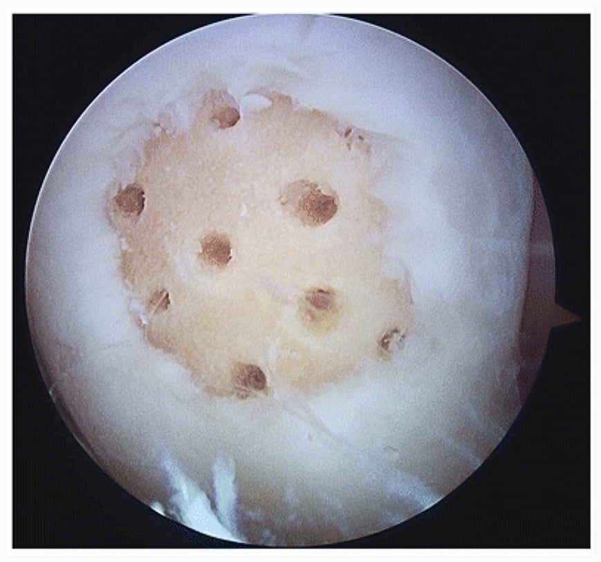
Principle. Holes made in the subchondral bone let marrow elements (mesenchymal stem cells, blood) fill the defect, which heals with fibrocartilage. It is first-line for small lesions, less than 2 cm² and ideally less than 1.5 cm², contained with a healthy shoulder.
Technique. Debride the edges to stable margins, then create holes 3-4 mm apart and 3-4 mm deep. Stable vertical margins retain the marrow clot, and the spacing preserves bridges of subchondral bone and reduces tunnel convergence.
Outcome. Results are good in the short term (2-5 years) but deteriorate at 5+ years, since fibrocartilage is less durable than hyaline cartilage.
ACI and MACI
Principle. Autologous chondrocyte implantation (ACI) and matrix-induced autologous chondrocyte implantation (MACI) repair the defect with the patient's own cultured chondrocytes, and produce hyaline-like cartilage (Type II collagen). It is a two-stage procedure:
- Harvest chondrocytes arthroscopically
- Culture and expand the cells (3-4 weeks)
- Implant them, injected under a periosteal patch (ACI) or on a collagen membrane (MACI)
Indications. Larger lesions (greater than 2-4 cm²), failed microfracture and younger patients.
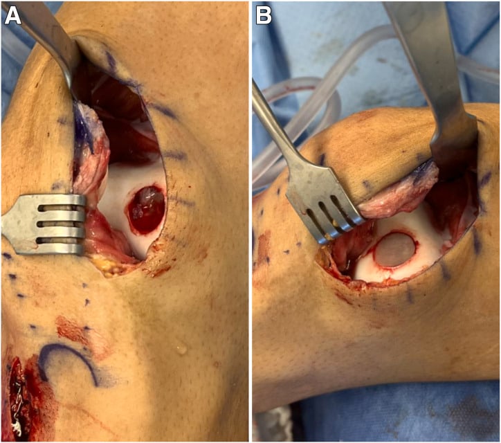
Bone loss. Where an osteochondral defect has lost bone, as in the trochlear defect below, autologous bone graft restores the subchondral plate before the MACI scaffold is placed cell-side toward bone and secured with fibrin glue. Bone and cartilage are addressed as separate reconstructive layers.
Outcome. Good results are reported beyond 10 years, and MACI/ACI is better than microfracture for larger lesions. The trial behind that claim is SUMMIT, in which MACI was superior on pain and function at 2 and 5 years for defects 3 cm² or larger. At 14-15 years, however, first-generation ACI showed no advantage over microfracture (Knutsen), a result that cannot be transferred directly to MACI.
Osteochondral autograft transfer (OAT, mosaicplasty)
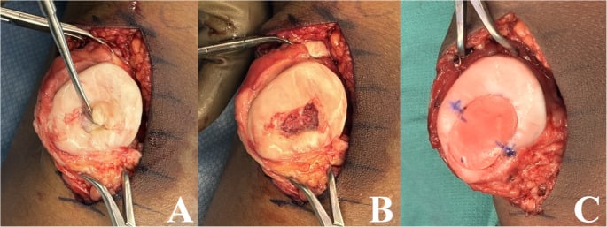
Principle. Osteochondral plugs are harvested from a non-weight-bearing area (trochlear edge, intercondylar notch) and transplanted into the defect in a single stage, bringing true hyaline cartilage with its bone. It suits small-to-moderate lesions (1-4 cm²) and is limited by donor-site morbidity.
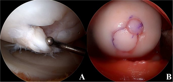
Technique. Congruent plug height and minimal interplug gaps are critical; proud, recessed or widely spaced plugs increase contact stress. The example below is a small structural lateral femoral condyle defect replaced with two plugs.
Osteochondral allograft (OCA)
Principle. A fresh allograft replaces bone and true hyaline cartilage together, like OAT, but without donor-site morbidity. It is used for large lesions (greater than 4 cm²) and is good for young patients with large defects. The price is disease-transmission risk, availability and cost.
AMIC and Scaffold-Augmented Marrow Stimulation
What it is. Autologous matrix-induced chondrogenesis (AMIC) augments marrow stimulation with a protective scaffold. Standard marrow stimulation (microfracture or drilling) is combined with a collagen type I/III membrane or scaffold laid over the defect and sealed, usually with fibrin glue.
What the membrane does. It protects and stabilises the marrow clot, which is otherwise prone to washout and uneven fill. It also provides a three-dimensional scaffold that organises the marrow-derived mesenchymal cells as they differentiate.
Why it exists. AMIC targets microfracture's two weaknesses, an unstable clot with variable fill and mechanically inferior fibrocartilage, while staying single-stage, cell-culture-free and lower-cost. ACI/MACI is two-stage, regulated and expensive, so AMIC is attractive where cell therapy is unavailable or unaffordable.
Where it sits. It is usually offered for medium (roughly 2-4 cm²) contained lesions, where microfracture alone is unreliable but a two-stage cell therapy is not warranted or accessible. Some surgeons extend it to larger contained defects.
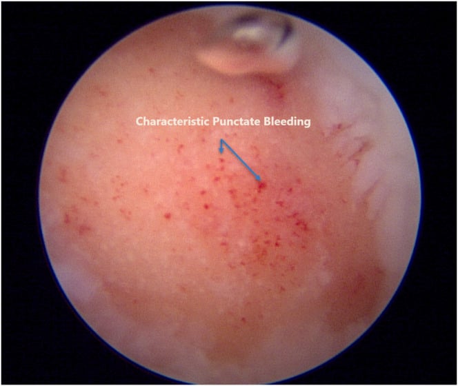
Technique. Punctate bleeding appears across the prepared bed as irrigation pressure falls, confirming access to the subchondral vascular bed. The scaffold is secured with fibrin glue and then tested under wet arthroscopy; a stable construct should remain flush through joint motion without lifting at the edge. The patellar and trochlear defects below show the sequence.
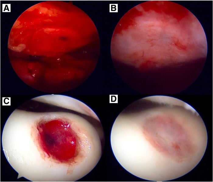
Evidence. Medium-term series report better and more durable defect fill than microfracture alone, but there is little high-level randomised evidence against MACI/ACI, so its precise place in the algorithm remains an area of active study.
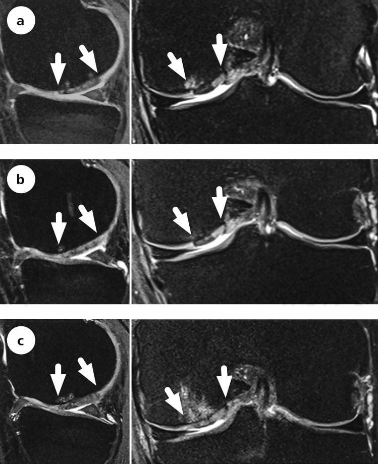
Follow-up imaging. MRI after AMIC must assess both defect fill and the subchondral plate. Structural appearance can evolve despite durable clinical improvement, and it does not alone define success; the series below follows a medial femoral condyle defect to ten years.
Complications
Failure is the dominant concern. Procedure failure with persistent symptoms (graft delamination, incomplete fill or progression to osteoarthritis) ultimately leads to revision restoration or arthroplasty. Each technique has its own ways of failing:
- Microfracture - subchondral bone overgrowth, intralesional osteophyte, subchondral cysts, and fibrocartilage breakdown with symptom recurrence at 2-5 years
- OAT / mosaicplasty - donor-site morbidity, plug subsidence or proud plugs, surface incongruity, dead space between plugs ("valleys"), limited graft availability for large lesions
- ACI / MACI - graft hypertrophy or delamination, periosteal patch overgrowth (older ACI), prolonged rehabilitation, two procedures and cost; possible incomplete integration
- OCA - graft non-incorporation or collapse, immunological and (rare) disease-transmission risk, and dependence on chondrocyte viability that falls with storage time
- General - infection, arthrofibrosis or stiffness, VTE, and accelerated degeneration if concurrent malalignment, meniscal deficiency or instability is left uncorrected
GRAFTCauses of Cartilage Procedure Failure
Hook:A cartilage repair fails for GRAFT reasons - the biggest avoidable ones are missed Realignment and Absent meniscus.
Guidelines, Registries & Global Practice
Global Epidemiology
- Full-thickness chondral lesions are found in roughly 5-11% of knee arthroscopies, and some degree of cartilage damage in up to 60% — most are incidental, only a minority are symptomatic, focal and treatable.
- Symptomatic defects cluster in active patients in their 20s-40s; the medial femoral condyle is the most common symptomatic site, followed by the patellofemoral joint.
- Traumatic and sport-related chondral injury predominates in younger patients, while degenerative/diffuse lesions in older patients are not restoration candidates.
Side-by-Side Guidance
- Position on cartilage repair of the knee
- Emphasises matching technique to lesion size and addressing concurrent malalignment, meniscal and ligament pathology; acknowledges limited high-level comparative evidence between techniques
- Position on cartilage repair of the knee
- ACI/MACI supported for selected symptomatic defects in patients without significant OA who have not had prior cartilage repair, in specialist centres; microfracture positioned for small lesions
- Position on cartilage repair of the knee
- Stresses staged correction of alignment and meniscal deficiency before/with cartilage restoration; subchondral bone preservation
- Position on cartilage repair of the knee
- Provide the ICRS grading framework and decision algorithms driven by lesion size, depth, containment and patient demand
Registry & Outcome Notes
- Dedicated cartilage-repair registries are less mature than arthroplasty registries, but national joint registries (NJR, AJRR, AOANJRR, Swedish/Norwegian) capture the key downstream endpoint: conversion of a prior cartilage procedure to arthroplasty, informing real-world failure and survivorship.
- Long-term RCT data (Knutsen 2016) is more sobering than early single-arm series — both microfracture and first-generation ACI show substantial failure and radiographic OA at 14-15 years.
High- vs Limited-Resource Practice Variation
- Well-resourced settings: full menu available — microfracture/AMIC, OAT, two-stage MACI/ACI and fresh OCA, with combined osteotomy/MAT where indicated. Cell-therapy products (e.g. MACI) require regulatory approval and tissue-bank/lab infrastructure that is not universal.
- Limited-resource settings: marrow stimulation (microfracture/drilling) and OAT/mosaicplasty dominate because they are single-stage, low-cost and require no cell culture or allograft banking. Cultured-chondrocyte and fresh-allograft options are often unavailable, making patient selection and concurrent biomechanical correction even more important.
Exam Relevance (global)
- A core topic across fellowship curricula worldwide.
- Vivas test the ICRS/Outerbridge grading, the size-based treatment algorithm, and — most importantly — the principle that alignment, meniscal and ligament status must be assessed and corrected for any cartilage procedure to succeed.
Related pages: Articular Cartilage Injuries for the general treatment of chondral defects across joints, of which this page is the knee-specific case — the two share most of their evidence base because cartilage restoration trials are overwhelmingly knee trials; Osteochondritis Dissecans of the Knee for the subchondral-bone-driven lesion that presents in the same compartment and is managed differently; Osteochondral Lesion of the Talus for the same techniques applied where lesion access and containment differ; Cartilage Healing and Repair for why the tissue cannot restore itself and what fibrocartilage actually is; MRI Cartilage Assessment for the imaging that sizes and stages these defects; and ACL Injuries, Meniscal Transplant and Patellofemoral Instability for the instability, meniscal deficiency and maltracking that must be corrected or any cartilage procedure fails.
Controversies & Areas of Uncertainty
Does cartilage restoration prevent osteoarthritis? The honest answer is uncertain. In the Knutsen 14-15 year RCT roughly half of survivors had early radiographic osteoarthritis whether they had ACI or microfracture, and there was no difference in total knee replacement. Restoration improves symptoms, but its disease-modifying claim is unproven; counsel patients accordingly.
The 2-4 cm² grey zone. Microfracture, OAT and MACI all have advocates as first-line treatment here. Microfracture is cheapest and single-stage but produces fibrocartilage, OAT gives true hyaline bone and cartilage but is donor-limited, and MACI gives hyaline-like tissue but is two-stage and costly. No single technique is definitively superior across this whole range.
Microfracture's declining reputation. Concerns about subchondral bone overgrowth, intralesional osteophytes and subchondral cysts after marrow stimulation have narrowed its indications toward truly small (under 2 cm²) contained lesions. The same concerns have driven interest in augmented, scaffold-assisted marrow stimulation such as AMIC.
Patellofemoral lesions remain the hardest subgroup. Historically poorer ACI results (Brittberg 1994) have improved with modern matrix techniques and concurrent patellofemoral realignment or unloading, but the evidence base is thinner than for the femoral condyle.
Allograft access. Availability and regulation of fresh osteochondral allograft vary enormously between countries, a major practical determinant of which option a surgeon can actually offer.
Biologics (PRP, BMAC, stem-cell injections). These are widely marketed as cartilage "regenerators" but lack high-level evidence for true cartilage restoration. Their current role is adjunctive symptom management at best.
MCQ Practice Points
Q: What lesion size thresholds guide treatment selection for osteochondral defects?
A: Less than 2 cm²: Microfracture or drilling preferred. 2-4 cm²: OATS (osteochondral autograft) or ACI (autologous chondrocyte implantation). Greater than 4 cm²: ACI or osteochondral allograft (fresh). Microfracture produces fibrocartilage (Type I collagen), while ACI/OATS produce hyaline-like cartilage (Type II collagen).
Q: What is the ICRS classification for cartilage lesions?
A: Grade 0: Normal. Grade 1: Superficial lesions (soft, fissures). Grade 2: Less than 50% depth. Grade 3: Greater than 50% depth, not reaching subchondral bone. Grade 4: Full thickness with exposed subchondral bone. Guides treatment: Grade 3-4 lesions with symptoms are candidates for cartilage restoration procedures.
Q: What are the advantages and disadvantages of OATS versus ACI?
A: OATS advantages: Single-stage, immediate hyaline cartilage, structural bone support. OATS disadvantages: Donor site morbidity, limited graft availability, plug mismatch. ACI advantages: Larger lesions, no donor morbidity. ACI disadvantages: Two-stage, requires periosteal flap or collagen membrane, expensive.
Q: What MRI findings indicate an unstable osteochondritis dissecans lesion?
A: Unstable OCD signs: High T2 signal rim surrounding fragment (fluid interface), cystic changes in subchondral bone, breach of articular cartilage, loose body formation. Unstable lesions require surgical fixation or fragment removal. Stable lesions may be treated nonoperatively in skeletally immature patients.
Q: What is the preferred harvest site for OATS in the knee?
A: Lateral femoral trochlea (superolateral, above sulcus terminalis) and intercondylar notch. These areas are non-weight-bearing. Harvest plugs perpendicular to surface. Maximum 2-3 plugs to avoid significant donor morbidity. Plug diameter typically 6-10mm. Match recipient site curvature.
Exam Viva Scenarios
Practise clinical reasoning and management decisions out loud
“A 28-year-old has a 3cm² full-thickness cartilage defect on the medial femoral condyle. Knee is stable with normal alignment. What are your treatment options?”
“You are seeing a 32-year-old recreational footballer in your clinic 3 years after he underwent arthroscopic microfracture for a 1.8cm² ICRS grade IV cartilage defect on the medial femoral condyle. At the time, microfracture was appropriate given the lesion size (under 2cm²) and he was a suitable candidate - young, active, stable knee with normal alignment. He did well initially, returning to football at 9 months post-operatively with significant improvement in his knee pain. However, over the past 6-8 months, his medial knee pain has gradually returned and is now limiting his ability to play football. He has tried physiotherapy, activity modification, and NSAIDs with minimal benefit. On examination, he has medial joint line tenderness, a small effusion, and pain with deep knee flexion. Range of motion is full (0-135°). His knee is stable to ligamentous examination and there is no malalignment on standing alignment. You order an MRI which shows deterioration of the previously treated cartilage lesion - the microfractured area now has poor fill with irregular surface and underlying bone marrow edema. The lesion measures approximately 2.2cm² (slightly larger than original). There is no evidence of other cartilage damage, meniscal pathology, or ligamentous injury. The patient is frustrated and asks what can be done - he wants to continue playing football. How do you counsel him and what is your management plan?”
“You are seeing a 35-year-old woman in your knee reconstruction clinic who was referred by a colleague. She has severe medial knee pain following a skiing injury 4 years ago where she sustained an ACL rupture which was reconstructed at the time. However, her pain has progressively worsened despite a stable ACL reconstruction. She has tried conservative management including physiotherapy, weight loss (BMI now 28, down from 32), activity modification, and multiple courses of injections with minimal sustained benefit. She is now unable to walk more than 500 meters without severe pain and has had to stop her active lifestyle completely. On examination, she has marked medial joint line tenderness, moderate effusion, full range of motion (0-130°), stable ACL reconstruction (negative Lachman and pivot shift), but varus thrust during gait. Standing alignment films show 6° of mechanical varus with the mechanical axis passing well medial to the knee center, loading the medial compartment. MRI shows a large ICRS grade IV cartilage defect on the medial femoral condyle measuring 5cm² with significant bone marrow edema. Additionally, the MRI shows that she had a previous total medial meniscectomy (you review the notes - at the time of her ACL reconstruction 4 years ago, the medial meniscus was found to be complex and degenerative with extensive tearing, and the surgeon performed a total meniscectomy). There is no evidence of significant arthritis in other compartments - the lateral compartment is pristine. The patient is desperate for help and wants to avoid knee replacement as long as possible. A previous surgeon told her nothing could be done and she would need a knee replacement within 5 years. How do you counsel her and what is your surgical plan if you proceed?”
Treatment by Size
- Small (less than 2cm²): Microfracture, OAT
- Medium (2-4cm²): OAT, MACI
- Large (greater than 4cm²): MACI, OCA
Cartilage Quality
- Microfracture: Fibrocartilage (Type I)
- MACI/ACI: Hyaline-like (Type II)
- OAT/OCA: True hyaline with bone
Address Concurrent
- Malalignment (HTO/DFO)
- Meniscal deficiency (MAT)
- ACL instability (ACLR)
MACI
- Two-stage procedure
- Larger lesions (greater than 2cm²)
- Better than microfracture long-term
- Hyaline-like cartilage
Evidence Base
Brittberg et al (first ACI series)
- 23 patients, full-thickness defects 1.6-6.5 cm², cultured autologous chondrocytes under periosteal flap
- 14 of 16 femoral condylar transplants good-to-excellent at 2 years
- Patellar lesions did far worse (only 2 of 7 good/excellent)
- Biopsy: hyaline-like cartilage in 11 of 15 femoral grafts vs 1 of 7 patellar
Saris et al (SUMMIT, 2-year)
- 144 patients, symptomatic Outerbridge III-IV defects 3 cm² or larger (mean lesion 4.8 cm²)
- MACI vs microfracture; co-primary endpoint KOOS pain + function at 2 years
- MACI significantly better for both pain and function (P=.001)
- Fewer treatment failures with MACI (12.5%) than microfracture (31.9%)
Brittberg et al (SUMMIT Extension, 5-year)
- 5-year follow-up of the SUMMIT cohort (128 of 144 patients)
- MACI superiority in KOOS pain + function maintained at 5 years (P=.022)
- MRI defect fill improved in both groups with no significant difference
- No unexpected safety signals
Knutsen et al (ACI vs microfracture RCT)
- 80 patients, single femoral condyle defect, 14-15 year follow-up
- No significant difference between first-generation ACI and microfracture on any clinical score
- Failures: 17 (ACI) vs 13 (microfracture); more TKRs in the ACI group (6 vs 3)
- Radiographic OA (Kellgren-Lawrence 2 or greater) in roughly half of survivors in each group