Night Pain | Aspirin Relief | Nidus Under 2cm | RFA vs Excision
- Night pain relieved by NSAIDs/aspirin - classic presentation
- Nidus under 2cm - larger is osteoblastoma (same histology)
- CT is gold standard - shows nidus within reactive sclerosis
- RFA first-line treatment - 90%+ success, day case procedure
- Surgical excision if RFA fails or for specific locations
- “Nidus under 2cm differentiates from osteoblastoma
- “Bone scan hot with double-density sign
- “Pain mechanism: prostaglandins from nidus
- “May cause scoliosis in spine (painful type)
Overview and Epidemiology
Osteoid osteoma is a benign osteoblastic bone tumour: a small nidus of osteoid tissue, under 2cm, surrounded by reactive bone sclerosis. It is histologically identical to osteoblastoma and is distinguished from it by size and clinical behaviour.
Who. It accounts for 10-12% of benign bone tumours, with an annual incidence of approximately 3 per million, and it outnumbers osteoblastoma 3:1. The peak age is 10-25 years, with 80% of patients under 30; males outnumber females 2:1 to 3:1, and all ethnic groups are equally affected.
Where. The lower extremity carries 50-60% of lesions, the femur and tibia most commonly, and the upper extremity 20%.
- Percentage
- 30-35%
- Clinical Features
- Proximal femur/femoral neck common
- Percentage
- 20-25%
- Clinical Features
- Diaphysis most frequent
- Percentage
- 10-15%
- Clinical Features
- Posterior elements, painful scoliosis
- Percentage
- 10%
- Clinical Features
- Phalanges, atypical features
- Percentage
- 5-10%
- Clinical Features
- Similar to femur/tibia
- Percentage
- 10-20%
- Clinical Features
- Any bone possible
Natural history. Untreated, an osteoid osteoma typically causes persistent symptoms for 3-5 years before it resolves spontaneously, which it does in most cases. Approximately 5-10% resolve within 2 years and most take 3-6 years, while some persist beyond 6 years without resolution. Regression occurs as the nidus matures into inactive sclerotic bone and its prostaglandin production gradually ceases.
Risk factors. No clear risk factor has been identified. The tumour is not associated with trauma, infection or inherited conditions, and it is a true neoplasm rather than a reactive process.
Anatomy and Pathophysiology
The nidus. A central zone of osteoid and woven bone in various stages of maturation, laid down as haphazardly interconnecting trabeculae. The trabeculae are rimmed by a single layer of plump but cytologically bland osteoblasts and sit in a loose, richly vascular fibrous stroma, which carries the unmyelinated nerve fibres that mediate the pain. Mineralisation is variable, and central calcification is common.
The host bone. The nidus is sharply demarcated from the dense reactive (sclerotic) bone around it, and there is no permeation or entrapment of the pre-existing lamellar bone; that circumscription is a key feature. In the cortical type the reactive sclerosis is extensive and disproportionate to the small nidus, which gives the classic radiographic appearance.
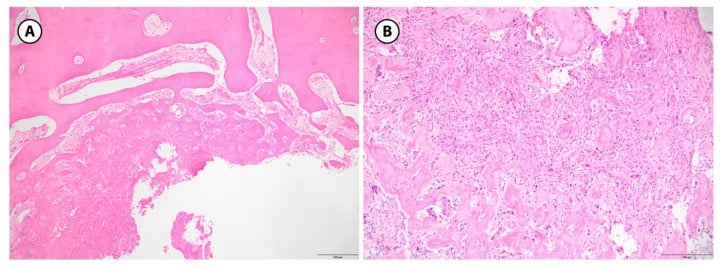
Biopsy. Tissue is not routinely needed when the clinical and CT findings are classic, but it is valuable when the diagnosis is uncertain.
On a small or fragmented specimen the osteoid and osteoblasts can be over-called as osteosarcoma. The discriminators are the lack of cytologic atypia, the organised architecture, sharp circumscription and the absence of permeative growth, together with clinico-radiological correlation: a small nidus under 2cm with reactive sclerosis. Never let the pathologist report a bone-forming lesion without the clinical and CT context.
Why it hurts. The nidus produces high levels of prostaglandin E2 (PGE2) through the COX pathway. PGE2 causes local vasodilation and oedema and sensitises nerve fibres, and it produces the characteristic nocturnal pain.
Why at night, and why aspirin works. The night pattern is attributed to the circadian rhythm of prostaglandin production, the fall at night in cortisol, which is anti-inflammatory, and venous stasis during recumbency increasing pressure. COX inhibitors block prostaglandin synthesis, which is why NSAIDs relieve the pain, and aspirin is particularly effective because its COX inhibition is irreversible.
Classification Systems
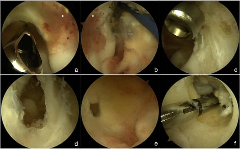
Osteoid osteoma is classified by where the nidus sits in the bone, and the site has important treatment implications. Cortical lesions are ideal for RFA, while intra-articular and subperiosteal lesions may require arthroscopic or open excision to protect the articular cartilage.
- Location
- Diaphyseal cortex
- Percentage
- 75%
- Clinical Features
- Classic presentation, dense sclerosis, night pain
- Location
- Metaphysis/epiphysis
- Percentage
- 15%
- Clinical Features
- Less sclerosis, harder to diagnose
- Location
- Under periosteum
- Percentage
- 10%
- Clinical Features
- Joint effusion, synovitis, minimal sclerosis
- Location
- Within joint capsule
- Percentage
- Subset of above
- Clinical Features
- Hip most common, mimics synovitis
Intra-articular and subperiosteal lesions. These sites have less sclerosis and more synovitis, and they often present as synovitis and stiffness rather than classic night pain, so delayed diagnosis is common. CT must interrogate the articular surface carefully.
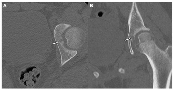
Osteoid osteoma or osteoblastoma. Nidus size is the only reliable differentiator: under 2cm is osteoid osteoma, over 2cm is osteoblastoma. The distinction matters because osteoblastoma is more aggressive, less likely to resolve spontaneously, and requires more extensive surgical excision rather than minimally invasive RFA.
- Osteoid Osteoma
- Under 2cm (usually under 1.5cm)
- Osteoblastoma
- Over 2cm (often 2-10cm)
- Osteoid Osteoma
- Night pain, dramatic NSAID relief
- Osteoblastoma
- Dull ache, less NSAID responsive
- Osteoid Osteoma
- Benign, may spontaneously resolve
- Osteoblastoma
- Locally aggressive, can recur
- Osteoid Osteoma
- Long bone diaphysis
- Osteoblastoma
- Spine (posterior elements)
- Osteoid Osteoma
- Same as osteoblastoma
- Osteoblastoma
- Same as osteoid osteoma
- Osteoid Osteoma
- RFA usually sufficient
- Osteoblastoma
- En bloc excision preferred
- Osteoid Osteoma
- Under 10% with complete ablation
- Osteoblastoma
- 10-20% after excision
Both osteoid osteoma and osteoblastoma harbour recurrent rearrangements of the FOS gene (and, less commonly, the related FOSB gene), driving over-expression of the FOS transcription factor. This shared genetic driver confirms that the two lesions are a single neoplastic spectrum of genuine clonal neoplasms, separated by size and behaviour rather than biology.
It is also of diagnostic value: FOS immunohistochemistry or molecular testing can help confirm an osteoblastic lesion and distinguish it from mimics, such as the bone-forming areas of osteosarcoma, supporting the histological assessment.
Clinical Assessment
History. The classic complaint is night pain that wakes the patient from sleep and is relieved by aspirin or NSAIDs within 20-30 minutes. The pain is dull and aching, gradually increasing over months, and well localised: the patient can point to it with one finger. Ask specifically about the time of the pain, the NSAID response, the duration of symptoms (usually under 2 years) and any prior imaging.
Atypical presentations. The site of the lesion changes the story:
- Intra-articular - joint effusion and stiffness, mimicking synovitis
- Spinal - painful scoliosis in adolescents, the concavity towards the lesion
- Near a growth plate - growth disturbance is possible
- Hand and foot - swelling, with fusiform soft-tissue enlargement
Examination. Expect point tenderness over the lesion, possibly with soft-tissue swelling or warmth, and there may be muscle wasting from chronic pain. The range of motion is usually normal unless the lesion is intra-articular, and each site has its own signs.
- Examination Findings
- Hip pain, limited internal rotation, antalgic gait
- Examination Findings
- Localised tenderness, may feel cortical thickening
- Examination Findings
- Paravertebral muscle spasm, painful scoliosis
- Examination Findings
- Effusion, limited ROM, synovitis signs
Differential diagnosis. The lesions that share the history or the imaging, and what separates them:
- Key Differentiating Features
- Larger nidus over 2cm, less pain relief with NSAIDs, can be aggressive
- Investigation Findings
- CT: nidus over 2cm, may have expansion
- Key Differentiating Features
- History of infection, systemic symptoms possible, different pain pattern
- Investigation Findings
- MRI: rim enhancement, sequestrum possible
- Key Differentiating Features
- Activity-related pain, improves with rest, recent increase in activity
- Investigation Findings
- MRI: linear signal, cortical involvement
- Key Differentiating Features
- Age 5-25, systemic symptoms, permeative pattern, soft tissue mass
- Investigation Findings
- MRI: large lesion, aggressive features
- Key Differentiating Features
- Night pain, NSAID relief, nidus under 2cm on CT
- Investigation Findings
- CT: small nidus with reactive sclerosis
Investigations
Radiographs come first, then CT. MRI is supplementary, and a bone scan is kept for when the diagnosis is uncertain.
Radiographs. These may be normal in early cases or cancellous locations. The typical finding is cortical thickening with a central lucency, the nidus, and the amount of sclerosis varies with the location and duration of the lesion.
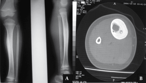
CT is the gold standard. Always request CT if osteoid osteoma is suspected. A thin-slice acquisition (1-2mm) through the affected area defines the nidus, its size, location and calcification, and CT is essential for treatment planning, including the RFA trajectory.
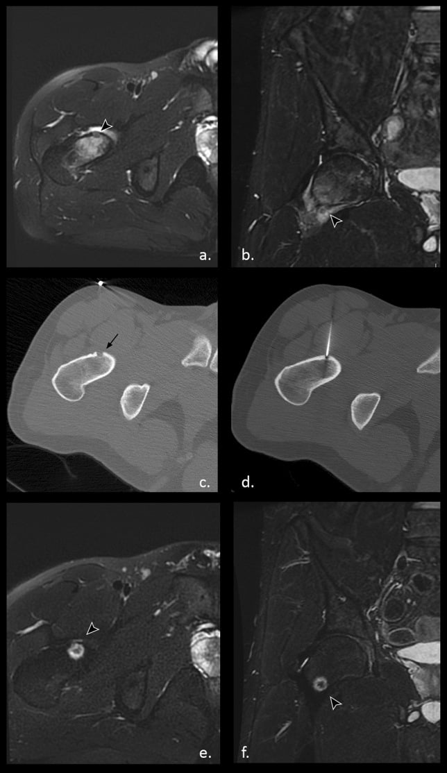
MRI can mislead. It shows extensive marrow and soft-tissue oedema, which may overestimate the extent of the lesion and appear aggressive enough to mimic infection or malignancy, while the nidus itself may be obscured by the reactive changes. It is still useful for intra-articular and spinal lesions.
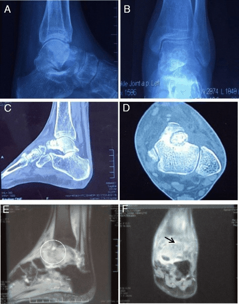
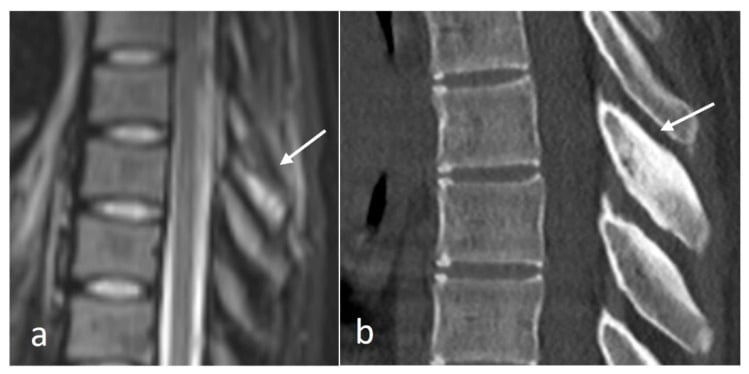
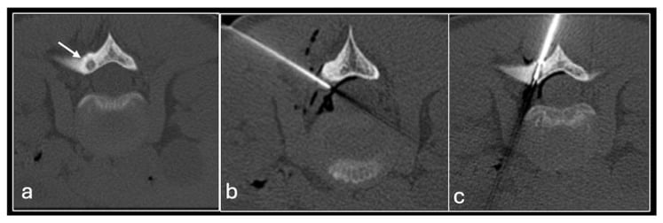
The spine. Unilateral posterior-element pain can drive a painful scoliosis and demands targeted thin-slice CT. MRI emphasises the reactive marrow oedema; CT localises the sclerotic posterior-element lesion better, but a conspicuous lucent nidus may be absent.
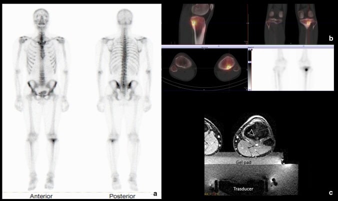
Bone scan. A technetium bone scan is very sensitive, nearly 100%, and shows the double-density sign: focal uptake at the nidus within a larger area of reactive uptake. Use it when the diagnosis is uncertain, for instance when symptoms are classic but radiographs and initial CT are non-diagnostic, and to screen multiple sites or find an occult lesion. SPECT/CT localises the focus precisely.
- Sensitivity
- 75%
- Key Findings
- Cortical thickening, central lucency
- Limitations
- May miss early/cancellous lesions
- Sensitivity
- Over 95%
- Key Findings
- Nidus with variable calcification, reactive sclerosis
- Limitations
- Radiation exposure
- Sensitivity
- 65-85%
- Key Findings
- Extensive oedema, nidus low signal
- Limitations
- May obscure nidus, appears aggressive
- Sensitivity
- Over 95%
- Key Findings
- Double-density sign, intense focal uptake
- Limitations
- Non-specific, poor anatomical detail
Laboratory tests. These are typically normal: FBC, ESR, CRP and alkaline phosphatase are usually within normal limits. They mainly serve to exclude infection, with WCC, CRP and ESR raised in a Brodie abscess, rather than to diagnose osteoid osteoma.
Management Algorithm
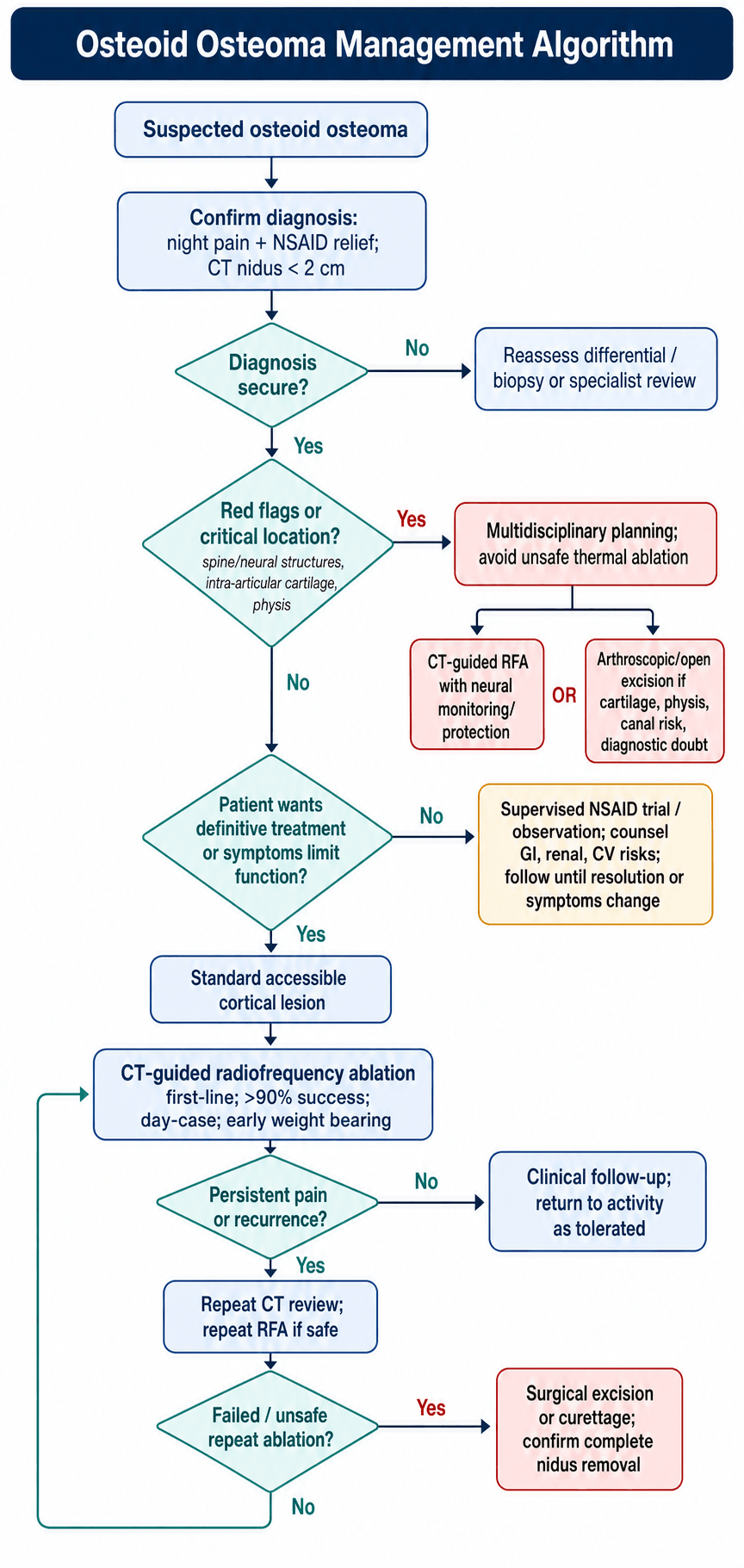
The decision. Confirm the diagnosis from the clinical history (night pain, NSAID relief) and a CT showing a nidus under 2cm. Then assess the location: a lesion in a standard location proceeds to RFA, while the spine, an intra-articular site or the physis calls for the alternatives to be considered. Last come the patient's wishes: one who prefers non-surgical treatment can have a trial of NSAIDs, as the lesion may resolve in 2-5 years, and one who requires definitive treatment has RFA or surgical excision.
- First Choice
- CT-guided RFA
- Alternative
- Surgical excision (en bloc)
- Special Considerations
- Most common location, excellent RFA access
- First Choice
- RFA with nerve monitoring
- Alternative
- Surgical excision
- Special Considerations
- Risk of thermal injury to neural structures
- First Choice
- Arthroscopic excision
- Alternative
- RFA with chondroprotection
- Special Considerations
- Cartilage damage risk with RFA
- First Choice
- Surgical excision
- Alternative
- RFA
- Special Considerations
- Risk of fracture, limited bone stock
- First Choice
- Surgical excision
- Alternative
- Cryoablation
- Special Considerations
- Thermal damage to physis with RFA
Non-operative management. It suits the patient who prefers conservative treatment, has a contraindication to anaesthesia, or has a lesion in a location that is difficult to access. The regimen is aspirin 650mg-1g twice daily or naproxen 500mg twice daily, continued until spontaneous resolution (average 3-4 years), with monitoring for GI side effects; its success is variable and symptoms continue until the lesion resolves.
Long-term NSAID use has GI, renal and cardiovascular risks and is not suitable for all patients. Definitive treatment is usually preferred; conservative management is a valid option for patients who cannot undergo, or who decline, intervention.
Indications for intervention. Operative intervention is the standard approach for most patients seeking symptom relief. The absolute indications are a patient who desires definitive treatment, symptoms affecting quality of life, and a spinal lesion causing neurological symptoms. The relative indications are failed conservative management, NSAID intolerance or contraindication, and proximity to the growth plate requiring excision.
Radiofrequency ablation is the first-line treatment in most cases. It is a CT-guided percutaneous procedure with over 90% success and lower morbidity than open surgery.
Surgical excision, en bloc or by curettage, is indicated when RFA is contraindicated or has failed. Its morbidity is higher, but it is definitive.
Other ablation techniques. Besides RFA, the nidus can be ablated by:
- Cryoablation - an alternative near growth plates; it allows real-time monitoring of the treatment margin and may be useful near sensitive structures
- Laser ablation - similar to RFA
- MR-guided focused ultrasound - an emerging technique
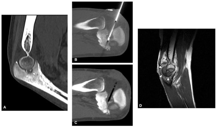
Surgical Technique
Planning. Review the CT for the optimal trajectory, identify a safe corridor that avoids neurovascular structures, and position the patient for access to the lesion.
Anaesthesia. General anaesthesia is preferred for patient comfort. Local anaesthesia is possible in a cooperative patient, and if it is used a periosteal injection of local anaesthetic is essential.
The procedure. Fifteen to twenty minutes, with heating at 90 degrees for 4-6 minutes:
- Position the patient to allow CT scanner access and a comfortable probe trajectory, and mark the skin entry point from the planning CT.
- Prepare with standard sterile technique, draping to allow CT imaging during the procedure.
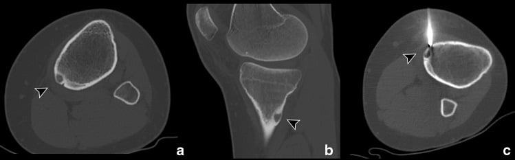
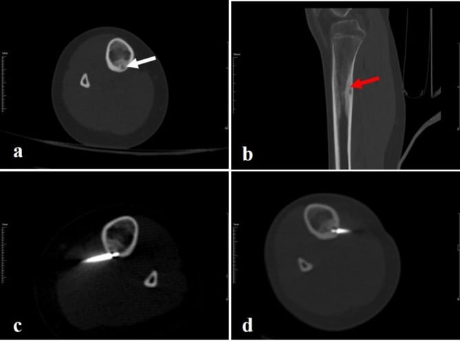
- Under intermittent CT guidance, advance a coaxial needle to the cortex overlying the nidus and confirm its position on CT.
- Penetrate the cortex with a hand drill or powered drill, advancing to the nidus but not through it, and confirm the position on CT.
- Optionally, take a core biopsy through the needle for histological confirmation if the diagnosis is uncertain.
- Insert the RFA probe through the coaxial system with its tip at the centre of the nidus, confirmed on CT.
- Heat to 90°C for 4-6 minutes, monitoring temperature and impedance.
- Allow the probe to cool before removing it, remove the coaxial system and apply a simple dressing.
- Image after the procedure to confirm complete ablation.
Getting it right. The entire nidus must be ablated for cure: incomplete ablation causes recurrence, and off-centre placement is a major preventable cause of persistence. The technique achieves complete nidus destruction while preserving the surrounding bone.
Near neural structures. Spinal ablation requires a safe thermal distance, active neuroprotection and temperature or neurological monitoring; otherwise open or navigated excision is safer. Air dissection creates an insulating margin before RFA.
Complications
- Incidence
- Under 5%
- Prevention/Management
- Maintain adequate distance from skin, use cooling
- Incidence
- Under 2%
- Prevention/Management
- Maintain over 1cm from nerves, use monitoring
- Incidence
- Under 1%
- Prevention/Management
- Avoid excessive cortical destruction, protect post-procedure
- Incidence
- 5-10%
- Prevention/Management
- Ensure complete nidus ablation, repeat RFA if needed
- Incidence
- Under 2%
- Prevention/Management
- Avoid RFA near articular surface, use chondroprotection
- Incidence
- Under 1%
- Prevention/Management
- Sterile technique, prophylactic antibiotics
Open excision. The complications of open excision and the factors that raise their risk are set out below.
- Incidence
- 5-10%
- Risk Factors
- Large cortical window, weight-bearing bone
- Incidence
- 5-15%
- Risk Factors
- Incomplete excision
- Incidence
- Under 2%
- Risk Factors
- Standard surgical risk
- Incidence
- Under 2%
- Risk Factors
- Deep location, poor visualisation
- Incidence
- Variable
- Risk Factors
- Intra-articular location
Recurrence. Recurrence typically occurs within 2 years of treatment. Assessment looks for the return of the typical night pain pattern and repeats the CT to confirm a residual or recurrent nidus.
Treating it. Repeat RFA is successful in most cases, 85-90%, and is effective when a safe trajectory remains. Surgical excision follows if RFA fails twice, and en bloc excision offers a definitive cure.
Persistent or recurrent pain after treatment requires repeat imaging. Do not assume treatment failure without confirming residual nidus on CT.
Postoperative Care
After RFA. Rapid recovery is one of the major advantages of RFA: most patients return to normal activity within 1 week and are back to full activity within 1-2 weeks, compared to 6-12 weeks for surgical excision. They are discharged the same day or the next morning, bear weight as tolerated immediately, and take simple analgesia (paracetamol, NSAIDs) for 24-48 hours.
RFA follow-up. The wound is checked at 1 week, and there are no activity restrictions. Clinical follow-up is at 6 weeks, CT is repeated only if symptoms persist or recur, and the patient is discharged if asymptomatic at 6 months.
After surgical excision. Recovery takes 4-8 weeks, and the return to work and sport varies with the size of the excision (table below). Weight bearing depends on the extent of resection: as tolerated after a minor cortical window, protected for 4-6 weeks after a significant resection.
Excision rehabilitation. Activity is restricted for 4-6 weeks for bone healing, wound management is standard, and physiotherapy is added if needed.
Excision follow-up. Monitor for fracture if significant bone has been removed. Clinical review is at 6 weeks, 3 months and 6 months, with imaging only if symptoms recur and assessment of the bone-graft site if applicable.
- Return to Work
- 1-3 days
- Return to Sport
- 1-2 weeks
- Return to Work
- 1-2 weeks
- Return to Sport
- 4-6 weeks
- Return to Work
- 2-4 weeks
- Return to Sport
- 8-12 weeks
Outcomes and Prognosis
Results of treatment. Both treatments achieve over 90% success. RFA has a primary success rate of 90-95%, and surgical excision succeeds in 90-95%, within a range of 88-100%. After successful treatment long-term cure is expected in over 95%, and the prognosis is excellent: osteoid osteoma is benign, with no risk of malignant transformation.
Prognostic factors. Outcomes are better with a cortical location (the best RFA access), complete ablation or excision of the nidus, and a small nidus under 1cm. A cancellous or intra-articular location, a large nidus approaching 2cm and a spinal location, with its technical challenges, are less favourable.
Function. After RFA patients make a full return to pre-morbid function, with no long-term restrictions and excellent satisfaction. After surgical excision recovery is generally full; a residual bone defect may remain on imaging, but it rarely causes functional limitation.
Guidelines, Registries & Global Practice
Global Epidemiology
Osteoid osteoma accounts for roughly 10-12% of benign bone tumours and an estimated 2-3% of all primary bone tumours, with a consistent young-male predominance (peak 10-25 years, M:F approximately 2:1) reported across European, North American, and Asian series. There are no significant ethnic differences in incidence. The lower limb (femur and tibia) dominates, with the spine involved in roughly 10-15% of cases. Large multicentre and meta-analytic cohorts confirm this distribution: the European multicentre spinal series of Beyer et al. (Neuroradiology 2019, DOI) and the 749-patient meta-analysis of Sangiorgio et al. (Eur Spine J 2023, DOI) both report spinal lesions concentrated in the lumbar and thoracic posterior elements of adolescents and young adults.
Guideline & Society Positions
There is no single dedicated international clinical practice guideline for osteoid osteoma; practice is shaped by interventional-radiology and orthopaedic-oncology society consensus. The table below synthesises the dominant positions.
- First-line stance
- CT-guided thermal ablation (RFA) endorsed as standard of care for symptomatic OO
- Evidence basis
- Consensus / Level III cohort evidence
- First-line stance
- Percutaneous image-guided ablation first-line; surgery reserved for failure or inaccessible/critical sites
- Evidence basis
- Cohort and registry-type data
- First-line stance
- Ablation preferred; en bloc/curettage excision for diagnostic doubt, intra-articular cartilage risk, or physeal proximity
- Evidence basis
- Level III-IV expert consensus
- First-line stance
- No OO-specific guideline; suspected bone tumour referred via sarcoma/bone-tumour pathway to a specialist centre
- Evidence basis
- Referral-pathway guidance
Registry & Evidence Synthesis
Osteoid osteoma is benign and is not captured by arthroplasty joint registries; the closest equivalent to registry-level evidence is pooled multicentre data and meta-analysis. The strongest synthesis is Sangiorgio et al. (Eur Spine J 2023, DOI): across 31 studies and 749 spinal cases, RFA and surgery achieved similar success (88.6% vs 85.6%) and recurrence (6.7% vs 5.6%), with fewer complications after RFA (4.4% vs 7.8%). The original comparative landmark, Rosenthal et al. (J Bone Joint Surg Am 1998, DOI), reported equivalent recurrence (12% RFA vs 9% surgery) with dramatically shorter hospital stay (0.2 vs 4.7 days).
Practice Variation
- Typical pathway
- CT-guided RFA as day-case via interventional radiology; surgery for critical-site or failed ablation
- Typical pathway
- Open surgical excision or supervised NSAID trial; referral to tumour unit
- Typical pathway
- Transfer to metropolitan tertiary centre for CT-guided ablation; diagnosis often delayed
Referral Pathway
Across high-income settings, CT-guided RFA is delivered through interventional radiology in metropolitan tertiary centres, with rural and remote patients commonly transferred for treatment. Suspected osteoid osteoma should be referred to an orthopaedic surgeon or musculoskeletal oncology unit, with interventional radiology involved for ablation and spinal-surgery or neurosurgery input for spinal lesions.
MCQ Practice Points
Q: What is the key size criterion distinguishing osteoid osteoma from osteoblastoma?
A: Osteoid osteoma nidus is under 2cm while osteoblastoma is over 2cm. Both lesions are histologically identical (osteoid and woven bone surrounded by vascular connective tissue), so size is the primary distinguishing feature. Osteoblastoma has higher recurrence rate and potential for malignant transformation.
Q: What is the gold standard imaging for osteoid osteoma diagnosis?
A: CT scan is the gold standard - it shows the nidus with surrounding reactive sclerosis clearly. MRI can obscure the nidus due to extensive bone marrow oedema. Bone scan shows "double density" sign but is non-specific. Always order CT if clinical suspicion is high, even with negative MRI.
Q: What is the first-line treatment for osteoid osteoma?
A: CT-guided radiofrequency ablation (RFA) is first-line treatment with over 90% success rate. It is minimally invasive, performed as day case, and allows immediate weight bearing. Recurrence rate is 5-10%. Surgical excision is reserved for RFA failure, intra-articular lesions near cartilage, or spinal lesions near neural structures.
Q: An adolescent presents with painful scoliosis that is worse at night. What diagnosis should be excluded?
A: Osteoid osteoma in the posterior spinal elements. Painful scoliosis in adolescence is osteoid osteoma until proven otherwise (idiopathic scoliosis is painless). The lesion is typically on the concave side of the curve. Early treatment allows curve resolution; curves present over 18 months may become structural.
Q: What is the classic clinical presentation of osteoid osteoma?
A: The classic triad is: (1) Night pain that wakes the patient from sleep, (2) NSAID relief - pain significantly improves with aspirin or ibuprofen (due to prostaglandin inhibition), (3) Young male aged 10-25 years. The mechanism is prostaglandin production by the nidus causing pain and local vasodilation.
Clinical Decision Scenarios
Practise clinical reasoning and management decisions out loud
“A 16-year-old male presents with 6 months of left thigh pain. Pain is worse at night and wakes him from sleep. He finds that ibuprofen helps significantly. Examination shows point tenderness over the mid-femur. X-rays show cortical thickening with a central lucency.”
“A 22-year-old female presents with 9 months of right hip pain and stiffness. She has been treated for hip synovitis with NSAIDs which provided significant relief. MRI shows extensive bone marrow oedema in the femoral neck with joint effusion. No clear lesion is identified.”
“A 14-year-old female presents with progressive painful scoliosis over 18 months. Pain is worse at night. Examination shows a 20-degree left thoracic curve with paravertebral muscle spasm. There is no neurological deficit.”
Key Diagnosis
- Nidus under 2cm differentiates from osteoblastoma
- Night pain relieved by NSAIDs (prostaglandin mechanism)
- CT is gold standard - MRI may obscure nidus
- Painful scoliosis = search for nidus on concave side
Treatment Algorithm
- RFA first-line treatment - over 90% success rate
- Spontaneous resolution possible in 3-5 years
- Surgical excision if RFA fails or near critical structures
RFA Surgical Steps
- CT guidance for probe trajectory planning
- Coaxial needle to cortex, drill through
- Position RFA probe at nidus centre
- Ablate at 90 degrees for 4-6 minutes
- Confirm position on post-procedure CT
Common Mistakes
- Relying on MRI alone - always get CT
- Confusing with osteoblastoma (size over 2cm)
- Performing RFA near cartilage without protection
- Missing spinal lesion in painful scoliosis
Exam Triggers
- Night pain in young patient = think osteoid osteoma
- NSAID-responsive bone pain = prostaglandin source
- Painful scoliosis = search for nidus on concave side
- CT showing nidus under 2cm = osteoid osteoma (not osteoblastoma)
Evidence and Guidelines
Landmark: RFA vs Operative Excision (Rosenthal)
- Consecutive series: 38 percutaneous RFA vs 87 operative excision (extraspinal lesions)
- Recurrence requiring re-intervention: 12% (4/33) after RFA vs 9% (6/68) after surgery for primary lesions - no significant difference
- Mean hospital stay 0.2 days for RFA vs 4.7 days for open surgery
- No complications in the RFA group; two patients required further operations after open excision
RFA vs Surgery for Spinal OO (Meta-analysis)
- 31 studies, 749 patients with spinal osteoid osteoma pooled
- Mean success: 88.6% RFA vs 85.6% surgical excision
- Recurrence: 6.7% (RFA) vs 5.6% (surgery)
- Complication rate: 4.4% (RFA) vs 7.8% (surgery)