Osteoclast Hyperactivity | Bone Remodelling Disorder | Bisphosphonate Responsive
- Pathophysiology: Abnormal osteoclast activity leads to disorganised bone remodelling with woven bone
- Biochemistry: Elevated alkaline phosphatase (ALP) with normal calcium and phosphate
- Radiology triad: Bone expansion, cortical thickening, trabecular coarsening
- First-line treatment: Bisphosphonates (zoledronic acid single dose highly effective)
- Orthopaedic complications: Pathological fracture, deformity, arthritis, sarcomatous change
- “Normal calcium differentiates from hyperparathyroidism and malignancy
- “Flame-shaped or V-shaped lytic lesions on X-ray are pathognomonic
- “Sarcomatous transformation presents as sudden pain increase with soft tissue mass
- “Prophylactic bisphosphonates reduce perioperative bleeding for elective surgery
Overview and Epidemiology
Paget's disease of bone is a chronic disorder of excessive and disorganised bone remodelling, and the second most common metabolic bone disease after osteoporosis. Most patients have no symptoms: in 70-80% the disease is an incidental finding on a radiograph or in the blood tests.
Who. Prevalence is 3-4% over the age of 55, with a male predominance (M:F 3:2). The disease is common in the UK, Australia and New Zealand, in people of Anglo-Saxon descent, and rare in Asia and Africa. It clusters in families, and 15-30% of patients have an affected relative.
Why it matters to the surgeon. Pagetic bone fractures and deforms, the joints beside it develop secondary arthritis, and at operation it bleeds more and makes implant fixation harder.
Pathophysiology and Bone Biology
The cell at fault. The disease is driven by abnormal osteoclast activity. Pagetic osteoclasts are abnormally large and multinucleated, with up to 100 nuclei against the normal 3-5.
Three phases. The lesion moves from resorption to formation, and each phase has its own radiographic signature:
- Lytic (osteoclastic). The initial phase. Excessive osteoclast activity resorbs bone faster than it is replaced, and radiographs show an advancing lytic front.
- Mixed (active remodelling). Osteoblasts attempt repair but lay down disorganised woven bone instead of lamellar bone, giving a mosaic pattern on histology. Most symptomatic disease occurs in this phase, and most lesions show this mixed pattern.
- Sclerotic (burnt-out). The late phase. Osteoblasts predominate and turnover falls, leaving dense sclerotic bone with a cotton-wool appearance on radiographs. The bone remains structurally abnormal and may still cause complications.
The histology. Woven bone replaces lamellar bone, marked by mosaic cement lines that record multiple resorption-formation cycles, with the giant osteoclasts described above.
Why the bone fails. Woven bone is structurally weaker than normal lamellar bone. Pagetic bone is therefore mechanically weak despite its increased mass and despite looking denser on imaging, and it is susceptible to stress fractures and pathological fractures with minimal trauma.
Note where these fractures appear: pagetic cortical fissures form on the convex surface of the bowed bone - the lateral femoral cortex and the anterior tibial cortex (Roy, J Radiol 1983, PMID 6663545) - because that is the surface loaded in tension. This is the mirror image of osteomalacia, whose Looser zones classically sit on the concave, compression side such as the medial femoral neck, and the contrast is a clean way to separate two diseases that both produce lucent cortical bands in a bowed long bone.
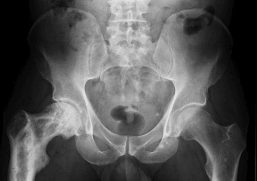
Aetiology. Multifactorial, and not settled. Genetic contributors include SQSTM1 gene mutations, found in 15-30% of familial cases, and chromosome 18q abnormalities. The paramyxovirus (measles) "slow virus" hypothesis remains unproven and contested. Geographic clustering and declining incidence suggest an environmental trigger; with the SQSTM1 genetics, they point to a gene-environment interaction whose trigger is still unidentified.
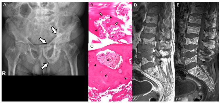
Clinical Assessment
History. When the disease does declare itself, the complaints are skeletal:
- Bone pain - deep, aching, worse at night
- Deformity - bowing of long bones, skull enlargement
- Fracture - after minimal trauma, or a stress fracture
- Arthritis pain - in the hip or knee next to pagetic bone
Systemic features are rare and are developed below: high-output cardiac failure with extensive disease, hearing loss and cranial nerve involvement from the skull base, spinal stenosis, and hypercalcaemia during immobilisation.
Examination. Look for the enlarged bone: an increased skull circumference, a bowed tibia or femur, and a gait disturbed by leg length discrepancy or joint deformity. Active lesions are tender, the skin over pagetic bone is warm from its hypervascularity, and secondary arthritis brings a joint effusion. Examine the cranial nerves when the skull base is involved, and look for spinal cord signs when vertebral disease brings stenosis.
Patients may report needing larger hat sizes due to progressive skull enlargement. This is a classic history finding in advanced skull Paget's. Similarly, tibial bowing may require shoe modifications.
Biochemical and Imaging Investigations
Paget's disease is defined by three cardinal features: a biochemical hallmark (elevated ALP with normal calcium), a radiological one (bone expansion with cortical thickening) and a histological one (mosaic woven bone).
Biochemistry. The alkaline phosphatase is raised, 10-15 times normal in active disease, and reflects osteoblast activity and disease extent. Calcium is normal, which separates Paget's from malignancy and hyperparathyroidism, and normal phosphate and PTH confirm an isolated disorder of bone remodelling.
- Finding
- Elevated (10-15x normal)
- Interpretation
- Reflects osteoblast activity and disease extent
- Differential
- Liver disease, bone metastases, hyperparathyroidism
- Finding
- Normal
- Interpretation
- Distinguishes from malignancy and hyperPTH
- Differential
- Elevated if immobilised or rare hypercalcaemia
- Finding
- Normal
- Interpretation
- Normal bone mineralisation
- Differential
- Abnormal in renal osteodystrophy
- Finding
- Normal
- Interpretation
- Excludes primary hyperparathyroidism
- Differential
- Elevated in hyperPTH
- Finding
- Elevated (urine N-telopeptide, serum CTX)
- Interpretation
- Reflects osteoclast activity, monitors treatment
- Differential
- Used to assess treatment response
Elevated ALP with normal calcium and phosphate is diagnostic. If calcium is elevated, consider immobilisation hypercalcaemia, malignancy, or coexistent hyperparathyroidism. Check PTH and 25-OH vitamin D to differentiate.
Radiographs. Plain radiographs are diagnostic in most cases. The pagetic bone is expanded (increased diameter), its cortex thickened and its trabeculae coarsened, and active disease shows a mixed lytic-sclerotic pattern. Each site adds its own signs.
The lytic phase produces osteoporosis circumscripta, a geographic lytic area. In the mixed and sclerotic phases the vault takes on the cotton-wool appearance of patchy sclerosis, with cortical thickening of the calvarium and skull base and basilar invagination (platybasia). Narrowing of the cranial nerve foramina and thickening of the inner table are the complications to look for.
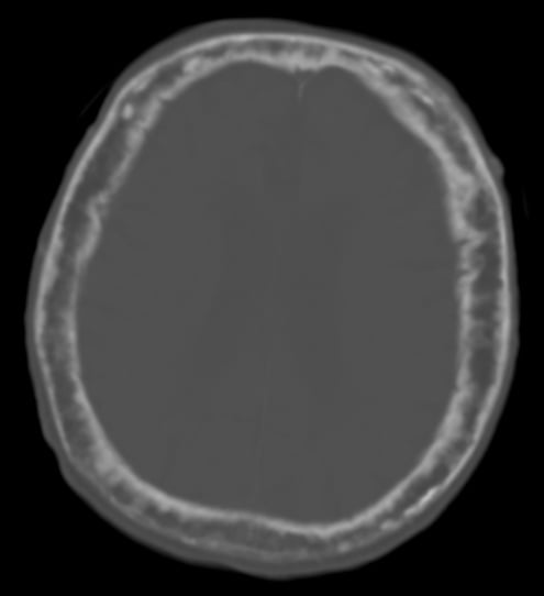
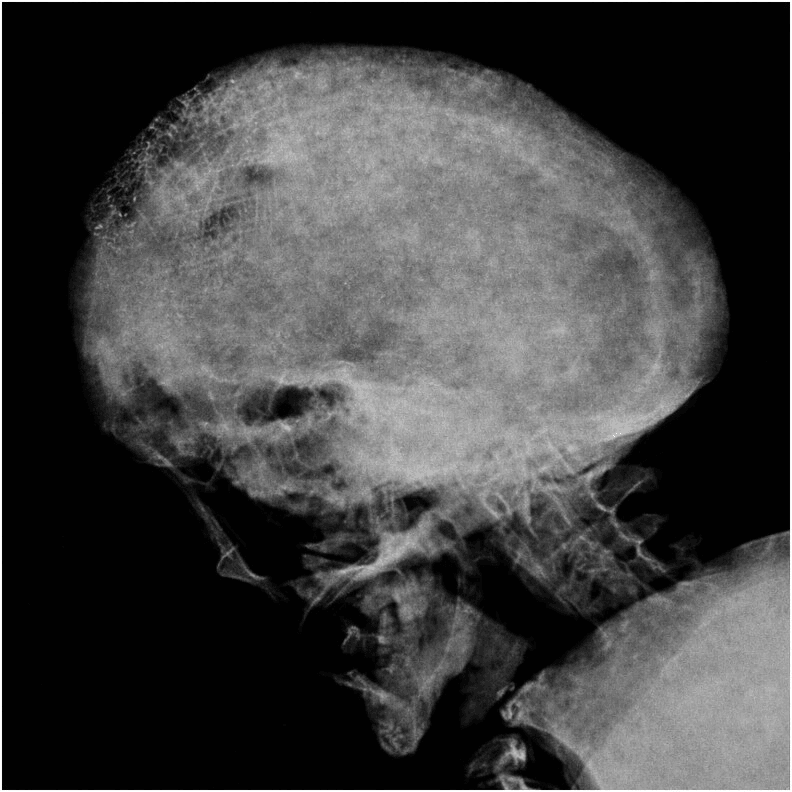
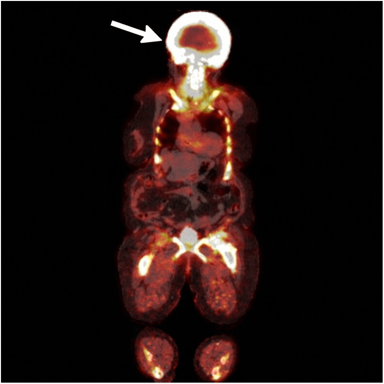
Bone scintigraphy. A Tc-99m MDP bone scan is highly sensitive for active lesions, which show increased uptake. It maps disease extent before treatment, detects polyostotic disease, and helps with surgical planning and monitoring response. It cannot distinguish Paget's from metastases, so the hot spots need radiographic correlation.
Differential Diagnosis
An elevated ALP with a sclerotic or expanded bone has several mimics. The discriminators are serum calcium, PTH, the pattern of bone change, and whether multiple bones are involved.
- Calcium / PTH
- Normal calcium, normal PTH
- ALP
- Elevated (10-15x)
- Distinguishing features
- Bone expansion, cortical thickening, coarse trabeculae; flame/blade-of-grass front; spares hands/feet
- Calcium / PTH
- Calcium normal or high; PTH low/normal
- ALP
- Elevated
- Distinguishing features
- Multiple ill-defined sclerotic foci WITHOUT bone expansion; known primary; PSA may be raised
- Calcium / PTH
- HIGH calcium, HIGH PTH
- ALP
- Mildly elevated
- Distinguishing features
- Subperiosteal resorption, brown tumours, osteopenia - not bone expansion
- Calcium / PTH
- Low/normal calcium, HIGH PTH, HIGH phosphate
- ALP
- Elevated
- Distinguishing features
- Rugger-jersey spine, CKD context, abnormal phosphate
- Calcium / PTH
- Normal calcium/PTH
- ALP
- Normal or mildly elevated
- Distinguishing features
- Younger patient; ground-glass lytic lesions, shepherd's crook; no cortical thickening
- Calcium / PTH
- Normal calcium/PTH
- ALP
- May rise disproportionately, but not reliably elevated
- Distinguishing features
- New lytic destructive lesion, cortical breach, soft-tissue mass on a background of Paget's
Bone expansion is the feature that most reliably separates Paget's from metastatic disease: pagetic bone enlarges and thickens its cortex, whereas metastases deposit within bone of normal size. Combined with normal calcium and PTH, this distinguishes Paget's from the metabolic mimics.
Management Algorithm
Bisphosphonates. First-line treatment. They inhibit osteoclast activity, reduce bone turnover, normalise the ALP and reduce bone pain. A single 5mg infusion of zoledronic acid is the standard of care, and its remission lasts 2 years or more.
- Dosing
- 5mg single infusion
- Efficacy
- 90% ALP normalisation at 6 months, durable response years
- Indications
- First-line, convenient, high efficacy
- Dosing
- 40mg daily × 6 months
- Efficacy
- 70-80% ALP normalisation
- Indications
- Alternative if IV not tolerated
- Dosing
- 30mg daily × 2-3 months
- Efficacy
- 75-85% ALP normalisation
- Indications
- Oral option, shorter course
- Dosing
- 60-90mg infusions
- Efficacy
- Effective but multiple doses required
- Indications
- Second-line if zoledronic acid unavailable
Who to treat. Symptomatic disease is treated: bone pain attributable to Paget's, neurological complications from nerve compression, and the rare high-output cardiac failure. Treatment is also given before planned surgery on pagetic bone, and for high-risk lesions even when they are asymptomatic, although whether the last group needs treatment is unresolved (see Controversies):
- Weight-bearing long bones - risk of fracture and deformity
- Skull base - risk of cranial nerve compression
- Spine - risk of stenosis
- Juxta-articular disease - risk of secondary arthritis
Acute phase reaction (fever, myalgia) occurs in 30% after IV bisphosphonate, managed with paracetamol. Rare complications: osteonecrosis of jaw (dental hygiene critical), atypical femoral fractures (long-term use).
Calcium and vitamin D. Ensure adequate calcium and vitamin D before treatment, and always co-administer calcium 1000-1200mg and vitamin D 800-1000 IU daily. They prevent hypocalcaemia after bisphosphonate treatment and optimise bone health.
Monitoring. Check the ALP at 3, 6 and 12 months. Normalisation or a 75% reduction marks a response, and a rising ALP means reactivation; bone turnover markers (NTX, CTX) are more sensitive. Normalising the ALP is a marker of response, not in itself a treatment goal worth chasing (see Controversies).
Surgical Management and Orthopaedic Complications
Indications. Surgery deals with the mechanical consequences of the disease:
- Pathological fractures - fixation often required
- Stress fractures - prophylactic fixation if high-risk (femoral neck)
- Nonunion - may require bone graft and revision fixation
- Secondary arthritis - joint arthroplasty, the hip most commonly
- Severe deformity affecting function - corrective osteotomy
- Spinal stenosis or nerve root compression - decompression
Preoperative optimisation. Give a bisphosphonate 3-6 months before elective surgery. It reduces bone vascularity, normalises turnover and improves fixation quality, and the reduction in operative blood loss is quoted at 40-50%, or up to 50%. Pagetic bone is highly vascular, so cross-match and have blood products available for significant intraoperative bleeding.
At operation. Fractures need long implants that bypass the pagetic bone, and deformity correction is complicated by the altered anatomy. Each of the recurring problems has its answer:
- Mechanism
- Hypervascularity of pagetic bone
- Management Strategy
- Preop bisphosphonate, tourniquet, cell saver, controlled hypotension
- Mechanism
- Bone expansion, deformity
- Management Strategy
- Preop CT for templating, custom implants if needed
- Mechanism
- Soft woven bone
- Management Strategy
- Cement augmentation, long-stem components, prophylactic cerclage
- Mechanism
- Weak bone during manipulation
- Management Strategy
- Gentle handling, prophylactic fixation extensions
Total hip arthroplasty. Femoral anatomy is altered by bowing and coxa vara, and acetabular protrusion is common. Classic teaching is a cemented long-stem femoral component for better fixation in soft bone, with a cementless acetabular component and screw fixation considered. Heterotopic ossification occurs at a higher rate, so consider prophylaxis with indomethacin or radiation.
Cemented femoral stems provide better fixation in the soft woven bone of Paget's disease compared to press-fit cementless stems. Long-stem components bypass deformity and stress risers. Acetabular components can be cementless if bone quality adequate.
Cemented or not. The cemented long stem is classic teaching, yet modern registry data (Di Martino 2021) show good survival with various fixation types. In that data the dominant problems are bleeding and heterotopic ossification, not fixation failure.
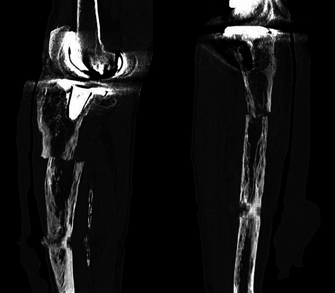
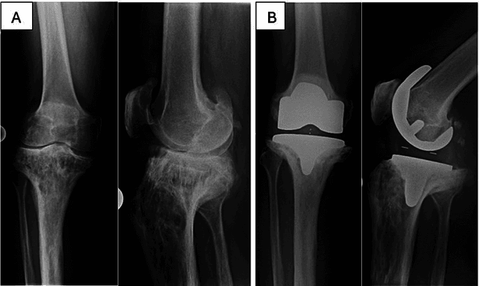
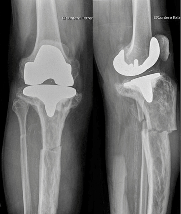
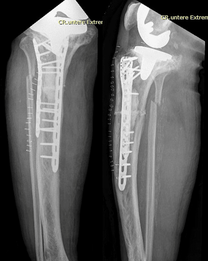
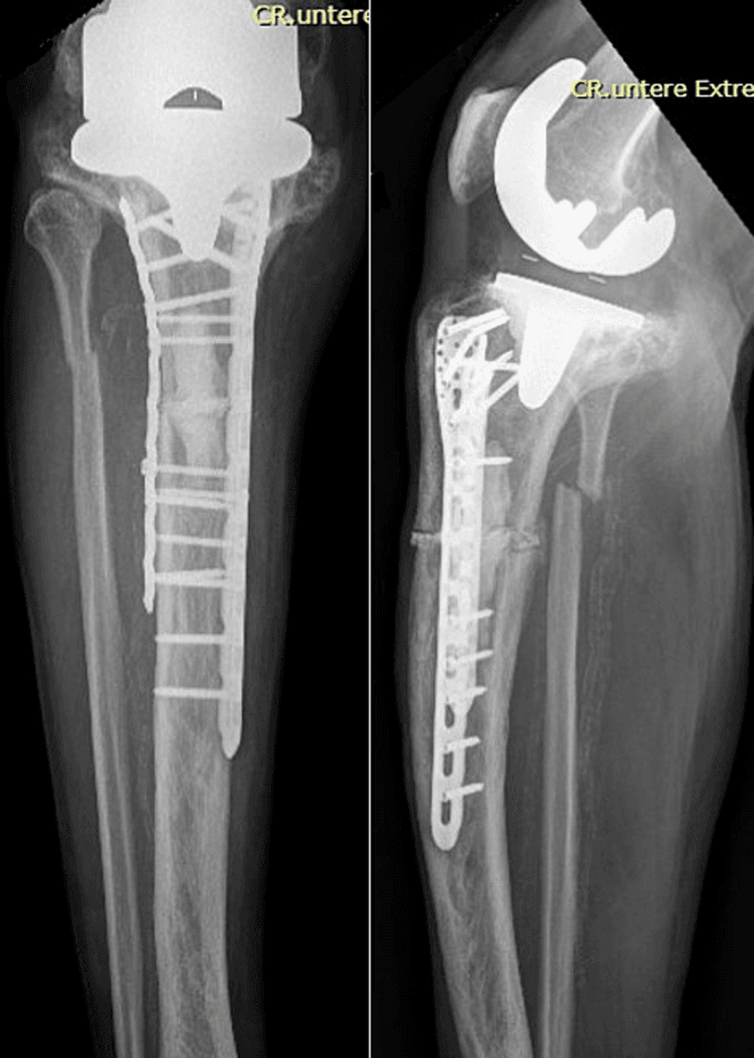
The four figures below show a pagetic proximal tibia at knee arthroplasty, after fracture, and after fixation.
Complications
- Incidence
- 10-30% lifetime risk
- Presentation
- Minimal trauma fracture, stress fracture (femur, tibia)
- Management
- Fixation (often requires long plates/nails), bisphosphonates
- Incidence
- Common in hip/knee
- Presentation
- Progressive joint pain, stiffness, juxta-articular disease
- Management
- Arthroplasty (THA most common), preop bisphosphonate
- Incidence
- ~0.3% lifetime risk
- Presentation
- Sudden pain increase, soft tissue mass, rapid lytic expansion
- Management
- Biopsy, staging, wide resection or amputation + chemo
- Incidence
- Common (tibia, femur)
- Presentation
- Progressive bowing, leg length discrepancy, gait abnormality
- Management
- Corrective osteotomy if severe functional impairment
- Incidence
- Skull base, spine
- Presentation
- Deafness (CN VIII), spinal stenosis, radiculopathy
- Management
- Bisphosphonates, surgical decompression if progressive
- Incidence
- Rare (immobilisation)
- Presentation
- Polyuria, confusion, immobilisation triggers
- Management
- Hydration, bisphosphonates, mobilise patient
Osteosarcoma (most common), fibrosarcoma, or malignant fibrous histiocytoma can arise in pagetic bone. Lifetime risk is about 0.3% and falling - the older "1%" figure predates the modern registry data. Suspect when sudden increase in pain, soft tissue mass, rapid radiological change. Do NOT require a sharply rising alkaline phosphatase: it is not reliably elevated in pagetic sarcoma. Prognosis is poor (5-year survival under 10%). Requires biopsy, staging MRI/CT, wide resection or amputation with chemotherapy. The full staging pathway, histology and reconstruction are developed in the dedicated Paget's sarcoma topic.
Cardiovascular Complications and High-Output Cardiac Failure
Why output rises. Actively remodelling pagetic bone is intensely hypervascular. Dilated marrow sinusoids and increased blood flow through the affected bone and overlying skin behave functionally like a low-resistance arteriovenous shunt, felt clinically as warmth over the bone. Where a large fraction of the skeleton is involved, the summed increase in resting blood flow raises cardiac output and preload.
Who decompensates. True high-output failure is rare and generally requires extensive polyostotic disease; monostotic or limited disease does not cause it. The increased demand most often decompensates pre-existing cardiac disease (ischaemic or valvular) in an elderly population rather than causing failure in a healthy heart. Warmth and bounding pulses over pagetic limbs are supportive signs.
The valve. Paget's is associated with an excess of calcific aortic valve disease and generalised vascular calcification, which compounds cardiac risk and can coexist with the high-output physiology.
Treatment. Suppressing bone turnover with a bisphosphonate reduces pagetic bone vascularity and blood flow, and can improve the high-output state. The same hypervascularity that drives the cardiac load is what makes preoperative bisphosphonate reduce surgical bleeding.
If asked why an asymptomatic patient with very widespread active Paget's might still be treated, the cardiovascular argument is a strong answer: extensive hypervascular bone (classically more than 15% of the skeleton) can raise cardiac output and decompensate an ageing heart, and bisphosphonate therapy that reduces bone blood flow can relieve it. Reserve this explanation for extensive polyostotic disease - it does not apply to monostotic Paget's.
Neurological Complications: Deafness, Basilar Invagination and Spinal Stenosis
Hearing loss is the commonest neurological complication, and is typically mixed sensorineural and conductive. The dominant contributor is thought to be pagetic remodelling and demineralisation of the cochlear (otic) capsule and cochlear structures, rather than simple compression of the eighth nerve at the internal auditory meatus; a conductive component can arise from involvement of the ossicles and temporal bone. Established hearing loss responds poorly, so treatment aims to slow progression.
Basilar invagination. Softening of the pagetic skull base allows the odontoid to migrate upward into the foramen magnum (basilar invagination, platybasia). The result can be brainstem and upper cervical cord compression, vertebrobasilar symptoms, and obstructive hydrocephalus from aqueduct or outflow compromise. It is a serious, though uncommon, complication of extensive calvarial disease.
Spinal stenosis. Vertebral Paget's with bony expansion narrows the canal and neural foramina, causing spinal stenosis, myelopathy, radiculopathy or cauda equina syndrome. In pagetic spinal syndrome some of the deficit is due not only to mechanical compression but to a vascular steal, with blood diverted from the spinal cord into the adjacent hypervascular bone.
Because of the vascular steal, medical therapy that reduces bone blood flow (bisphosphonates; historically calcitonin) can improve neurology without surgery. A trial of medical treatment is therefore reasonable first-line for slowly progressive pagetic myelopathy, reserving urgent surgical decompression for rapidly progressive, severe or refractory deficits.
Guidelines, Registries & Global Practice
Global epidemiology: Strong geographic and ethnic gradient. Highest prevalence in populations of Anglo-Celtic / Western European descent (UK, Australia, New Zealand, North America, and emigrant communities), where it affects roughly 1-4% over age 55. Rare in Scandinavia, much of Asia and sub-Saharan Africa. Incidence and severity have declined markedly over recent decades across high-prevalence regions, supporting an environmental contribution alongside SQSTM1-related genetic susceptibility.
Side-by-Side Guideline Comparison
- When to treat
- Active disease at risk of complications; before pagetic-bone surgery
- First-line agent
- Single 5mg IV zoledronate
- Treatment target
- Symptom control; ALP/turnover marker response
- When to treat
- Bone pain attributable to disease; pre-surgery; selected high-risk sites
- First-line agent
- Zoledronate (potent, durable)
- Treatment target
- Symptom control rather than universal ALP normalization (PRISM-EZ)
- When to treat
- Mechanical complications: impending/actual fracture, deformity, arthritis, stenosis
- First-line agent
- Bisphosphonate plus surgery as indicated
- Treatment target
- Restore alignment, stable fixation, reduce perioperative bleeding
older guidance emphasised normalizing ALP in all active disease; the PRISM-EZ RCT showed no clinical benefit from intensive turnover suppression in established disease, so contemporary UK/European practice favours treating symptoms and high-risk sites rather than every elevated ALP.
Arthroplasty registries (NJR England & Wales, AOANJRR Australia, AJRR US, Swedish/Norwegian/NZ registries) and dedicated pagetic cohorts report 10-year hip implant survival around 90%, with bleeding/transfusion and heterotopic ossification as the dominant complications rather than early loosening.
Where IV zoledronate and DXA/bone scan are available, a single infusion with biochemical monitoring is standard. In limited-resource settings, oral risedronate/alendronate and serial ALP (a cheap, widely available marker) are pragmatic alternatives; plain radiographs alone are usually sufficient for diagnosis and surgical planning.
Related pages: Paget's Sarcoma for the staging, histology and reconstruction of the transformation summarised here, and Osteosarcoma for the disease it becomes - noting that pagetic sarcoma behaves nothing like the adolescent tumour and is not managed by the same neoadjuvant protocol; Osteomalacia for the mirror-image cortical lucency, whose Looser zones sit on the compression side while pagetic fissures sit on the tension side; Renal Osteodystrophy and Hyperparathyroidism for the other causes of a raised alkaline phosphatase with abnormal bone turnover, separated here by the normal calcium and phosphate; Fibrous Dysplasia for the other expansile disorder that deforms and bows long bones in an older skeleton; Bisphosphonates for the drug class whose Paget's dosing is unlike its osteoporosis dosing - a single 5mg zoledronate infusion, not an annual course; Osteoclasts and Bone Resorption for the RANKL-driven cell that starts the pagetic lesion, and Bone Remodeling for the coupled cycle that is disordered here; Total Hip Arthroplasty Indications and Heterotopic Ossification for the arthroplasty considerations the Di Martino data above informs; and Osteoporosis for the disease most often confused with this one on a report of "abnormal bone density".
Controversies and Areas of Uncertainty
Treating to an ALP target. The PRISM-EZ RCT found that intensively suppressing bone turnover to normalise ALP gave no improvement in pain, quality of life or fractures over symptomatic treatment, and a non-significant trend to more fractures.
Treating asymptomatic disease. Guidelines suggest treating active disease "at risk of complications", but robust evidence that bisphosphonates prevent fracture, deformity, deafness or arthritis is lacking (Cochrane). Whether asymptomatic high-risk sites still warrant treatment remains unresolved, and practice varies from treat-all-active to treat-symptoms-only.
MCQ Practice Points
Q: What is the characteristic biochemical profile of Paget's disease of bone?
A: Elevated alkaline phosphatase (ALP) with NORMAL serum calcium and phosphate. This distinguishes Paget's from hyperparathyroidism (elevated calcium with elevated PTH) and malignancy (elevated calcium with suppressed PTH). ALP can be elevated 10-15x normal, reflecting high bone turnover.
Q: What are the three radiological phases of Paget's disease and their characteristic appearances?
A: Lytic phase: Osteoporosis circumscripta (skull), advancing V-shaped, flame-shaped or "blade of grass" lytic lesion (long bones). Mixed phase: Active remodeling. Sclerotic phase: Dense cortical thickening, trabecular coarsening, "cotton wool" skull appearance. Most lesions show mixed pattern.
Q: What is the most commonly affected site in Paget's disease?
A: Pelvis (70% of polyostotic cases). Order of frequency: pelvis, spine (lumbar more than thoracic), femur, skull, tibia. Paget's typically affects the axial skeleton but spares hands and feet. Monostotic disease occurs in 15-30% of cases.
Q: What clinical features suggest sarcomatous transformation in a patient with known Paget's disease?
A: Sudden increase in pain with new soft tissue mass and rapid lytic expansion on imaging. The lifetime risk is about 0.3%, not the 1% of older texts - the proportion has fallen steadily since Paget's original description, and the decline began before effective therapy existed. Prognosis is devastating (median survival about 8 months). Two traps: alkaline phosphatase is not reliably elevated in pagetic sarcoma, so a modest ALP does not exclude it, and most patients have no prior diagnosis of Paget's - in the Birmingham registry series only 42% were known to have the disease, so the sarcoma is often how the Paget's presents.
Exam Viva Scenarios
Practise clinical reasoning and management decisions out loud
“A 68-year-old man has routine blood tests showing alkaline phosphatase of 850 U/L (normal 30-120 U/L). Calcium, phosphate, and PTH are normal. He has no bone pain or symptoms. Pelvic X-ray shows cortical thickening and trabecular coarsening of the left hemipelvis. How do you assess and manage this patient?”
“A 72-year-old woman with known Paget's disease affecting her right femur presents with sudden onset thigh pain after a minor fall. X-ray shows a complete transverse fracture through pagetic bone in the mid-femoral shaft. Discuss your management approach.”
“A 70-year-old man with a 12-year history of Paget's disease of the femur, previously stable on bisphosphonates, presents with a 6-week history of new, severe, constant thigh pain and a palpable soft-tissue swelling. His ALP has risen sharply. What is your concern and how do you proceed?”
Key Pathophysiology
- Abnormal osteoclast activity (up to 100 nuclei) leads to excessive bone resorption
- Osteoblasts lay down disorganized woven bone (not lamellar)
- Mosaic pattern on histology, mechanically weak despite increased mass
- Three phases: Lytic, Mixed (active), Sclerotic (burnt-out)
Biochemistry
- ALP elevated 10-15x normal (reflects osteoblast activity)
- Calcium NORMAL (key distinguishing feature)
- Phosphate NORMAL
- PTH NORMAL (excludes hyperparathyroidism)
Radiology Triad
- Bone expansion (increased diameter)
- Cortical thickening (widened cortex)
- Trabecular coarsening (thickened trabeculae)
- Flame/V-shaped lytic front (advancing edge), Cotton-wool sclerosis (skull)
Treatment Algorithm
- First-line: Zoledronic acid 5mg single IV dose
- Indications: Symptoms, high-risk sites (pelvis/long bones/skull/spine), preoperative
- Coadminister calcium 1000-1200mg + vitamin D 800-1000 IU daily
- Monitor ALP at 3, 6, 12 months as a marker of response and of reactivation - but do NOT re-treat purely to normalise it: PRISM-EZ showed chasing biochemical targets gave no benefit in pain, quality of life or fracture
Surgical Pearls
- Preop bisphosphonate 3-6 months before elective surgery (reduces bleeding 40-50%)
- Cross-match blood (pagetic bone highly vascular)
- Long-stem cemented implants for arthroplasty (better fixation in soft bone)
- Gentle handling (risk of fracture propagation)
Complications
- Pathological fracture 10-30% (transverse stress fractures common)
- Secondary arthritis (hip/knee juxta-articular disease)
- Sarcomatous transformation about 0.3% (sudden pain increase, soft tissue mass)
- Deformity (tibial/femoral bowing, skull enlargement)
Evidence Base and Key Studies
Single-Infusion Zoledronic Acid vs Risedronate (Landmark RCT)
- Two pooled identical double-blind RCTs, 347 evaluable patients: one 5mg IV zoledronic acid infusion vs oral risedronate 30mg daily for 60 days
- Therapeutic response at 6 months 96.0% (zoledronic acid) vs 74.3% (risedronate) (P less than 0.001)
- ALP fully normalized in 88.6% vs 57.9%; shorter median time to response (64 vs 89 days)
- During post-trial follow-up (median 190 days) loss of response occurred in 1/113 zoledronic acid vs 21/82 risedronate patients
Intensive vs Symptomatic Bisphosphonate Strategy (PRISM-EZ)
- 3-year extension of PRISM RCT, 502 patients: intensive treatment to normalize bone turnover vs treating symptoms only
- Despite significantly lower ALP in the intensive arm, no difference in quality of life or bone pain
- THREE harm signals all pointing the same way, none individually significant: fractures HR 1.90 (0.91-3.98, p=0.087), orthopaedic procedures HR 1.81 (0.71-4.61, p=0.214), and serious adverse events RR 1.28 (0.96-1.42)
- No clinical benefit from chasing biochemical normalization in established disease
Bisphosphonates for Paget's Disease (Cochrane Review)
- 20 RCTs, 3168 participants
- Bisphosphonates tripled the proportion achieving pain resolution vs placebo (31% vs 9%; RR 3.42), moderate-quality evidence
- Zoledronate gave better pain relief than pamidronate or risedronate in head-to-head data - but the review grades this VERY LOW quality and its authors state they are uncertain about the head-to-head comparisons
- Insufficient evidence that biochemical control reduces fractures, orthopaedic surgery or improves quality of life; NONE of the placebo-controlled trials reported hearing thresholds, quality of life or orthopaedic surgery at all