Radial neck | olecranon | elbow dislocation | medial epicondyle | Monteggia check
- Do the neurovascular exam before and after reduction. Document median, ulnar and posterior interosseous nerve function.
- A reduced elbow is not a finished assessment. Recheck the medial epicondyle, radiocapitellar line and ulna after reduction.
- Radial neck open reduction is a last resort. It is associated with higher stiffness and vascular complications than closed or percutaneous strategies.
- Olecranon fractures need an extensor mechanism decision. Undisplaced plus intact extension is different from displaced articular disruption.
- Look for Monteggia variants. A radial neck injury with ulna deformity or radiocapitellar malalignment is not an isolated radial neck fracture.
- “Finger MCP and thumb extension test posterior interosseous nerve function.
- “A missing medial epicondyle after dislocation may be trapped in the joint.
- “Radial neck management depends on angulation, translation, rotation block, age, associated injury and success of closed reduction.
- “Olecranon fixation choice is driven by fracture pattern, comminution, skeletal maturity and hardware prominence risk.
Overview/Epidemiology
These three injuries sit close together, anatomically and clinically. A child presents with a radial neck fracture, an olecranon fracture or an elbow dislocation, and the surgeon has to go looking for the combined pattern. A radial neck fracture can accompany an elbow dislocation or a Monteggia injury, an olecranon fracture can be part of proximal ulna instability, and a reduced dislocation can hide an incarcerated medial epicondyle or an entrapped nerve.
Mechanism points to the pattern. Each mechanism has a likely injury and a check it should trigger:
- Fall on the outstretched hand with valgus load - radial neck fracture. Measure the angulation and check forearm rotation.
- Posterolateral dislocation mechanism - elbow dislocation with medial epicondyle avulsion, capsuloligamentous injury or nerve traction. Find the medial epicondyle and document median, ulnar and posterior interosseous nerve function.
- Direct blow to the posterior elbow - olecranon fracture, often with swelling over the extensor surface. Assess active extension and articular displacement.
- Forceful triceps contraction or avulsion - olecranon apophyseal or proximal ulna avulsion. Displacement and the state of the extensor mechanism are the decision points.
- High-energy fall or sport injury - biepicondylar fracture-dislocation, radial neck fracture with dislocation, proximal ulna fracture, or open injury. Do not call it isolated until the whole elbow and forearm are imaged.
Surgically Relevant Anatomy
The radial neck. The radial head ossifies during childhood, and the paediatric radial neck behaves differently from the adult radial head. The periosteal sleeve and the physis contribute to remodelling, but severe angulation, translation and a mechanical block to rotation are poorly tolerated. The posterior interosseous nerve passes close to the proximal radius as it enters the supinator, so its function must be documented before and after any manipulation.
The olecranon. It is the triceps insertion and part of the greater sigmoid notch, so treatment turns on whether the articular surface and the extensor mechanism still work. In a child the olecranon apophysis and proximal ulna alignment can make the injury look deceptively small. Any proximal ulna deformity should raise concern for radiocapitellar instability.
The medial epicondyle. In children the medial collateral ligament attaches to the medial epicondylar apophysis, so a dislocation can avulse the medial epicondyle rather than rupture the ligament in its midsubstance. During spontaneous or closed reduction the fragment can become trapped in the ulnohumeral joint. The ulnar nerve runs posterior to the medial epicondyle, and the median nerve can, rarely, be entrapped after dislocation.
Pathophysiology
The common theme is failure of the paediatric elbow ring, and the visible fracture may be only one part of the instability pattern.
Radial neck. Valgus and axial load compress the radial head against the capitellum. The cartilaginous head and physis make displacement harder to appreciate on plain films, so painful rotation and a mechanical block matter more than the radiographic label alone.
Olecranon. The triceps pulls the proximal fragment, and the olecranon forms the articular notch of the ulnohumeral joint. Displacement therefore threatens both the joint surface and active elbow extension.
Dislocation. The ulna and radius dislocate relative to the distal humerus, often through posterolateral instability.
Monteggia variants. Proximal ulna deformity changes the relationship of the radial head to the capitellum. Restoring only the radial neck, or only the dislocation, misses the driver of the radiocapitellar malalignment.
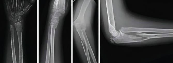
What remodelling will not fix. Young children remodel some angulation. Remodelling does not reliably correct severe translation, blocked forearm rotation, joint incongruity, incarcerated fragments or extensor mechanism failure, and these are the findings that move treatment from observation to reduction or fixation.
Classification
Classification should help communication and treatment, not replace clinical judgement. The practical groups for each injury, and the treatment each implies, are set out under Treatment Thresholds. For the radial neck, examiners expect a named formal classification as well.
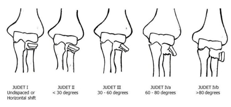
Judet grades the radial neck fracture by angulation. Higher Judet types are progressively harder to reduce closed and more likely to need percutaneous or intramedullary assistance.
- Type I - undisplaced
- Type II - angulation under 30 degrees
- Type III - angulation about 30-60 degrees
- Type IV - angulation over 60 degrees, up to complete displacement, sometimes subdivided IVa and IVb
O'Brien is a simpler angulation tier and a commonly quoted shorthand: Type I under 30 degrees, Type II 30-60 degrees, Type III over 60 degrees. It maps onto the same treatment ladder.
Type I and many low Type II injuries are immobilised. The common reduction threshold sits around 30 degrees of angulation, or any block to rotation, modified by age and translation. High Judet or O'Brien grades enter the closed, then percutaneous, then intramedullary ladder, with open reduction last.
Clinical Assessment
History. Ask enough to establish the injury pattern and its urgency:
- Mechanism - fall, valgus load, direct posterior blow, dislocation event or high-energy trauma
- Time from injury, and whether the elbow was reduced before you saw it
- Where it hurts - radial head and neck, olecranon, medial epicondyle, or diffusely
- Mechanical block - inability to pronate or supinate suggests radial neck displacement or an intra-articular block
- Neurological symptoms - numbness, paraesthesia, weakness, clawing, inability to extend the fingers or thumb
- Vascular symptoms - cold hand, colour change, severe swelling or an escalating analgesic requirement
- Previous elbow injury, hyperlaxity, sporting demand and hand dominance
Look. Inspect the elbow from the front, the side and behind for swelling, deformity, bruising, skin puckering, wounds and the carrying angle. Gross deformity suggests a dislocation or displaced fracture, and posterior swelling localises to the olecranon. Threatened skin or an open wound escalates urgency.
Feel. Palpate the distal humerus, medial epicondyle, lateral condyle, radial head and neck while gently rotating the forearm, then the olecranon, proximal ulna and wrist. Focal radial neck tenderness with painful rotation supports a radial neck injury, and medial tenderness after a dislocation suggests epicondyle avulsion. Ulna tenderness raises concern for a Monteggia injury.
Move. If it is safe, assess active flexion-extension and pronation-supination, and do not force movement in a displaced injury. Blocked rotation suggests radial neck displacement, an incarcerated fragment or a radiocapitellar problem.
The extensor mechanism. Ask the child to extend the elbow actively against gravity if pain and the fracture pattern allow. Loss of active extension with an olecranon fracture suggests extensor disruption and supports fixation.
Stability after reduction. Reassess congruity clinically and radiographically, and avoid aggressive stress testing in an awake child. Persistent instability or blocked motion suggests a complex dislocation, an incarcerated fragment or an associated fracture.
Nerves and vessels. Test and record each structure before and after reduction.
- How To Test
- Radial pulse, capillary refill, hand colour, temperature and pulse oximetry if uncertain.
- Why It Matters
- A cold, pale or poorly perfused hand needs urgent escalation.
- How To Test
- Light touch to index finger pulp; thumb opposition or thenar activation if cooperative.
- Why It Matters
- Median neuropathy may occur with dislocation; persistent or delayed symptoms require urgent reassessment.
- How To Test
- Ask for the OK sign: thumb IP flexion and index DIP flexion.
- Why It Matters
- AIN deficit is a motor median-nerve branch problem; document separately from sensation.
- How To Test
- Small-finger sensation, finger abduction, crossing fingers or first dorsal interosseous activation.
- Why It Matters
- At risk with medial epicondyle avulsion, fixation and dislocation.
- How To Test
- Finger MCP extension and thumb extension with wrist supported; wrist extension alone does not test it.
- Why It Matters
- PIN palsy can accompany radial neck injury or be iatrogenic during percutaneous/open procedures.
SAFESAFE - First Assessment
Hook:SAFE is the order before any reduction.
Differential diagnosis. The painful, swollen paediatric elbow has several overlapping causes, and their management differs sharply. Separate them with a systematic radiograph read rather than a single glance.
- Discriminating Features
- Lateral elbow tenderness, painful or blocked forearm rotation, valgus mechanism.
- How to Confirm
- AP/lateral plus a view at maximum deformity; measure angulation and translation.
- Discriminating Features
- Posterior swelling and tenderness, possible loss of active extension.
- How to Confirm
- True lateral showing the trochlear notch; CT if articular surface unclear.
- Discriminating Features
- Gross deformity, fixed posture, rapid relief after reduction.
- How to Confirm
- Pre- and post-reduction AP/lateral confirming congruity.
- Discriminating Features
- Medial tenderness, ulnar symptoms, absent medial epicondyle for age.
- How to Confirm
- Confirm the epicondyle is in position using CRITOE; CT or comparison films if doubtful.
- Discriminating Features
- Ulna deformity or plastic bowing with a malaligned radiocapitellar line.
- How to Confirm
- Image the whole forearm; trace the radiocapitellar line on every view.
- Discriminating Features
- Distal humeral tenderness, gross swelling, high neurovascular risk.
- How to Confirm
- AP/lateral assessing the anterior humeral line. A different pathway; AIN and brachial artery are the priority.
- Discriminating Features
- Lateral tenderness, subtle metaphyseal fleck, late displacement risk; an articular fracture prone to non-union if displacement is missed.
- How to Confirm
- Internal-oblique view; consider CT or short-interval films.
- Discriminating Features
- Toddler, axial-traction history, arm held pronated, no swelling, normal films.
- How to Confirm
- Clinical diagnosis; films are normal. Treated by simple reduction manoeuvre, never surgery.
Investigations
What to request. Order imaging that answers the surgeon's questions, not just "elbow X-ray":
- AP and true lateral elbow radiographs - mandatory first-line views
- Radiocapitellar or oblique view - helpful if radial neck alignment or the relationship to the capitellum is unclear
- Forearm AP and lateral including wrist and elbow - when there is ulna pain, deformity, plastic bowing or any concern for a Monteggia variant
- Post-reduction AP and lateral radiographs - mandatory after reduction of an elbow dislocation
- Comparison elbow radiographs - useful when the ossification centres make the medial epicondyle or trochlea difficult to interpret
- CT - complex fracture-dislocation, articular olecranon fracture planning, uncertainty about an incarcerated fragment, or surgical planning
- MRI or ultrasound for nerve entrapment - not routine, but useful when neurological symptoms persist or median nerve entrapment is suspected
The radiocapitellar line. A line drawn down the centre of the radial shaft and neck should pass through the capitellum. Failure suggests a radial head dislocation, a Monteggia pattern or an inadequate reduction. Draw it on both the AP and the lateral: apparent reduction on one projection is insufficient.
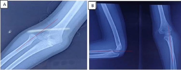
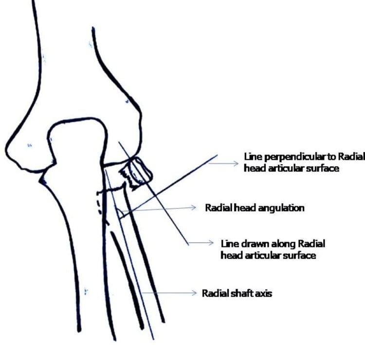
Radial neck angulation and translation. Measure the angle between the radial shaft axis and the radial head and neck axis on the view showing maximum deformity, never from a single suboptimal view. Then assess percentage displacement and head-neck contact. Translation plus angulation predicts reduction difficulty and may require percutaneous or intramedullary assistance.
The olecranon and the ulna. Inspect the greater sigmoid notch on the true lateral and AP, and get a CT if the joint line is unclear. Look along the ulna for a proximal fracture, plastic deformation or bowing, and image the forearm if uncertain.
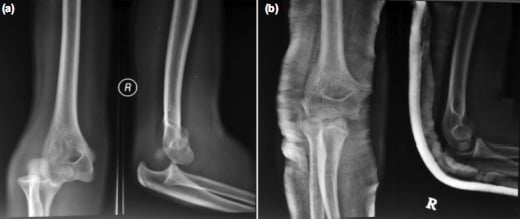
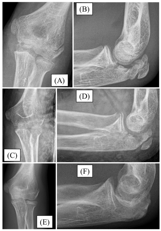
The medial epicondyle. After a dislocation, confirm that the medial epicondyle is visible in its normal position for age. An absent or intra-articular fragment suggests entrapment and operative extraction and fixation. A trapped medial epicondyle can mimic another ossification centre and should not be dismissed as normal development.
CRITOE. The ossification centres appear in the order Capitellum, Radial head, Internal (medial) epicondyle, Trochlea, Olecranon, External (lateral) epicondyle, at roughly 1, 3, 5, 7, 9 and 11 years (a useful approximation; girls slightly earlier than boys). The order is far more reliable than the exact ages. After a dislocation, run the sequence on the film: an absent medial epicondyle for age, or an extra fragment in the joint line, means search for and extract an incarcerated medial epicondyle, with comparison films or CT if unsure.
The medial epicondyle always ossifies before the trochlea. A trochlear ossification centre should never be present without a medial epicondyle already present. So if there is an ossific fragment in the region of the trochlea and no separate medial epicondyle can be identified, the fragment is the displaced medial epicondyle sitting in the joint - an entrapped medial epicondyle until proven otherwise.
Treatment Thresholds
Each injury has practical groups, and the group sets the treatment direction.
Operate for unacceptable angulation or translation, blocked rotation, failed closed reduction or associated instability. The goal is to restore radiocapitellar alignment and forearm rotation while avoiding open reduction if possible.
- Typical Features
- Low angulation, minimal translation, painless or acceptable rotation and no associated instability.
- Treatment Direction
- Above-elbow immobilisation, early review and motion when safe.
- Typical Features
- About 30-60 degrees angulation (around the common reduction threshold), translation, or clinically important loss or block of rotation.
- Treatment Direction
- Closed reduction first; accept reduction only if motion and alignment are satisfactory.
- Typical Features
- Marked angulation, major translation, mechanical block or unsuccessful closed manoeuvre.
- Treatment Direction
- Percutaneous leverage, joystick technique or intramedullary reduction/fixation.
- Typical Features
- Soft-tissue interposition, complete displacement, delayed presentation or failed minimally invasive methods.
- Treatment Direction
- Open reduction only when required.
- Typical Features
- Elbow dislocation, olecranon or proximal ulna injury, or Monteggia variant.
- Treatment Direction
- Changes the plan: treat the whole pattern.
Management
Initial care. Pain control and a splint come first, because a calm, supported child is easier and safer to assess. Open injury, threatened skin, vascular compromise or compartment concern override routine fracture pathways and are escalated. Document median, ulnar and posterior interosseous nerve function before any reduction, so that a new deficit after manipulation is recognised.
Reduction. Reduce a dislocated elbow promptly if there is no contraindication; a congruent joint protects the soft tissues and nerves. Then repeat the neurovascular examination and the AP and lateral radiographs.
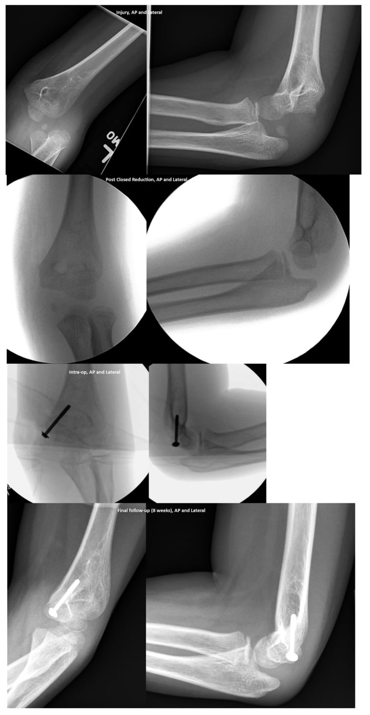
Do not reduce a paediatric elbow dislocation, see a congruent joint, and stop. The post-reduction film must show the medial epicondyle in the correct place, the radiocapitellar line through the capitellum, a satisfactory ulnohumeral joint, and no missed radial neck, olecranon, lateral condyle or proximal ulna injury.
Non-operative care is appropriate when the joint is congruent, fracture displacement is acceptable, extensor function is preserved where relevant, and there is no nerve, vascular, open or mechanical-block concern.
Immobilisation and early films. An above-elbow splint or cast, with the forearm position chosen for comfort and stability, protects the fracture or the reduced joint while swelling settles. Repeat the films early for fractures at risk of displacement, to detect loss of position before it becomes established.
Motion. The paediatric elbow stiffens quickly after trauma, so avoid prolonged immobilisation once stability allows motion. Encourage hand, wrist and shoulder movement while the elbow is protected, which reduces stiffness and swelling outside the injured elbow.
Safety-net advice. Parents should bring the child back for increasing pain, numbness, cold fingers, swelling, fever, a wound concern or loss of finger extension. These symptoms may signal a nerve, vascular, infective or compartment problem.
Operative Technique
Radial neck reduction ladder
The principle is to reduce the radial neck with the least soft-tissue stripping that gives a stable, congruent, mobile elbow.
Closed reduction. Traction, varus or valgus correction as appropriate, forearm rotation and direct pressure over the radial head and neck, under the image intensifier. Do not force the fracture repeatedly; recheck rotation and radiocapitellar alignment.
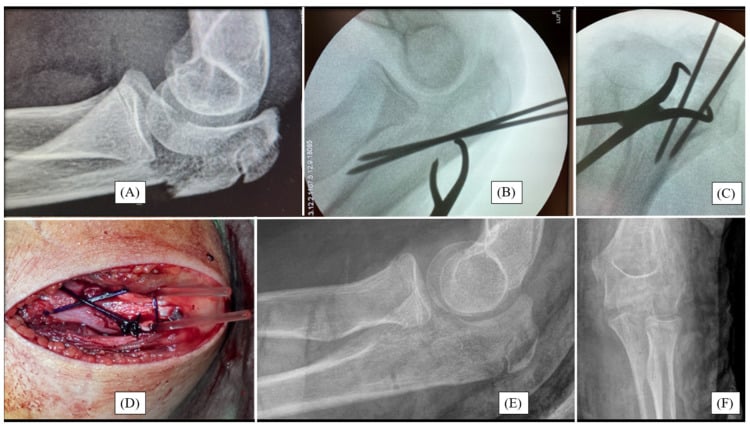
Percutaneous leverage. A small K-wire or elevator is used as a joystick or lever under fluoroscopy. Stay aware of the posterior interosseous nerve: make controlled passes and avoid multiple blind attempts. In the rotation-prying manoeuvre a prebent wire is advanced to the fragment and rotated to elevate the radial head, with controlled thumb pressure added. Every pass is image-guided to protect the posterior interosseous nerve and the physis.
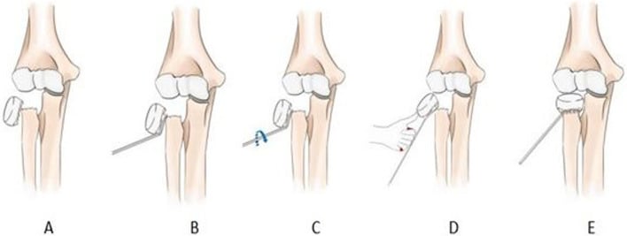
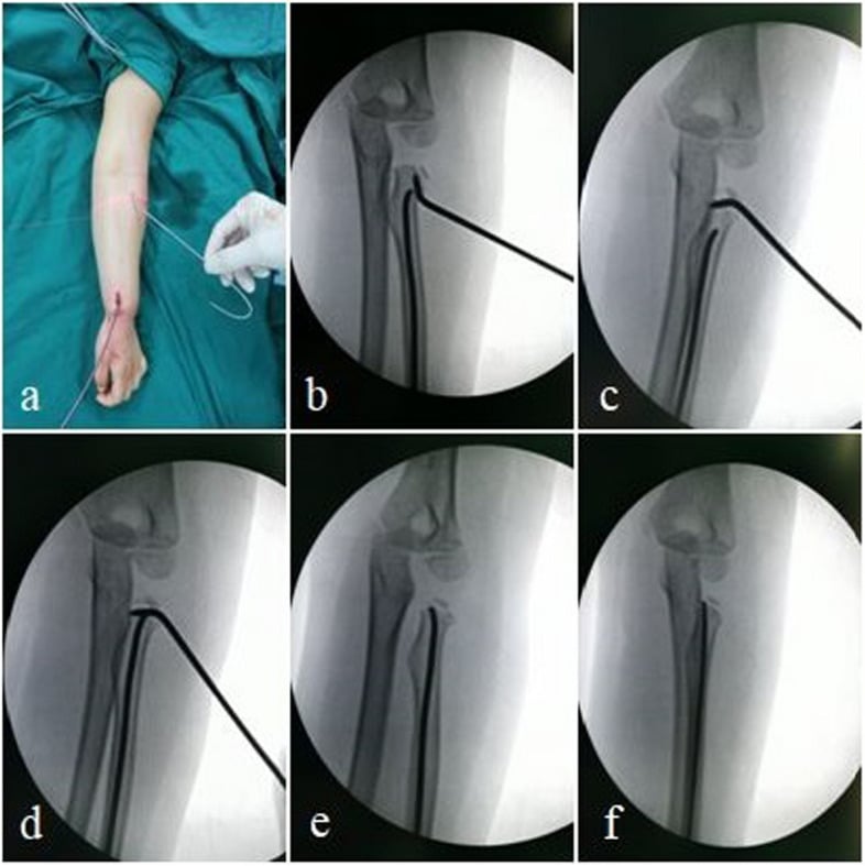
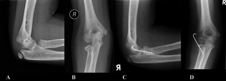
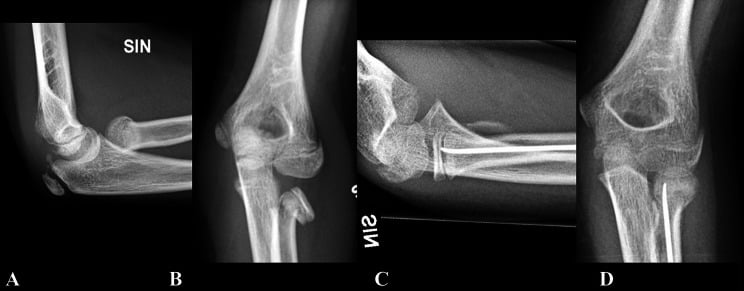
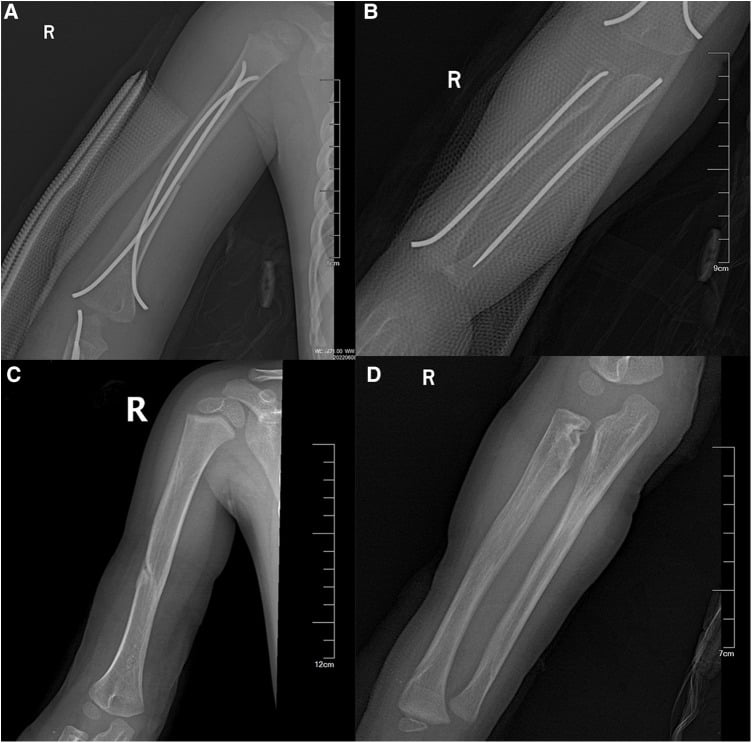
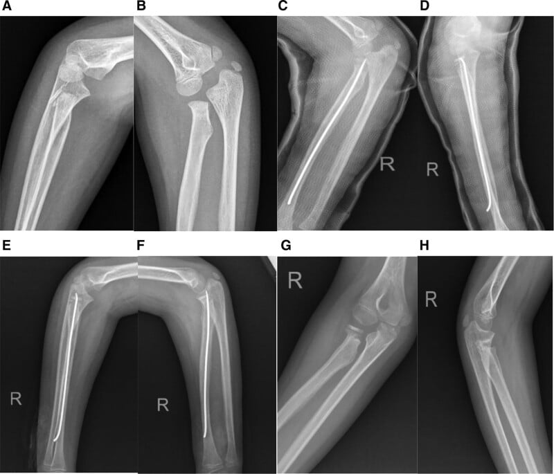
Intramedullary reduction and fixation. A prebent elastic nail is passed from the distal radius region and rotated to elevate and reduce the radial head fragment. The nail reduces and supports the fragment while limiting direct disruption of the radial head blood supply. It is useful for displaced fractures when closed reduction is inadequate: avoid physeal injury, and confirm head-neck alignment and free forearm rotation before accepting the construct.
Open reduction. A lateral approach, only when closed and percutaneous methods fail or the fracture is irreducible. Preserve the periosteum and soft tissue, and warn about stiffness, avascular necrosis and synostosis.
Olecranon fixation
A posterior approach centred on the olecranon, with the operation directed by the fracture pattern:
- Simple transverse displaced fracture - reduce the articular surface anatomically. Fixation may be a suture tension band, wire tension band, intramedullary screw or combined construct, depending on size and maturity.
- Comminuted or oblique pattern - a plate may give better control than a tension band alone.
- Skeletally immature child - minimise physeal and apophyseal injury, and choose fixation that avoids unnecessary hardware prominence.
- Associated instability - confirm radiocapitellar congruity and ulna alignment after fixation.
The steps:
- Position supine or lateral with image intensifier access.
- Mark the olecranon, proximal ulna, medial epicondyle and the course of the ulnar nerve.
- Posterior incision; protect the skin flaps and avoid unnecessary ulnar nerve handling unless the approach extends medially.
- Clear the fracture haematoma without stripping soft tissue unnecessarily.
- Reduce the articular surface and confirm on AP and lateral fluoroscopy.
- Fix according to the pattern above, whether tension band, screw, plate or hybrid.
- Test elbow motion and radiocapitellar alignment before closure.
- Immobilise initially, then start motion when fixation and soft tissues allow.
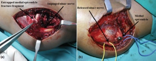
Elbow dislocation with incarcerated medial epicondyle
This is a surgical problem because the fragment blocks restoration of the joint and carries the medial collateral ligament attachment.
- Position supine with the arm on a hand table.
- Mark the medial epicondyle, olecranon and ulnar nerve.
- Use a medial approach centred over the medial epicondyle.
- Identify and protect the ulnar nerve; transpose only if needed for exposure, instability or nerve symptoms.
- Open the joint enough to extract the incarcerated fragment.
- Reduce the medial epicondyle to its origin and fix with a screw, K-wire or suture construct according to fragment size and skeletal maturity.
- Confirm ulnohumeral and radiocapitellar congruity on AP and lateral images.
- Check elbow stability through a safe arc.
- Repeat and document median, ulnar and posterior interosseous nerve function post-operatively.
Complications and Failure Management
- Where It Occurs
- All paediatric elbow trauma, especially delayed or open radial neck reduction.
- Prevention and Management
- Avoid unnecessary prolonged immobilisation; start supervised motion when stable; treat established stiffness with therapy and selective surgery.
- Where It Occurs
- Radial neck malunion, radioulnar synostosis or over-manipulation.
- Prevention and Management
- Restore radial neck alignment, avoid repeated soft-tissue trauma, monitor rotation early.
- Where It Occurs
- Severe radial neck displacement or open reduction; risk rises with displacement and vascular disruption.
- Prevention and Management
- Preserve periosteal blood supply and attachments; reserve open reduction for irreducible cases.
- Where It Occurs
- Severe proximal radius injury or open surgery.
- Prevention and Management
- Limit soft-tissue stripping and repeated attempts; manage established synostosis only when mature and functionally limiting.
- Where It Occurs
- Post-reduction elbow dislocation.
- Prevention and Management
- Always find the medial epicondyle; CT or comparison films if uncertain; open extraction/fixation if incarcerated.
- Where It Occurs
- Medial epicondyle injury or fixation.
- Prevention and Management
- Document before surgery, protect during approach, reassess after fixation.
- Where It Occurs
- Radial neck injury or percutaneous/open reduction.
- Prevention and Management
- Pre/post documentation, careful trajectory, observation for neurapraxia unless progressive or iatrogenic concern.
- Where It Occurs
- Olecranon tension band or wires.
- Prevention and Management
- Choose fixation carefully; remove symptomatic hardware after union when needed.
Implant migration. An elastic intramedullary nail used for a radial neck fracture can migrate into the joint. New pain, loss of motion or an altered implant position requires urgent imaging and timely removal or revision.
Post-Operative Care and Rehabilitation
Unstable fixation or an unrecognised associated injury is worse than a short period of protection, so rehabilitation depends on the stability achieved.
- Immobilisation
- Above-elbow cast or splint initially.
- Follow-up Focus
- Repeat radiographs, pain, rotation, extension and nerve function.
- Immobilisation
- Splint or cast until early healing; avoid prolonged immobilisation if stable.
- Follow-up Focus
- Radiocapitellar alignment, forearm rotation, PIN function and stiffness.
- Immobilisation
- Protect fixation, then progressive motion.
- Follow-up Focus
- Union, hardware symptoms, extension strength and articular congruity.
- Immobilisation
- Brief protection if stable; longer protection if complex.
- Follow-up Focus
- Stability, medial epicondyle healing, ulnar nerve symptoms and range of motion.
What recovery means. Parents should understand that bone healing is only part of it. The child also needs a congruent elbow, recovering motion, intact nerve function and no missed associated injury.
Guidelines, Registries & Global Practice
Paediatric elbow trauma is managed worldwide, and the broad principles are consistent across societies even though the supporting evidence is mostly low level. There is no implant registry for paediatric elbow fractures comparable to adult arthroplasty registries, so practice is guided by society statements, trauma networks and large series.
Global epidemiology
- Elbow fractures are among the most common paediatric fractures, with supracondylar fractures the largest group; radial neck fractures comprise roughly 5-10% of paediatric elbow fractures and about 1% of all paediatric fractures.
- Medial epicondyle fractures have an estimated incidence near 40-60 per 100,000 per year and are associated with elbow dislocation in about 60% of cases.
- Median nerve injury complicates approximately 3% of paediatric elbow dislocations, linked to medial epicondyle avulsion.
Side-by-side guidance
- Emphasis
- Least-invasive reduction first for radial neck; individualised medial epicondyle care.
- Practical Point
- Open radial neck reduction is reserved for failed closed and percutaneous methods.
- Emphasis
- Standardised paediatric trauma pathways; prioritised the medial epicondyle question (SCIENCE trial).
- Practical Point
- Displaced medial epicondyle management is treated as an evidence gap, not a fixed rule.
- Emphasis
- Pattern-based fixation principles and the least-invasive construct that restores stability.
- Practical Point
- Choose tension band, screw or plate by fracture pattern and maturity.
- Emphasis
- Recognition of associated injuries (Monteggia, medial epicondyle) and early motion.
- Practical Point
- Always trace the radiocapitellar line and avoid prolonged immobilisation.
High- versus limited-resource practice
- Typical Reality
- Fluoroscopy, CT, flexible nails and early specialist review available.
- Adaptation
- Use the full reduction ladder and image-guided percutaneous techniques.
- Typical Reality
- Variable fluoroscopy, delayed presentation, limited implants.
- Adaptation
- Prioritise safe closed reduction, K-wire constructs and reliable follow-up; refer complex fracture-dislocations early.
- Typical Reality
- More common where access is limited; chronic Monteggia or stiff radial neck malunion.
- Adaptation
- Plan reconstruction (ulnar osteotomy, annular ligament reconstruction) at a capable centre.
Controversies and Areas of Uncertainty
Much of paediatric elbow trauma rests on retrospective series rather than randomised trials. Examiners reward the candidate who can say where the evidence is genuinely unsettled.
- Arguments For
- Younger children remodel; thresholds near 30 degrees are widely quoted.
- Arguments Against
- Remodelling is unreliable for translation and rotation; older children remodel poorly.
- Practical Position
- Treat the rotation block and the child's age, not a single angle in isolation.
- Arguments For
- Higher union odds with fixation; helps high-demand athletes and unstable elbows.
- Arguments Against
- Similar pain and function in retrospective series; non-union is often asymptomatic.
- Practical Position
- Fix incarcerated or open fractures; individualise the rest while the SCIENCE trial reports.
- Arguments For
- Wire is familiar and strong; suture avoids prominent metal and removal.
- Arguments Against
- Wire carries higher hardware-symptom and removal rates.
- Practical Position
- Match the construct to pattern and maturity; suture is a legitimate option.
- Arguments For
- Missing it leads to chronic radial head dislocation and PIN palsy.
- Arguments Against
- Over-imaging every forearm is low yield.
- Practical Position
- Trace the radiocapitellar line on every elbow film and image the forearm if the ulna looks bowed.
- Arguments For
- Removes prominent or symptomatic hardware in active children.
- Arguments Against
- A second anaesthetic and surgical risk for asymptomatic implants.
- Practical Position
- Remove for symptoms or planned protocols, not reflexively.
Clinical Scenarios
Practise clinical reasoning and management decisions out loud
“A 9-year-old has a radial neck fracture with about 45 degrees angulation and painful blocked rotation. The hand is perfused and PIN function is normal.”
“A 12-year-old elbow dislocation is reduced in the emergency department. Post-reduction films show a congruent joint, but the medial epicondyle cannot be seen in its normal position.”
“A child has an undisplaced olecranon fracture and can actively extend the elbow. The radiocapitellar line is normal.”
Assessment
- Document skin, perfusion and median, ulnar and posterior interosseous nerve function.
- Order true AP and lateral elbow radiographs; add forearm views if Monteggia is possible.
- After dislocation reduction, find the medial epicondyle and recheck post-reduction films.
Treatment decisions
- Radial neck treatment depends on angulation, translation, rotation block, age and associated injury.
- Use the radial neck reduction ladder: closed, percutaneous leverage, intramedullary reduction, open only if needed.
- Olecranon treatment depends on displacement, articular congruity, extensor mechanism and proximal ulna alignment.
Do not miss
- Simple stable elbow dislocations need early protected motion; complex patterns need fixation or stabilisation.
- Warn about stiffness, loss of rotation, AVN, synostosis, nerve injury, hardware prominence and missed associated injury.
- A missing medial epicondyle or abnormal radiocapitellar line after reduction changes the diagnosis.
Evidence Signals
Paediatric olecranon fixation: suture vs wire tension band
- Nine studies were pooled, with three directly comparing tension-band suturing (TBS) with tension-band wiring (TBW).
- No statistically significant differences in pain, non-union, revision fixation or extension loss.
- TBS demonstrated fewer hardware-related complications than TBW.
Metaizeau closed intramedullary pinning technique
- Radial neck fractures account for about 1% of all paediatric fractures and 5-10% of paediatric elbow fractures.
- A retrograde nail is rotated 180 degrees to reduce the radial head while preserving the tenuous periosteal blood supply; the forearm is pronated during insertion to protect the posterior interosseous nerve.
- Across cited series, good or excellent Mayo Elbow Performance Scores were reported in roughly 80-100% of selected cases.
Percutaneous leverage technique
- A modified percutaneous leverage technique (Wallace technique) plus flexible intramedullary fixation reduced eight displaced paediatric radial neck fractures at a single Level I centre.
- The technique is minimally invasive compared with open reduction and permits early elbow mobilisation.
- No patient in the series developed synostosis or sustained a peripheral nerve injury.
Surgical radial neck series: predictors of poor outcome
- 151 surgically treated children; mean angulation improved from 43 to 13 degrees and displacement from 37% to under 1%.
- Among 131 with follow-up, 31% had an unsuccessful outcome. THE THREE INDEPENDENT PREDICTORS on logistic regression were age 10 years or more (OR 5.85), greater displacement (OR 1.25 per 10%) and - counter-intuitively - surgery within 2 days of injury (OR 4.73). Open reduction and fracture severity were ASSOCIATED with poor results but were not independent predictors.
- THE TIMING PARADOX IS SEVERITY CONFOUNDING, NOT A REASON TO DELAY: the worst fractures were operated on fastest, so early surgery marks a bad injury rather than causing a bad outcome - the same artefact seen in paediatric femoral neck data.
- Closed manipulation was predicted to fail in about half of fractures angulated 36 degrees or more, and about half displaced 65% or more needed open reduction; predicted unsuccessful outcomes reached 50% at 76% displacement.