Rare Sesamoid Carpal Bone | FCU Insertion | Ulnar Nerve at Risk
- Only sesamoid bone in the carpus - embedded in FCU tendon
- Direct blow mechanism - fall onto hypothenar eminence with wrist extended
- Guyon canal runs adjacent - ulnar nerve and artery at risk
- Pisotriquetral joint forms articulation with triquetrum
- Excision is effective treatment for symptomatic fractures
- “Pisiform is the most ulnar and volar of the proximal carpal row
- “Only carpal bone with a single articular surface (pisotriquetral)
- “FCU inserts onto pisiform - force transmitted to hook of hamate via pisohamate ligament
- “Carpal tunnel view best visualizes the pisiform
Overview and Epidemiology
Pisiform fractures are rare, only 1-2% of all carpal fractures, but clinically significant. The pisiform is the only sesamoid bone in the carpus, lying entirely within the tendon of flexor carpi ulnaris (FCU), and that anatomy shapes how it breaks, how it presents and how it is treated.
Who. More common in males, with a peak incidence at 20-40 years. The fracture is often associated with other wrist injuries.
Mechanism. A direct blow to the hypothenar eminence is the most common cause. The other routes are few enough to list:
- Fall onto the outstretched hand with the wrist extended
- Avulsion by FCU contraction (rare)
- Repetitive stress in cyclists and racquet sports, and tool use with vibration exposure; stress fractures are rare
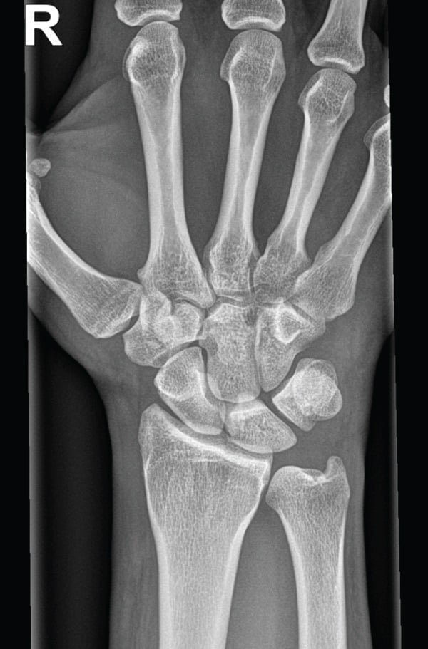
Why it is missed. Pisiform fractures are frequently missed on initial presentation. The pisiform overlaps the triquetrum on the standard PA view, and the patient may put the symptoms down to a "wrist sprain". Presentation may be delayed by weeks, and associated injuries can distract from the pisiform.
Always obtain dedicated carpal tunnel views if pisiform injury is suspected clinically.
Anatomy and Biomechanics
The bone. The pea-shaped pisiform is the most ulnar and volar bone of the proximal carpal row. It is the only carpal bone with a single articular surface, the pisotriquetral joint with the triquetrum, and that joint is where osteoarthritis develops and where the grind test is aimed.
Its attachments. FCU inserts onto the pisiform, and the bone passes the force on through two ligaments:
- Pisohamate ligament to the hook of hamate, transmitting force to the distal row
- Pisometacarpal ligament to the base of the 5th metacarpal, forming the base of the hypothenar eminence
Its function. The pisiform acts as a fulcrum (hypomochlion) that increases the mechanical advantage of FCU in wrist flexion. It also stabilises the ulnar column of the wrist, resisting triquetral subluxation (Beckers and Koebke, Clin Anat 1998).
Blood supply. The pisiform is supplied by branches of the ulnar artery, and its sesamoid status provides tendon-derived nutrition. Nonunion is uncommon because of this good vascularity, and unlike the scaphoid, AVN is rare.
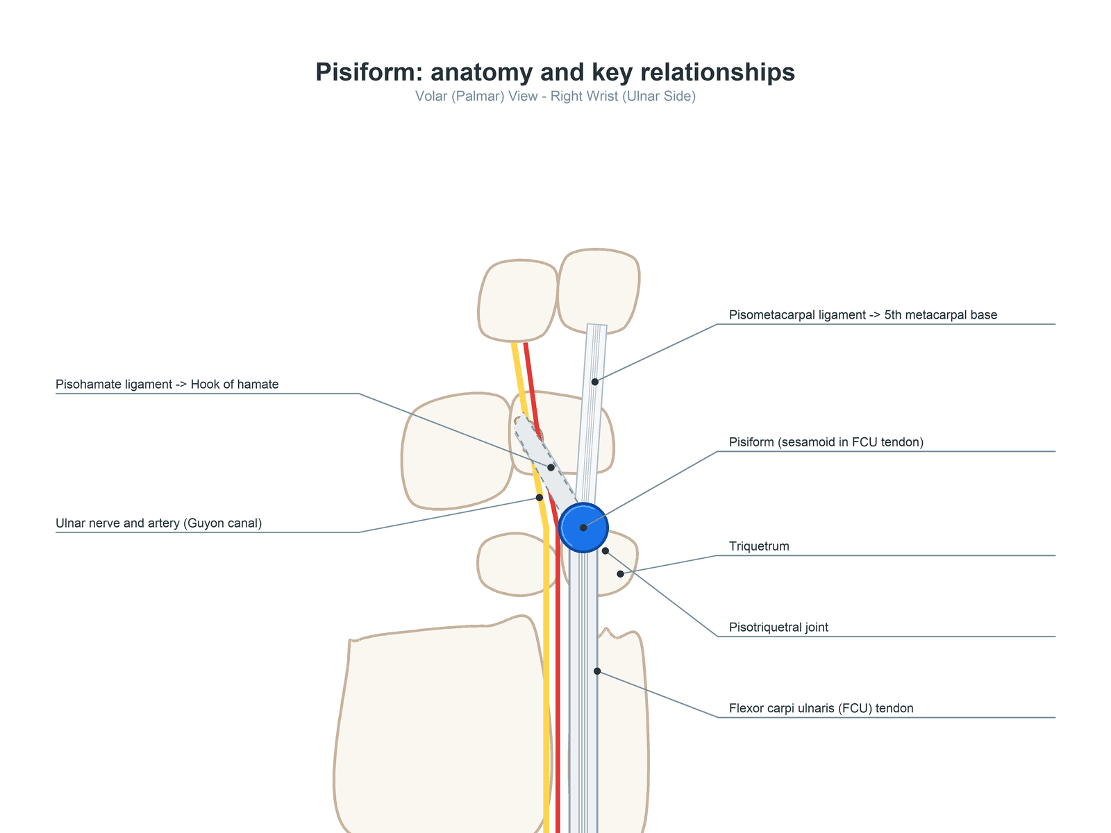
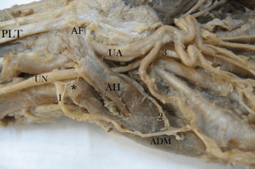
Guyon canal. A fibro-osseous tunnel on the ulnar side of the wrist, whose ulnar border and floor are formed by the pisiform and whose radial border is the hook of hamate. It carries the ulnar nerve and artery, which run immediately radial to the pisiform, so a pisiform fracture can cause compression neuropathy within the canal and every one needs the nerve and artery assessed.
Classification Systems
Pisiform fractures are described by pattern, and each pattern carries a default treatment. Whatever the type, the description must include displacement, comminution, pisotriquetral articular involvement, open injury and ulnar-neurovascular symptoms, because these features change treatment.
- Description
- Multiple fragments, crush injury
- Treatment
- Excision recommended
- Description
- Body fracture through middle
- Treatment
- Cast or excision based on displacement
- Description
- Small fragment at FCU insertion
- Treatment
- Conservative usually successful
- Description
- Repetitive microtrauma
- Treatment
- Activity modification, may need excision
Clinical Presentation and Assessment
History. A direct blow to the hypothenar eminence from a fall or sport, or repetitive handlebar pressure in a cyclist. The pain is localised to the ulnar palm and wrist and is aggravated by gripping and wrist flexion.
Examination. The pisiform is easily palpated at the base of the hypothenar eminence, at the proximal wrist crease on the ulnar side; flexing the wrist against resistance tenses FCU and makes it prominent. In a fracture, direct pressure over it reproduces the pain.
- Point tenderness over the pisiform at the hypothenar base
- Pain with resisted FCU contraction (wrist flexion plus ulnar deviation)
- Pisiform grind test: compress the pisiform against the triquetrum
- Ulnar nerve motor and sensory assessment
Always assess ulnar nerve function in pisiform fractures, and document it: hypothenar sensation, finger abduction (interossei) and thumb adduction (adductor pollicis). Delayed recognition of ulnar nerve compression can lead to permanent intrinsic muscle weakness and sensory deficits.
- Distinguishing features
- Direct blow/FOOSH; point tenderness over pisiform; positive grind test
- Confirmatory test
- Carpal tunnel/lateral view, CT
- Distinguishing features
- Same mechanism; tenderness 1-2 cm distal-radial to pisiform; pain on resisted ulnar grip
- Confirmatory test
- Carpal tunnel view, CT
- Distinguishing features
- Dorsal-ulnar tenderness; common dorsal chip
- Confirmatory test
- Lateral/oblique radiograph, CT
- Distinguishing features
- Gradual onset; positive grind; joint narrowing/osteophytes
- Confirmatory test
- Supinated oblique view, examination
- Distinguishing features
- Tenderness along FCU; pain on resisted flexion; no fracture line
- Confirmatory test
- Ultrasound, MRI
- Distinguishing features
- Hypothenar/ulnar 1.5-digit numbness, intrinsic weakness, NO dorsal sensory loss
- Confirmatory test
- Nerve conduction studies, MRI
- Distinguishing features
- Ulnar-sided pain with rotation; positive fovea sign; DRUJ ballottement
- Confirmatory test
- MRI/MR arthrography, wrist arthroscopy
Fracture or pisotriquetral arthritis. A fracture follows clear trauma, begins suddenly and swells acutely, and the radiograph shows a fracture line. Arthritis comes on gradually after repetitive use or a prior injury, with minimal swelling, and the radiograph shows joint space narrowing and osteophytes. The grind test may be positive in a fracture and is typically positive in arthritis.
Pisotriquetral instability. Not every symptomatic pisotriquetral problem is a fracture or arthritis: the pisiform can be dynamically unstable, subluxating on its triquetral facet. It arises from ligamentous laxity, an unbalanced FCU pull, or a healed avulsion or malunion that leaves the bone poorly seated, and presents as ulnar-palmar pain and a painful "clunk" with wrist flexion-extension or gripping, often with no fracture line on plain films.
The pisiform shuck (ballottement) test looks for it: grasp the pisiform between thumb and finger and translate it radial-ulnar and proximal-distal against the triquetrum, reproducing pain and excess glide compared with the other side. The grind test is typically positive too, and stress or dynamic imaging can demonstrate the abnormal translation. Management mirrors the fracture pathway, with activity modification, FCU rebalancing or physiotherapy and a diagnostic pisotriquetral injection first, and pisiform excision the definitive option for persistent symptomatic instability.
Investigations
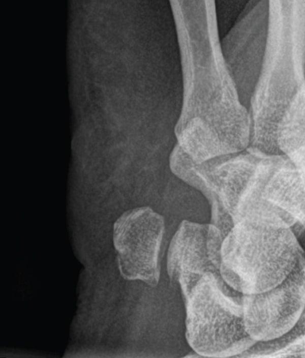
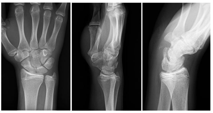
Radiographs. Ask for dedicated views rather than accepting a generic "wrist series" when pisiform tenderness is present. A normal AP film does not exclude injury, because the pisiform overlaps the triquetrum:
- PA view: the pisiform overlaps the triquetrum and the fracture is often missed
- Lateral view: the pisiform is visible as a separate structure anteriorly
- Carpal tunnel view: the best visualisation of the pisiform
- 30-degree supinated oblique: an alternative view
The carpal tunnel view is an axial view taken with the wrist in maximum extension and the beam angled 25-30 degrees to the palm. It gives the best view of the pisiform, the hook of hamate and the carpal tunnel, and is essential when pisiform pathology is suspected.
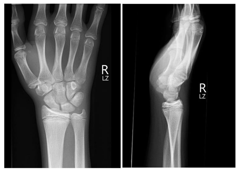
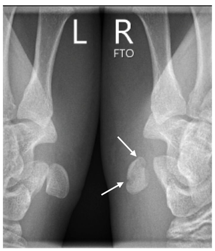
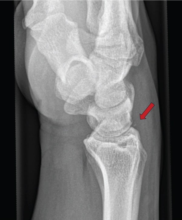
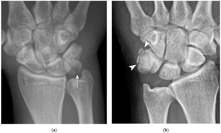
A small fragment. In acute ulnar-sided pain, a small ossicle beside the pisiform may be a fracture fragment rather than an incidental accessory ossicle. Correlate it with focal tenderness and use CT if uncertainty remains; comparison with the opposite wrist can help when a tiny fragment or a normal variant creates doubt.
The pisiform is the last carpal bone to ossify (ossification centre appearing around age 8-12) and frequently does so from more than one centre, so a bipartite (or multipartite) pisiform is a recognised normal variant. On a carpal tunnel or lateral view it can be mistaken for an acute fracture. The clues to a variant are smooth, well-corticated (rounded) margins on both fragments rather than a sharp lucent line, symmetry on the contralateral wrist (image the other side if unsure), and absence of point tenderness or grind matching the radiographic finding. In a child with ulnar wrist pain, remember the bone is still ossifying and irregular ossification can be normal. When genuinely uncertain, CT (a sharp, non-corticated fracture line) or MRI (marrow oedema) settles it: treat the patient and the corticated edges, not the X-ray appearance alone.
CT confirms the fracture pattern and defines its orientation, articular extension, comminution and displacement, the features that decide whether immobilisation is sufficient or excision, or rarely fixation, should be considered. Obtain thin-slice CT when focal pisiform tenderness persists despite equivocal standard views.
Two further findings change the plan. An associated hook of hamate fracture means both injuries are addressed, and pisotriquetral joint narrowing marks established arthritis.
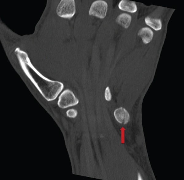
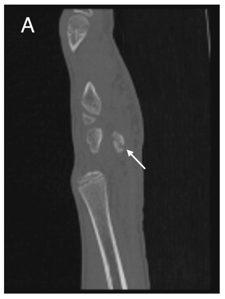
MRI shows occult fractures, bone marrow oedema and ligament injuries. Ultrasound allows dynamic assessment of FCU and the soft tissues.
Management Algorithm
First steps. Confirm the diagnosis with a carpal tunnel and a lateral view, assess the ulnar nerve, and splint the wrist in a neutral position with ice and elevation for the swelling. Ulnar nerve symptoms call for urgent decompression. The imaging is reviewed for the fracture pattern at 1-2 weeks, which sets the path.
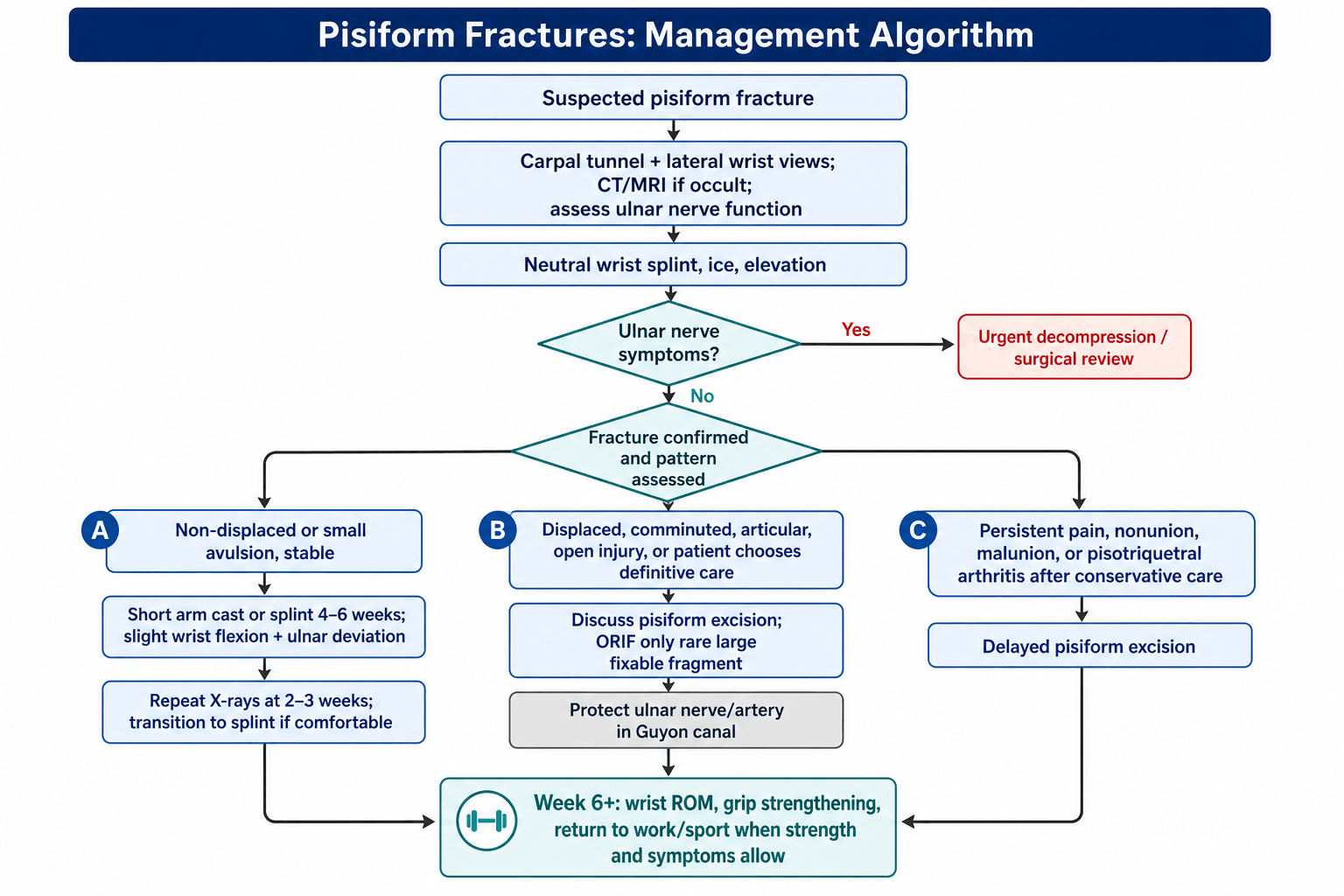
Conservative treatment suits a non-displaced simple fracture, a chip or avulsion fracture with minimal symptoms, and the patient who prefers a non-operative trial, and has a high success rate for stable patterns. The wrist goes in a short arm cast for 4-6 weeks, in slight flexion and ulnar deviation, with follow-up radiographs at 2-3 weeks to confirm position and transition to a splint if comfortable.
Primary excision is indicated for:
- Comminuted fracture pattern, where the fragments are unreconstructable
- Displaced fracture with articular involvement
- Open fracture with soft tissue injury
- Patient preference for definitive treatment
Delayed excision follows failed conservative care:
- Nonunion after conservative treatment
- Post-traumatic pisotriquetral arthritis
- Chronic pain unresponsive to conservative measures
- Malunion with symptomatic articular incongruity
- Pisotriquetral instability (subluxation)
- Key Features
- Small fragment, minimal symptoms
- Treatment
- Splint 3-4 weeks, symptomatic treatment
- Key Features
- Intact articular surface
- Treatment
- Short arm cast 4-6 weeks
- Key Features
- Articular incongruity
- Treatment
- Consider excision vs ORIF
- Key Features
- Multiple fragments, articular damage
- Treatment
- Pisiform excision
- Key Features
- Persistent pain, pisotriquetral OA
- Treatment
- Pisiform excision
Surgical Technique
Set-up. Supine with the arm on a hand table, under regional or general anaesthesia and an upper-arm tourniquet. The approach is volar and longitudinal, along the FCU tendon.
The ulnar nerve and artery run immediately radial to the pisiform in Guyon canal. Meticulous dissection and identification of these structures is essential before any bone work. Consider using loupe magnification.
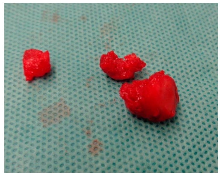
Excision. The key steps:
- Identify and protect the ulnar nerve in Guyon canal
- Incise the FCU tendon sheath longitudinally
- Shell out the pisiform from within the tendon
- Preserve distal FCU continuity to the 5th metacarpal
FCU function is preserved after pisiform excision because the tendon continues distally to insert on the hook of hamate (via the pisohamate ligament) and the base of the 5th metacarpal (via the pisometacarpal ligament). This is why excision is safe and effective.
ORIF. Rarely performed; excision is usually preferred. It is considered for:
- A large displaced fragment amenable to fixation
- A young, active patient wishing to preserve the anatomy
- An acute fracture with good bone quality
The approach is volar, similar to excision, with mini-fragment screws (1.5-2.0mm) and protected mobilisation after fixation. If fixation fails, excision may be needed.
Closure. Close the FCU tendon sheath if possible, then subcutaneous absorbable sutures and skin with nylon or absorbable sutures, under a bulky soft dressing with a wrist splint.
Complications
- Risk Factors
- Displaced fracture, haematoma, late OA
- Treatment
- Decompression, Guyon canal release
- Risk Factors
- Malunion, articular damage
- Treatment
- Conservative initially, excision if refractory
- Risk Factors
- Inadequate immobilisation, displacement
- Treatment
- Pisiform excision (definitive)
- Risk Factors
- Undiagnosed fracture, OA
- Treatment
- Injection trial, then excision
- Risk Factors
- Post-excision (mild)
- Treatment
- Rehabilitation, usually not limiting
- Risk Factors
- Direct trauma, iatrogenic
- Treatment
- Vascular repair if symptomatic
FCU weakness, if chronic, also reduces grip.
Guyon canal syndrome. Compression from a pisiform fracture weakens the intrinsic muscles (interossei, lumbricals 3-4 and the hypothenar muscles) and numbs the hypothenar eminence and the ulnar 1.5 digits. Pisiform fractures typically affect Zone 1 or 2 of the canal:
- Zone 1: motor and sensory, proximal to the bifurcation
- Zone 2: motor only (deep branch), with intrinsic weakness and no sensory loss
- Zone 3: sensory only (superficial branch), with numbness and no weakness
Guyon canal or cubital tunnel. Both weaken the interossei and lumbricals 3-4; the discriminator is the dorsal ulnar cutaneous branch. It leaves the nerve proximal to Guyon canal, so compression at the pisiform spares dorsal hand sensation, whereas cubital tunnel syndrome at the elbow does not.
Postoperative Care and Rehabilitation
Post-Excision Rehabilitation Protocol
Soft bulky dressing with wrist splint for 2 weeks. Finger ROM exercises encouraged from the start. Elevation for 48-72 hours to reduce swelling. Wound check at 10-14 days for suture removal.
Transition to removable wrist splint. Begin active wrist ROM exercises. Continue finger exercises. Scar massage when wound healed.
Wean from splint during day. Light grip strengthening exercises. Progress ROM to full. Avoid heavy lifting.
Full activity as tolerated. Progressive grip and wrist strengthening. Sport-specific rehabilitation. Most return to full activity by 8-12 weeks.
Progression. Motion and loading advance according to tenderness and radiographic union, and return to sport or work is judged on strength recovery and symptoms. Persistent focal pain should prompt CT for delayed union or pisotriquetral incongruity.
Goals. Rehabilitation aims for:
- Full wrist ROM by 6-8 weeks
- Functional grip strength by 8-12 weeks
- Pain-free activity by 12 weeks
- Return to sport by 3-4 months
Red flags. Watch for:
- Increasing pain after initial improvement
- Numbness or tingling in the ulnar distribution
- Weakness of the intrinsic muscles
- Signs of infection: redness, swelling, discharge
Outcomes and Prognosis
After conservative treatment. Non-displaced patterns have a high union rate and a low complication rate, and most patients make a full recovery (Athanasiou 2018).
After excision. Pain relief is reliable, with visual analogue scores falling from 6.8 to 1.1 in De Almeida's 2019 series. Grip typically recovers to about 86% of the contralateral side, a reduction of about 14%, leaving a minimal functional deficit and high patient satisfaction. Persistent symptoms after excision should be interpreted with examination, tenderness in the pisotriquetral bed and the ulnar-nerve findings, not the AP view alone.
Return to activity. Most patients return at 3-4 months, and full recovery is expected. Sport-specific rehabilitation guides the return, and occupational demands influence it.
- Favourable
- Non-displaced, simple
- Unfavourable
- Comminuted, displaced
- Favourable
- Acute presentation
- Unfavourable
- Delayed diagnosis
- Favourable
- Adherent to protocol
- Unfavourable
- Poor compliance
- Favourable
- Isolated fracture
- Unfavourable
- Multiple carpal injuries
- Favourable
- No ulnar symptoms
- Unfavourable
- Guyon canal compression
The long view. Pisiform fractures generally have favourable long-term outcomes: non-displaced fractures heal reliably with conservative treatment, and even when excision is required, function is excellent with minimal grip strength loss. The key to good outcomes is early diagnosis, appropriate treatment selection and attention to ulnar nerve status.
Guidelines, Registries & Global Practice
Global epidemiology. The pisiform is fractured uncommonly, accounting for roughly 1-2% of carpal fractures, with carpal injuries themselves being dominated by the scaphoid. Most pisiform and pisotriquetral injuries follow a direct blow or a fall onto the extended, ulnar-deviated wrist (commonly cyclists landing on a handlebar) and predominate in active adults (Athanasiou 2018). Pisotriquetral osteoarthrosis is described as the second most common degenerative wrist arthritis after scaphotrapezial disease (Beckers and Koebke 1998). There is no dedicated national registry for carpal sesamoid fractures; the evidence base is small retrospective series and case reports rather than randomised trials.
- Position relevant to pisiform
- Carpal fractures other than scaphoid managed by pattern: non-displaced bodies immobilised, comminuted/symptomatic pisiform treated by excision
- Evidence level
- Expert consensus
- Position relevant to pisiform
- Acute wrist trauma pathways prioritise excluding scaphoid and perilunate injury; rare carpal fractures referred to hand surgery for tailored care
- Evidence level
- Expert consensus / pathway
- Position relevant to pisiform
- No pisiform-specific guideline; managed under general carpal/hand trauma principles
- Evidence level
- Expert consensus
- Position relevant to pisiform
- No pisiform-specific guidance; covered by general non-complex fracture management (NG38) and physiotherapy-led rehabilitation
- Evidence level
- Guideline (indirect)
- Position relevant to pisiform
- Endorse pisiformectomy as treatment of choice for refractory pisotriquetral arthritis and nonunion
- Evidence level
- Level IV-V series
- What the evidence shows
- Pisiformectomy gives complete pain relief in 65 of 67 wrists with no loss of strength
- Source
- Carroll & Coyle 1985 (PMID 4045152)
- What the evidence shows
- VAS pain 6.8 to 1.1; grip 86% of contralateral; QuickDASH +40, PRWE +53 at 7.5 years
- Source
- De Almeida 2019 (PMID 30904496)
- What the evidence shows
- Non-displaced pisotriquetral fractures heal with 6 weeks casting (Mayo 100 at 1 year)
- Source
- Athanasiou 2018 (PMID 29619121)
- What the evidence shows
- Pisiformectomy preferred over pisotriquetral arthrodesis: easier, quicker mobilisation, good function
- Source
- De Almeida 2019 (PMID 30904496)
Practice variation. Because evidence is limited to small series, thresholds for excision versus a prolonged conservative trial differ between centres. The consistent international theme is reliable relief from excision for refractory or comminuted disease, and immobilisation for non-displaced patterns. There are no implant or device registry data because excision (the dominant operation) uses no implant; mini-fragment screws for the rare ORIF are universally available.
Be prepared to describe the unique anatomy of the pisiform (sesamoid, single articulation) and its relationship to Guyon canal. Know the imaging strategy (carpal tunnel view) and indications for excision vs conservative treatment. Understand why excision is safe (FCU continuity preserved) and effective (excellent outcomes).
MCQ Practice Points
- Pisiform is the only sesamoid in the carpus
- Single articulation with triquetrum only
- Forms ulnar border of Guyon canal
- Embedded within FCU tendon
- Acts as a fulcrum and ulnar-column stabiliser
- Best view: Carpal tunnel view
- PA view misses due to overlap with triquetrum
- Test: Pisiform grind test
- Must assess: Ulnar nerve function
- Mechanism: Direct blow to hypothenar
- Non-displaced: Cast 4-6 weeks
- Comminuted: Excision
- Nonunion: Excision (definitive)
- Post-excision grip loss: 10-20%
- FCU preserved via: Pisohamate/pisometacarpal ligaments
- Guyon canal compression: NO dorsal sensory loss
- Cubital tunnel (contrast): HAS dorsal sensory loss
- Zone 2 lesion: Motor only (intrinsic weakness)
- Zone 3 lesion: Sensory only (numbness)
- Late complication: Pisotriquetral OA
Q: Which carpal bone is the only sesamoid bone and has only a single articulation?
A: Pisiform. It is the only sesamoid bone in the carpus (embedded within the FCU tendon) and articulates only with the triquetrum at the pisotriquetral joint.
Q: A patient has hypothenar pain after a direct blow but PA wrist X-rays are normal. What additional view should be ordered?
A: Carpal tunnel view (axial view). This view profiles the pisiform and prevents overlap with the triquetrum seen on PA views. Alternatively, a true lateral view can also visualize the pisiform.
Q: How do you clinically differentiate Guyon canal syndrome from cubital tunnel syndrome?
A: Dorsal hand sensation. In Guyon canal compression, there is NO dorsal sensory loss (dorsal cutaneous branch comes off proximal to Guyon canal). Cubital tunnel syndrome affects the dorsal cutaneous branch, causing numbness over the dorsal ulnar hand.
Q: What is the definitive treatment for pisiform nonunion?
A: Pisiform excision. This is the treatment of choice for nonunion, comminuted fractures, and symptomatic pisotriquetral arthritis. FCU function is preserved through intact pisohamate and pisometacarpal ligaments with only 10-20% grip strength reduction.
Q: What happens to FCU function after pisiform excision?
A: FCU function is preserved with only modest grip strength loss (grip around 86% of the contralateral side; De Almeida 2019). The pisiform acts as a fulcrum, but excision does not disrupt the FCU tendon itself - it passes over where the pisiform was. Pisohamate and pisometacarpal ligaments maintain distal FCU attachment.
Q: What is the most common mechanism of pisiform fracture?
A: Direct blow to the hypothenar eminence. The pisiform is subcutaneous and vulnerable to direct trauma. Common scenarios include falls onto outstretched hands (landing on hypothenar), cycling handlebar injuries, and racquet sport impacts.
Clinical Decision Scenarios
Practise clinical reasoning and management decisions out loud
“A 35-year-old cyclist presents with ulnar-sided wrist pain after falling off their bike onto an outstretched hand. X-rays are reported as normal. How would you proceed?”
“A 45-year-old presents with chronic ulnar-sided wrist pain after a fall 6 months ago. CT shows a pisiform nonunion with early pisotriquetral arthritis. What are your treatment options?”
“What is unique about the pisiform bone's anatomy, and how does this influence injury patterns and treatment decisions?”
Key Anatomy
- Only sesamoid bone in carpus - embedded in FCU tendon
- Single articulation - pisotriquetral joint only
- Guyon canal - pisiform forms ulnar border (ulnar nerve at risk)
- Acts as a fulcrum (hypomochlion) and ulnar-column stabiliser
Clinical Features
- Direct blow to hypothenar eminence - most common mechanism
- Point tenderness at base of hypothenar eminence
- Pain with resisted FCU contraction
- Always assess ulnar nerve function
Imaging
- Carpal tunnel view - best visualization
- PA view misses pisiform (overlaps triquetrum)
- CT for fracture characterization
- MRI for occult fractures
Treatment Principles
- Non-displaced: Short arm cast 4-6 weeks
- Comminuted/displaced: Pisiform excision
- Nonunion or OA: Excision is definitive
- Excision preserves FCU function - minimal strength loss
Exam Pearls
- Only carpal bone with single articulation
- Excision outcomes excellent - pain relief with 10-20% grip strength reduction
- Guyon canal syndrome - weakness of intrinsics, NO dorsal hand sensory loss
- FCU continuity maintained after excision via pisohamate/pisometacarpal ligaments
Evidence Base
Pisiform Excision for Pisotriquetral Dysfunction (Landmark Series)
- 67 painful pisotriquetral joints treated by pisiform excision over 30 years; 42 had prior trauma. Excision gave complete relief of localised hypothenar pain in 65 of 67 wrists with no loss of wrist motion or strength.
- Ulnar neuropathy was present in 22 patients (especially with associated fractures/subluxation); neurolysis restored full sensation in all 22 and full motor function in 5 of 6. No late flexor carpi ulnaris dysfunction after excision.
Pisotriquetral Fracture Patterns and Diagnosis
- Reviews pisotriquetral injury morphology, distinguishing dorsal cortical (chip), main body, and volar lip avulsion fractures, and an additional intra-articular distal triquetral pattern.
- Cyclist fall-on-extended-wrist (FOOSH) mechanism; diagnosis required high suspicion plus oblique radiographs, CT and MRI. Short-arm cast for 6 weeks gave complete pain resolution and a Mayo score of 100 at one year.
Pisiform Kinematics In Vivo
- Spiral-CT kinematic study of healthy wrists: with extension the pisiform translates over and is pressed against the distal triquetrum; with flexion it moves away and translates proximally.
- These motion patterns explain why repetitive loading and extension predispose to pisotriquetral degenerative change.
Mechanical Strain at the Pisotriquetral Joint
- Anatomical and mechanical study of 112 pisotriquetral joints. The pisiform acts as a fulcrum (hypomochlion) transmitting forearm force to the hand and stabilises the ulnar column, holding the triquetrum against subluxation even in extreme extension.
- Pisotriquetral osteoarthrosis was noted as the second most common degenerative wrist arthritis after scaphotrapezial disease.
Long-Term Outcomes After Pisiformectomy
- 12 wrists treated by pisiformectomy for resistant pisotriquetral arthritis, mean follow-up 90 months. Pain on a 10-point visual analogue scale fell from 6.8 to 1.1.
- Mean grip strength was 86% of the contralateral side (about 14% reduction); QuickDASH improved by 40 points and PRWE by 53 points.
Carpal Osteoarthrosis Including Pisotriquetral Disease (Review)
- Review of post-traumatic intercarpal osteoarthrosis, including pisotriquetral arthritis, describing the common pathway from injury and altered kinematics to abnormal contact pressure and degeneration.