Ranawat Classification of Cervical Myelopathy in Rheumatoid Arthritis
Ranawat Class IIIB (non-ambulatory) carries a poor prognosis for neurological recovery even after surgical decompression and stabilisation. This is why early operative intervention is recommended at Class II or IIIA, before the patient loses ambulation. Examiners expect you to state that the single greatest predictor of post-operative neurological outcome is the pre-operative Ranawat class — timing matters more than technique.
Pathoanatomy: Why the Rheumatoid Cervical Spine Fails
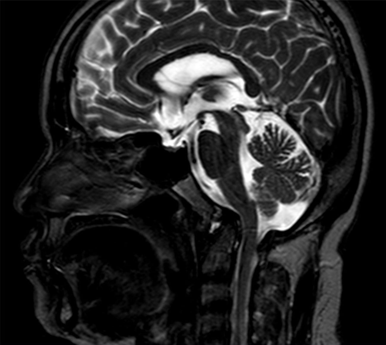
Three patterns of instability occur in the rheumatoid cervical spine, often coexisting. Understanding these is essential because the Ranawat class grades their neurological consequences, regardless of which pattern is dominant.
- Mechanism
- Transverse ligament destruction by pannus; C1 slides forward on C2
- Key Radiographic Feature
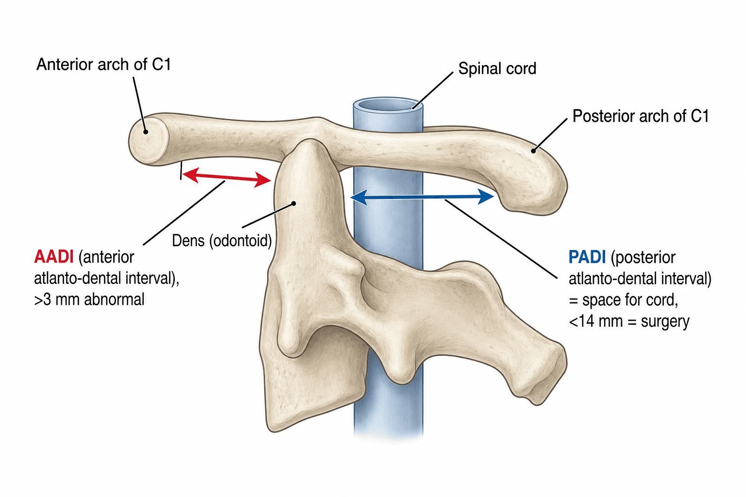
- Anterior atlantodental interval greater than 3 mm (or posterior atlantodental interval less than 14 mm)
- Clinical Concern
- Cord compression between odontoid and posterior arch of C1
- Mechanism
- Lateral mass destruction; odontoid migrates superiorly through foramen magnum
- Key Radiographic Feature
- Ranawat index less than 13 mm (male) or less than 12 mm (female); McRae line violated
- Clinical Concern
- Brainstem and upper cord compression; most dangerous pattern
- Mechanism
- Facet joint and disc destruction below C2
- Key Radiographic Feature
- Listhesis greater than 20 percent or 3 mm at any subaxial level
- Clinical Concern
- Cord compression at subaxial segments; can be multi-level
A · B · SThree patterns of rheumatoid cervical instability
Hook:They frequently coexist — always look for all three on imaging (MRI the whole cervical spine).
Basilar invagination carries the highest risk of sudden death from brainstem compression and vertebral artery compromise. A patient with worsening Ranawat class and radiographic basilar invagination needs urgent surgical consultation, not serial observation. The posterior atlantodental interval (space available for the cord) is more prognostic than the anterior atlantodental interval.
Basilar invagination (cranial settling) is hard to measure because the eroded odontoid tip is often invisible on plain films, so examiners expect you to know several lines and to combine them:
- Chamberlain line — hard palate to the posterior foramen magnum (opisthion); the odontoid tip more than roughly 3 mm above it suggests basilar invagination.
- McGregor line — hard palate to the lowest point of the occiput (used because the opisthion is often not visible); odontoid tip more than about 4.5 mm above it is abnormal.
- McRae line — across the foramen magnum (basion–opisthion); the odontoid tip should not cross above it, and if it does the patient is usually symptomatic.
- Ranawat index — the vertical height of C1 on C2 measured along the odontoid axis (centre of the C2 pedicle to the C1 transverse axis); reduced (about under 13 mm in men, under 12 mm in women) in cranial settling.
- Redlund-Johnell criterion — distance from the McGregor line to the midpoint of the C2 inferior endplate; abnormal under roughly 34 mm (men) / 29 mm (women).
- Clark station — divide the odontoid into thirds; the anterior arch of C1 sitting on the middle (station II) or caudal (station III) third signals progressive settling.
- Wackenheim clivus–canal line — drawn down the clivus; it should be tangential to the posterior odontoid, and an odontoid crossing it indicates invagination.
Use a combination (e.g. Clark station, Redlund-Johnell and the Ranawat index together) — relying on any single line misses cases, and MRI/CT is the definitive confirmation of cord/brainstem compromise.
The Ranawat Classification
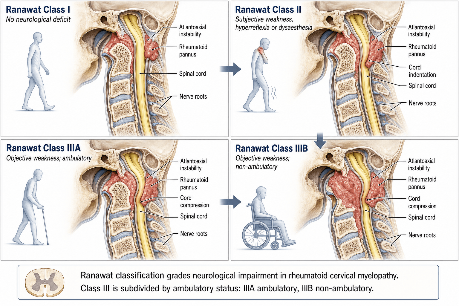
The Ranawat classification grades cervical myelopathy in rheumatoid arthritis into four classes based on the presence and severity of neurological deficit. It was originally described in the context of atlantoaxial subluxation and basilar invagination in RA but applies to all patterns of rheumatoid cervical spine involvement.
- Neurological Status
- No neurological deficit
- Key Features
- Pain only; no objective weakness, normal reflexes, no long-tract signs
- Functional Level
- Fully ambulatory
- Neurological Status
- Subjective weakness
- Key Features
- Hyperreflexia, dysesthesia, subjective weakness — but no objective motor deficit on examination
- Functional Level
- Fully ambulatory
- Neurological Status
- Objective weakness — ambulatory
- Key Features
- Objective motor deficit with long-tract signs (Babinski, clonus, Hoffman); patient can still walk
- Functional Level
- Ambulatory with difficulty
- Neurological Status
- Objective weakness — non-ambulatory
- Key Features
- Objective motor deficit with long-tract signs; patient cannot walk (quadriparetic or quadriplegic)
- Functional Level
- Non-ambulatory
Pain · Subjective · Objective-walk · Objective-no-walkRanawat classes
Hook:The key divide is IIIA vs IIIB (ambulatory vs not) — it determines surgical prognosis; operate before IIIB.
Class II is the most commonly tested boundary. The patient complains of weakness and has hyperreflexia or dysesthesia, but when you formally examine motor power, it is normal (5/5 throughout). The deficit is subjective — the patient feels weak, the signs suggest upper motor neuron involvement, but there is no demonstrable motor loss. Once objective weakness appears, the patient is Class III.
Workup and Monitoring in RA Patients
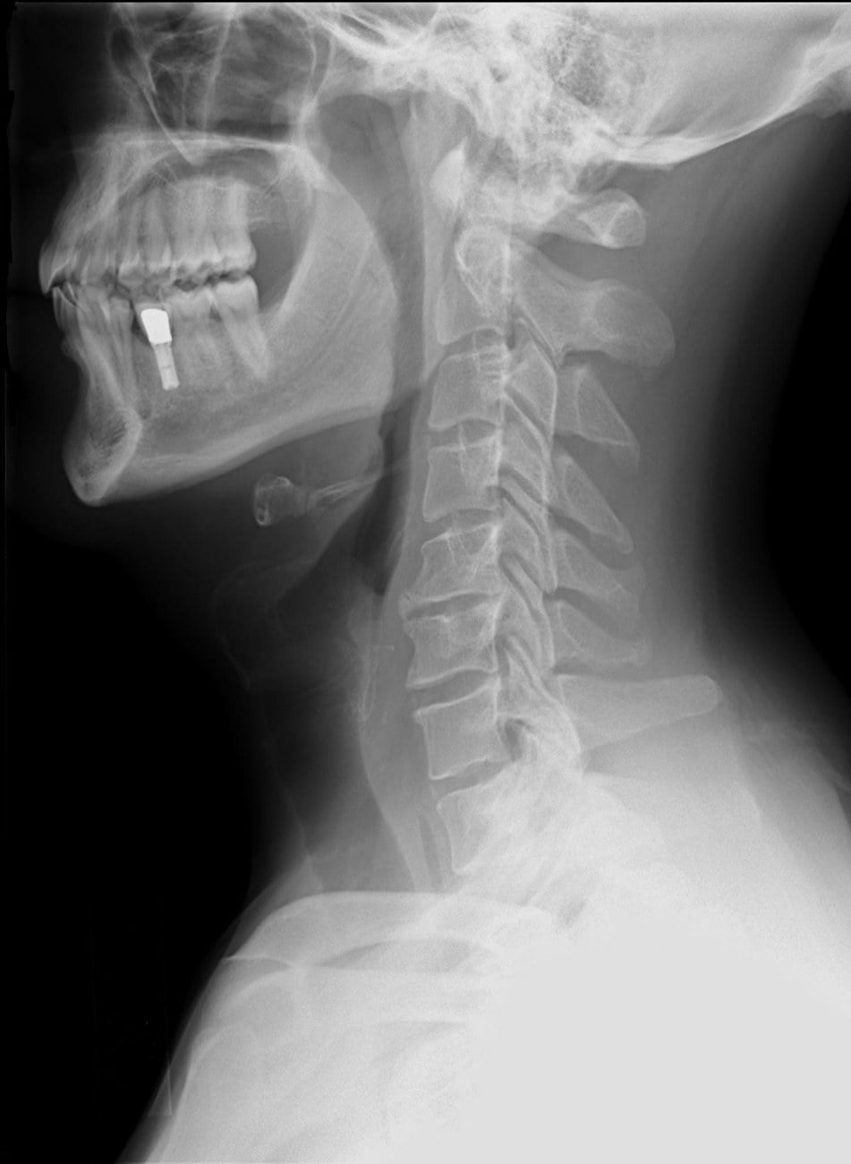
Three Ps — Progressive · Painful · PADI-lowWhen to refer the RA patient with cervical involvement for surgery
Hook:Do not wait for Class IIIB — refer and operate while the patient is still ambulatory.
Surgical Prognosis by Ranawat Class
The primary value of the Ranawat classification is prognostic: it predicts neurological recovery after surgical decompression and stabilisation.
- Likely Post-op Recovery
- Remains pain-free; no neurological deterioration
- Key Prognostic Point
- Excellent prognosis; surgery is for instability or progressive radiographic changes
- Recommendation
- Operate if progressive instability or pain refractory to conservative measures
- Likely Post-op Recovery
- Good — most patients improve or stabilise
- Key Prognostic Point
- Subjective symptoms often resolve; objective motor function is preserved
- Recommendation
- Operate once identified to prevent progression to Class III
- Likely Post-op Recovery
- Moderate — objective deficit may partially recover
- Key Prognostic Point
- Ambulation is preserved post-operatively in the majority; some residual deficit common
- Recommendation
- Operate without delay; further delay risks Class IIIB
- Likely Post-op Recovery
- Poor — few patients regain ambulation
- Key Prognostic Point
- Non-ambulatory patients rarely walk again even with successful decompression
- Recommendation
- Operate to prevent further deterioration and relieve pain; set realistic expectations
The single most important sentence for the exam: neurological recovery after surgery for rheumatoid cervical myelopathy is directly related to the pre-operative Ranawat class. A patient who is Ranawat IIIA has a meaningful chance of recovery; a patient who deteriorates to IIIB largely does not. This is why surveillance (serial neurological examination and flexion-extension radiographs or MRI) in RA patients is mandatory — you must detect deterioration and operate before the patient becomes non-ambulatory.
The Ranawat class decides whether to operate; reducibility decides the approach (the question the topic's vivas raise). Assess it on flexion-extension films and with pre-operative traction (halo or skeletal):
- Reducible deformity (realigns on extension or traction) → a posterior procedure alone. For isolated atlantoaxial subluxation, C1–C2 fusion — modern Goel-Harms (C1 lateral-mass + C2 pedicle screws) or Magerl transarticular screws, or the older Gallie/Brooks wiring. When there is basilar invagination / cranial settling or an incompetent C1, extend to an occipitocervical fusion (occiput to C2 or below).
- Irreducible ANTERIOR compression (a fixed odontoid/pannus that will not reduce and is indenting the cord) → anterior decompression first — a transoral (or endoscopic endonasal) odontoidectomy to remove the offending mass — followed by posterior fusion to stabilise.
- Pre-operative traction is the pivot: it can convert an apparently irreducible deformity into a reducible one, allowing a posterior-only operation (in Nannapaneni's IIIb series, the reducible cases had direct posterior fusion and only the fixed ones needed transoral decompression then posterior fusion).
- A retro-odontoid pannus often regresses after a stable posterior fusion alone (stopping the motion lets the pannus resorb), so a reducible lesion with anterior pannus can frequently be treated posteriorly without transoral surgery.
- Subaxial subluxation is addressed with fusion (posterior, or anterior) of the involved levels, and may be combined with the craniocervical construct when patterns coexist.
Always check the vertebral artery anatomy (CT angiography) before C1–C2 or occipitocervical instrumentation, especially with basilar invagination.
Limitations and Modern Context
- Ranawat class is a static snapshot. It does not capture the rate of neurological deterioration, which is itself prognostic — a patient who progresses from Class II to IIIA over two weeks is more urgent than one who progresses over two years.
- The classification was originally described for atlantoaxial disease but is applied to all patterns of rheumatoid cervical myelopathy. Its prognostic accuracy for isolated subaxial subluxation is less well validated.
- Modern biologics (TNF inhibitors, IL-6 blockers, JAK inhibitors) have reduced the incidence and severity of rheumatoid cervical spine involvement in countries with early access to these agents. However, cervical myelopathy still occurs, particularly in patients with long-standing, poorly controlled disease.
- MRI is essential alongside clinical classification. Cord signal change on T2-weighted MRI (myelomalacia) is an independent predictor of poor recovery and should influence surgical timing regardless of the Ranawat class.
- Ranawat class does not guide surgical technique (anterior versus posterior approach, extent of fusion, need for occipitocervical fusion). That decision is driven by the pattern of instability and the level of compression.
- Functional scoring systems (such as the Japanese Orthopaedic Association score and the Neck Disability Index) provide a more granular assessment of impairment and are increasingly used alongside Ranawat class in clinical research.
Guidelines, Registries and Global Practice
- No single guideline universally mandates a specific Ranawat threshold for surgery, but the consensus across the AO Foundation, BOA–BOAST (UK), and the Japanese Orthopaedic Association is that progressive neurological deficit (Class II evolving toward IIIA) is an indication for operative stabilisation.
- Global practice varies by access to biologics. In high-income countries with early biologic therapy, severe cervical myelopathy (Class IIIA and IIIB) is now less common. In resource-limited settings, late presentation with advanced myelopathy remains frequent, and the Ranawat classification retains high prognostic utility.
- Registry evidence (from national joint replacement and spine registries in Scandinavia and the UK) shows that the incidence of cervical spine surgery for rheumatoid arthritis has declined in parallel with biologic utilisation, but surgical outcomes in operated patients have not fundamentally changed — pre-operative Ranawat class remains the dominant predictor.
- The posterior atlantodental interval (PADI) less than 14 mm is a widely accepted radiographic threshold for surgical consideration, supplementing the clinical Ranawat class. PADI is more predictive of neurological deficit than the anterior atlantodental interval.
- Practice variation exists in surgical approach (anterior decompression versus posterior fusion versus combined) and in the use of occipitocervical fusion for basilar invagination. The Ranawat class guides the decision to operate, not the technique.
Viva practice
Exam Viva
Practise clinical reasoning and management decisions out loud
“A 58-year-old woman with long-standing seropositive rheumatoid arthritis presents with increasing neck pain, tingling in both hands, and difficulty fastening buttons. On examination she has brisk reflexes in the upper and lower limbs, bilateral Hoffman sign, but normal motor power (5/5 throughout). Flexion-extension radiographs show an anterior atlantodental interval of 7 mm. What is her Ranawat class and how would you manage her?”
“A 72-year-old man with rheumatoid arthritis has been increasingly unsteady over six months. He fell at home last week and is now unable to walk. Examination shows spastic quadriparesis (3/5 upper limbs, 2/5 lower limbs), bilateral Babinski signs, and a sensory level at C4. MRI shows basilar invagination with the odontoid tip above McRae line and severe cord compression with T2 signal change from the cervicomedullary junction to C4. Discuss his Ranawat class, prognosis, and surgical options.”
Exam cheat sheet
The four classes
- Class I: pain only, no deficit
- Class II: subjective weakness, hyperreflexia, dysesthesia — no objective motor loss
- Class IIIA: objective deficit with long-tract signs, still ambulatory
- Class IIIB: objective deficit, non-ambulatory (quadriparetic or quadriplegic)
Prognosis and surgical timing
- Pre-operative Ranawat class is the strongest predictor of surgical outcome
- Class II and IIIA: good-to-moderate recovery expected — operate without delay
- Class IIIB: poor ambulation recovery — surgery still indicated to prevent worsening
- The goal of surveillance is to operate before the patient becomes Class IIIB
Three patterns of rheumatoid cervical instability (A-B-S)
- Atlantoaxial subluxation: transverse ligament pannus destruction, AADI greater than 3 mm
- Basilar invagination: odontoid migrates through foramen magnum, Ranawat index low, highest mortality
- Subaxial subluxation: facet and disc destruction below C2, listhesis greater than 3 mm or 20 percent
- All three can coexist — MRI the entire cervical spine
Key thresholds and measurements
- AADI greater than 3 mm: abnormal (but PADI is more prognostic)
- PADI less than 14 mm: surgical consideration
- Ranawat index less than 13 mm (male) or less than 12 mm (female): basilar invagination
- T2 cord signal change on MRI: independent poor prognostic factor
Evidence
Cervical spine fusion in rheumatoid arthritis
- Reviewed 33 rheumatoid patients undergoing cervical fusion for atlantoaxial subluxation, superior odontoid migration and subaxial subluxation; devised the pain/neurological classification and a new method for measuring superior migration (the Ranawat index).
- Of 19 patients with neurological involvement, 8 improved, 7 were unchanged and 2 worsened — outcome tracked pre-operative neurological status.
- Defined the surgical strategy (Gallie C1-C2 fusion, occiput-to-C2 for superior migration, posterior fusion for subaxial disease).
A prospective study of the progression of rheumatoid arthritis of the cervical spine
- Prospective series documenting progressive cervical-spine involvement in rheumatoid arthritis over time.
- Atlantoaxial subluxation was the most common pattern; basilar invagination the most dangerous.
- Neurological deterioration tracked radiographic progression — supporting serial surveillance and early referral.
Rheumatoid atlantoaxial subluxation can be prevented by intensive use of traditional disease-modifying antirheumatic drugs
- FIN-RACo randomised trial: combination DMARD therapy from diagnosis versus single-DMARD; cervical radiographs at 5 years in 149 patients.
- Anterior atlantoaxial subluxation developed in 9% overall, but far less often in the combination group (2 vs 11 cases) — intensive early DMARD therapy prevented or retarded it.
- A subset still developed subluxation despite intensive treatment, so cervical surveillance remains necessary.
According to PubMed, the classification and index are from Ranawat et al. 1979 (J Bone Joint Surg Am 1979;61(7):1003-10; PMID 489640), with progression data from Pellicci et al. 1981 (PMID 7204429). The PADI < 14 mm threshold and the finding that severity (not duration) predicts recovery are from Boden et al. 1993 (DOI 10.2106/00004623-199309000-00004); Class-IIIb surgical benefit (one-grade improvement) from Nannapaneni et al. 2005 (DOI 10.1227/01.neu.0000156202.80185.32); and DMARD prevention of atlantoaxial subluxation from the FIN-RACo trial, Kauppi et al. 2009 (DOI 10.3899/jrheum.080429).