Regan-Morrey Classification of Coronoid Fractures
Anatomy & biomechanics
The coronoid process forms the anterior wall of the trochlear notch and is the principal anterior buttress against posterior ulnar translation. Several stabilisers attach around it:
- Attachment
- Coronoid (anterior surface, near tip/base)
- Stabilising role
- Dynamic anterior support
- Attachment
- Sublime tubercle (medial coronoid base)
- Stabilising role
- Primary valgus stability
- Attachment
- Coronoid tip/anterior margin
- Stabilising role
- Secondary restraint to posterior translation
- Attachment
- Anterior wall of the trochlear notch
- Stabilising role
- The anterior bony buttress against posterior subluxation
The anteromedial facet of the coronoid overhangs the medial ulna and is unsupported by the metaphysis, so even a small fracture here removes the medial buttress and produces varus posteromedial rotatory instability — a pattern Regan-Morrey (size-based) under-rates. This is why the O'Driscoll classification (which categorises by location — tip, anteromedial facet, base) is needed alongside it.
The terrible triad context
The terrible triad — posterior dislocation with fractures of the coronoid and radial head — is where Regan-Morrey is most used. The mechanism is a fall onto an outstretched hand with the forearm supinated, producing axial load, valgus and posterolateral rotatory force, disrupting the three primary stabilisers: the anterior buttress (coronoid), the lateral column (radial head) and the ligaments. Recurrent instability is the hallmark complication if any component is left unaddressed.
- 1Reduce and imageReduce the dislocation urgently; CT to size the coronoid (plain films underestimate it) and assess the radial head and any anteromedial facet involvement.
- 2Address the coronoidFix type II/III fragments (lag screw or suture-lasso). For a tip (type I) fragment, repair the anterior capsule/fragment — but recognise the soft-tissue injury, not the tiny bone, is the real problem.
- 3Address the radial headRepair if reconstructable; if comminuted, REPLACE with a metallic radial head — never excise in a terrible triad (excision invites recurrent instability).
- 4Repair the LCL, then reassessRepair the lateral collateral ligament complex off the lateral epicondyle. If the elbow still subluxates/dislocates, repair the MCL or apply a hinged external fixator.
In a terrible triad, never excise a comminuted radial head — replace it. Excision removes the lateral column and almost guarantees recurrent instability. And type the coronoid on CT, not plain films, which underestimate fragment size and can misclassify the injury.
The reconstruction exists to permit early motion, so the post-operative protocol is part of the answer. With a stable construct, begin supervised early active and active-assisted range of motion (within days to about a week), positioning the forearm to protect the repair - in pronation if the lateral collateral ligament was repaired (pronation tightens/protects the lateral side), in supination if the medial side was the concern - and use the overhead (supine, shoulder-flexed) motion programme so gravity compresses and stabilises the elbow during active extension. Avoid varus stress and unsupported/resisted extension early; a hinged elbow brace, sometimes with an initial extension block, protects the arc while allowing motion. Watch for re-subluxation, flexion contracture/stiffness (the commonest residual problem) and heterotopic ossification. If a stable arc cannot be achieved, a hinged external fixator maintains reduction while permitting motion.
Limitations & the O'Driscoll system
- Regan-Morrey is size-based only. It ignores fracture location and morphology; two type II fractures behave very differently depending on whether the line runs through the anteromedial facet or transversely.
- The O'Driscoll classification is more anatomically useful: it separates tip, anteromedial facet and basal fractures, mapping fracture lines to surgical approaches. Examiners increasingly expect familiarity with both.
- CT is essential for accurate typing and planning — a "type II tip" on plain film may be a type III, or a destabilising anteromedial facet fracture, on CT.
- It does not account for comminution — a comminuted type II can be harder to fix than a simple transverse type III.
- Isolated type I fractures without instability are benign; the classification is most actionable within a complex injury pattern.
Because Regan-Morrey ignores location, the companion O'Driscoll classification divides coronoid fractures into three anatomic types, each with subtypes that map to the surgical approach:
- Type 1 - tip (transverse): subtype 1 = 2 mm or less of coronoid height, subtype 2 = more than 2 mm. This is the terrible-triad coronoid.
- Type 2 - anteromedial facet: subtype 1 = anteromedial rim; subtype 2 = rim + tip; subtype 3 = rim + sublime tubercle (± tip). This is the varus posteromedial rotatory instability fracture, fixed through a medial approach with a buttress plate.
- Type 3 - basal: subtype 1 = coronoid body/base; subtype 2 = transolecranon basal coronoid. The large, unstable fragment needing buttress fixation.
The point: O'Driscoll tells you WHERE the fracture is (and therefore the approach); Regan-Morrey only tells you how big it is.
The Regan-Morrey classification
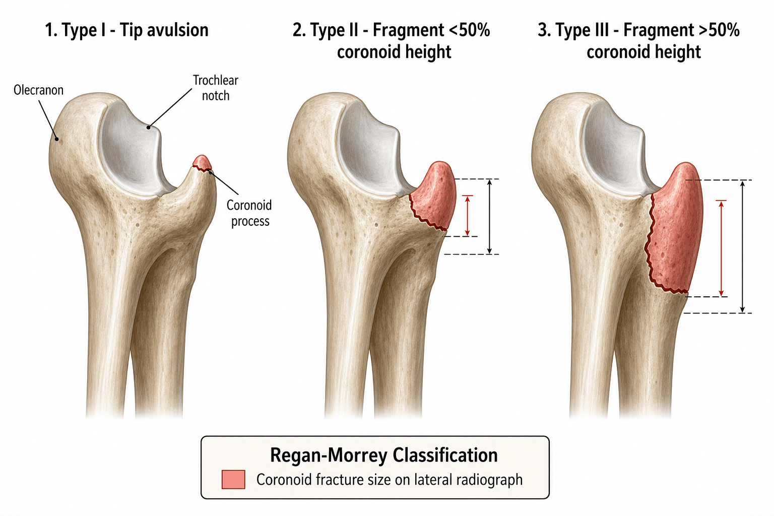
Described by Regan and Morrey in 1989 from lateral radiographs, the system grades the fracture by the proportion of coronoid height involved — and in their original series instability and poor outcomes rose steeply with the type.
- Description
- Avulsion of the coronoid tip only
- Proportion of coronoid
- Tip fragment only
- Original satisfactory result
- 92% (best; usually stable if isolated)
- Description
- Fragment involving half the coronoid or less
- Proportion of coronoid
- 50% or less of coronoid height
- Original satisfactory result
- 73% (moderate instability risk)
- Description
- Fragment involving more than half the coronoid
- Proportion of coronoid
- More than 50% of coronoid height
- Original satisfactory result
- 20% (significant instability; fixation required)
Tip • Half • Beyond HalfThe three types
Hook:Tip, Half, Beyond-half - the cut point is simply the halfway mark of the coronoid height; instability climbs and outcomes fall as the fragment grows.
The tip fragment (type I) is clinically important not for the bone but for what it signals: the brachialis inserts onto the coronoid, and the anterior capsule and anterior bundle of the MCL attach near it. A tip avulsion is a marker of capsuloligamentous disruption — especially within a terrible triad — even though the bone loss is small.
Viva practice
Exam viva
Practise clinical reasoning and management decisions out loud
“A 28-year-old man fell from a ladder onto an outstretched right hand. He presents with a posterior elbow dislocation, a coronoid fracture, and a comminuted radial head fracture. How do you classify the coronoid fracture, and what is the injury pattern called?”
“A 42-year-old woman presents after a fall with elbow pain and a sense of instability. Imaging shows an anteromedial facet fracture of the coronoid process — a small fragment on the medial side, not the tip. She has no dislocation and no radial head fracture. How does this challenge the Regan-Morrey system, and what is the mechanism?”
Exam & revision
Everything below condenses coronoid fracture classification for revision and viva practice.
- Coronoid = anterior buttress against posterior ulnar translation.
- Regan-Morrey by size: I tip, II ≤50%, III over 50% — instability and poor outcomes rise with type (satisfactory 92% → 73% → 20%).
- A tip fragment is a marker of capsuloligamentous injury, not just bone loss.
- Terrible triad = coronoid + radial head + dislocation; reconstruct coronoid → radial head (replace, don't excise) → LCL → reassess → MCL/hinged fixator.
- Pair Regan-Morrey with O'Driscoll (tip / anteromedial facet / base) — the anteromedial facet fracture (varus posteromedial rotatory instability) is small but destabilising.
- Type on CT, not plain films.
Coronoid • Radial Head • LCL • (MCL)Terrible triad priorities
Hook:Work lateral to medial: Coronoid → Radial head → LCL → reassess → MCL/hinged fixator. Restore enough stability for early motion.
Exam cheat sheet
The three types (by size)
- Type I: tip avulsion only - usually stable if isolated (92% satisfactory in the original series)
- Type II: 50% or less of coronoid height - moderate instability (73%)
- Type III: more than 50% - significant instability, fixation required (20% satisfactory)
- Always type on CT - plain films underestimate fragment size
Terrible triad reconstruction sequence
- Reduce the dislocation urgently
- Fix the coronoid (suture-lasso/lag screw for large; capsular/fragment repair for tip)
- Radial head: repair if possible, REPLACE if comminuted (never excise)
- Repair the lateral collateral ligament; reassess; add MCL repair or hinged fixator if still unstable
Beyond Regan-Morrey
- O'Driscoll: tip / anteromedial facet / basal - guides surgical approach by location
- Anteromedial facet fracture → varus posteromedial rotatory instability (small but destabilising)
- Regan-Morrey measures size; O'Driscoll measures location/morphology - both needed for planning
- Cadaveric data: type I coronoid SUTURE fixation alone does not restore kinematics - repair the MCL if still unstable
Evidence Base
Fractures of the coronoid process of the ulna
- Review of 35 patients defining three types by fragment size: tip (I), 50% or less (II), more than 50% (III).
- A concurrent dislocation/associated fracture was present in 14%, 56% and 80% of types I, II and III respectively - severity rose with type.
- Satisfactory results (32 patients, mean 50-month follow-up) were 92% (type I), 73% (type II) and 20% (type III); type III had the most residual stiffness. Early motion was recommended for I/II, reduction and fixation for III.
Standard surgical protocol to treat elbow dislocations with radial head and coronoid fractures
- 36 terrible-triad elbows managed with a standard protocol: radial head fixation/replacement, coronoid fixation if possible, and lateral ligament (± MCL/hinged fixator) repair.
- Mean Mayo Elbow Performance Score 88 (15 excellent, 13 good, 7 fair, 1 poor); concentric stability was restored in 34 of 36 elbows, allowing early motion.
- Eight patients needed reoperation (synostosis, instability, stiffness, infection).
According to PubMed: the size-based classification and its outcome gradient come from Regan & Morrey 1989 (35 patients; satisfactory 92/73/20% for types I/II/III, PMID 2793888); the systematic terrible-triad protocol from Pugh et al. 2004 (DOI); the anteromedial facet pattern from Doornberg & Ring 2006 (DOI); the suture-lasso-vs-ORIF comparison from Garrigues et al. 2011 (DOI); and the cadaveric caution that type I coronoid suture fixation alone does not restore kinematics (consider MCL repair) from Beingessner et al. 2007 (DOI). These are retrospective series and a cadaveric study (Level III-V), appropriate for a classification/technique topic. The O'Driscoll location-based classification is the established companion system.