Map bone | classify defect | restore fixation | prepare backups
Reconstruction Families
Critical Must-Knows
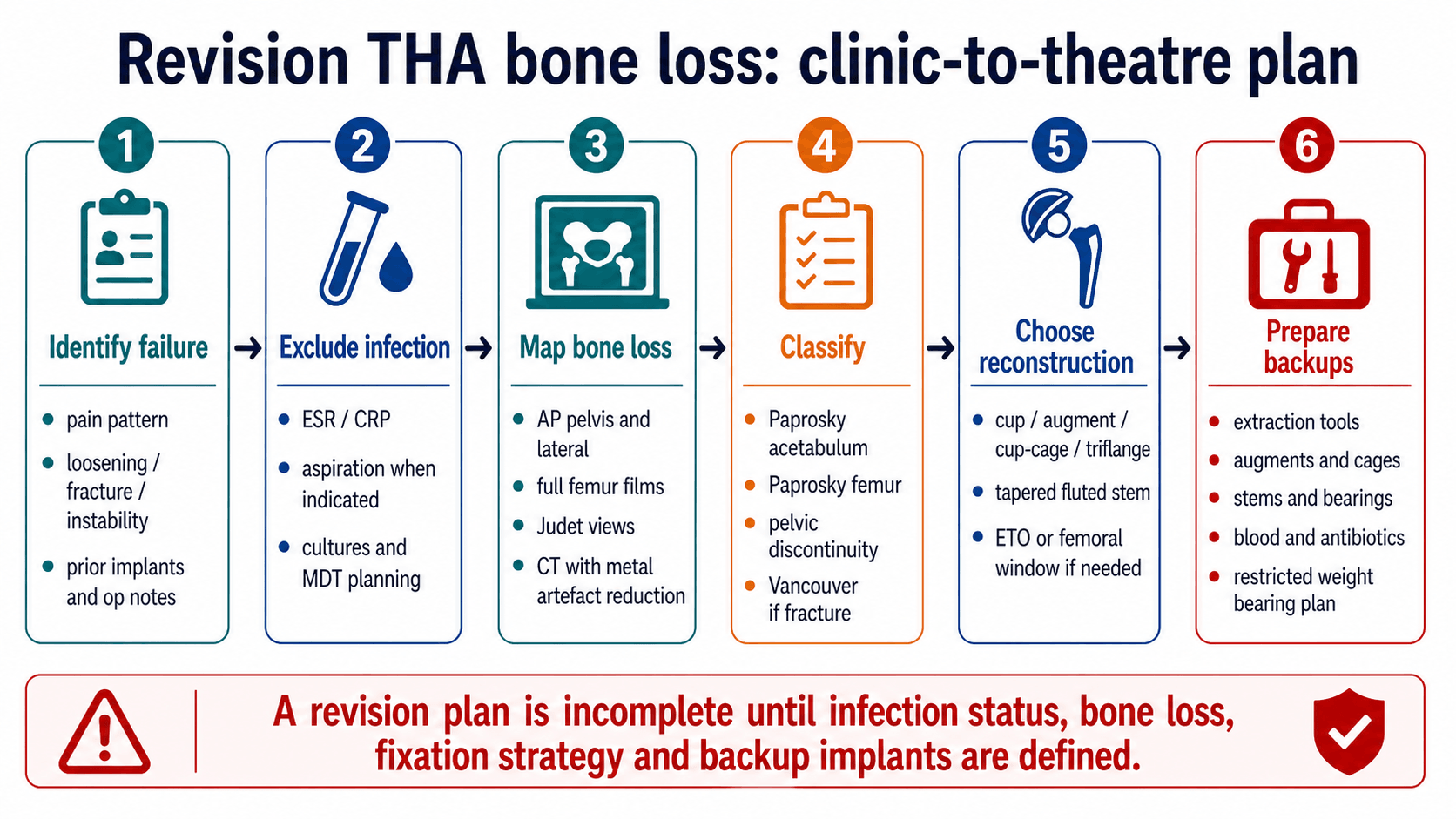
- Revision THA starts by defining failure and excluding infection. Bone loss reconstruction fails if the infection diagnosis is missed.
- Paprosky acetabular classification is useful because it estimates rim and column support. It is not just a memorised table.
- Pelvic discontinuity means the superior ilium and inferior ischiopubic segment are separated. Standard hemispherical fixation is often insufficient.
- Femoral revision succeeds by obtaining stable fixation beyond the defect. Diaphyseal/isthmus support matters more than proximal appearance alone.
- A revision plan is incomplete without backup implants. Extraction tools, augments, cages, stems, bearings and weight-bearing plan must be decided before theatre.
Clinical Pearls
- "Order AP pelvis, lateral hip, full femur films, Judet views and CT with metal artefact reduction when acetabular columns are uncertain.
- "Use Paprosky to decide if a cup can grip host bone, whether augments are required, and whether the construct must span or unitise the pelvis.
- "A custom triflange can be powerful for severe defects but has high complication and dislocation risk; counsel accordingly.
- "ETO is a controlled exposure tool for well-fixed stems or cement, not a failure of technique.
Do not plan revision THA from an AP pelvis alone
The AP pelvis gives the first impression. It does not reliably define posterior column support, pelvic discontinuity, femoral isthmus fixation, version, occult fracture or infection. The operation should not start until the surgeon has mapped the bone and planned extraction plus reconstruction.
Revision THA Mental Models
MAPPre-op workup | HOSTAcetabulum | STEMFemur |
|---|---|---|
M Mechanism of failure Loosening, infection, fracture, instability, wear or adverse local tissue reaction. | H Host contact Porous shell needs enough supportive host bone. | S Support at isthmus Diaphyseal support enables tapered fluted fixation. |
A Anatomy of bone loss Columns, rim, medial wall, isthmus, cortical tube and fracture lines. | O Offset and hip centre Reconstruction must restore biomechanics. | T Trochanteric osteotomy ETO preserves bone during difficult extraction. |
P Plan extraction and backups Removal tools, ETO, augments, cages, stems and weight-bearing restrictions. | S Structural support Augments, cup-cage or triflange when columns are deficient. | E Endoprosthesis Consider when host femur cannot support a revision stem. |
T Test discontinuity CT/Judet views and intra-operative stability determine strategy. | M Measure length/version Restore leg length, offset and version, not just fixation. | |
Map before cutting. | Host bone decides fixation. | Fix beyond the defect. |
BACKUPTheatre Readiness
| B | Blood and biology Optimise haemoglobin, infection status, bone quality and soft tissues. |
| A | Augments and cages Have modular porous metal, cage, cup-cage or triflange plan available. |
| C | Component extraction Plan cup, stem, cement and screw removal before incision. |
| K | Known implants Identify prior implant sizes, bearings, tapers and approach where possible. |
| U | Unexpected fracture Have cables, plates, struts, long stems and fracture strategy ready. |
| P | Post-op restrictions Weight bearing and precautions depend on construct stability. |
| B | Blood and biology Optimise haemoglobin, infection status, bone quality and soft tissues. | C | Component extraction Plan cup, stem, cement and screw removal before incision. | U | Unexpected fracture Have cables, plates, struts, long stems and fracture strategy ready. |
| A | Augments and cages Have modular porous metal, cage, cup-cage or triflange plan available. | K | Known implants Identify prior implant sizes, bearings, tapers and approach where possible. | P | Post-op restrictions Weight bearing and precautions depend on construct stability. |
Hook:Revision THA without BACKUP is not planned surgery.
Overview and Definitions
Revision THA bone loss is a reconstruction problem, not simply a component exchange. The surgeon must identify why the arthroplasty failed, whether infection is present, how much host bone remains, and which construct can achieve durable fixation.
The practical definitions are:
Definitions That Change Treatment
| Term | Meaning | Treatment Implication |
|---|---|---|
| Contained defect | Bone loss surrounded by a rim or cortical shell. | May be filled with morselised graft or cement/augment depending size and fixation. |
| Segmental defect | A rim, wall or column segment is missing. | Needs structural support: augment, cage, structural graft or custom component. |
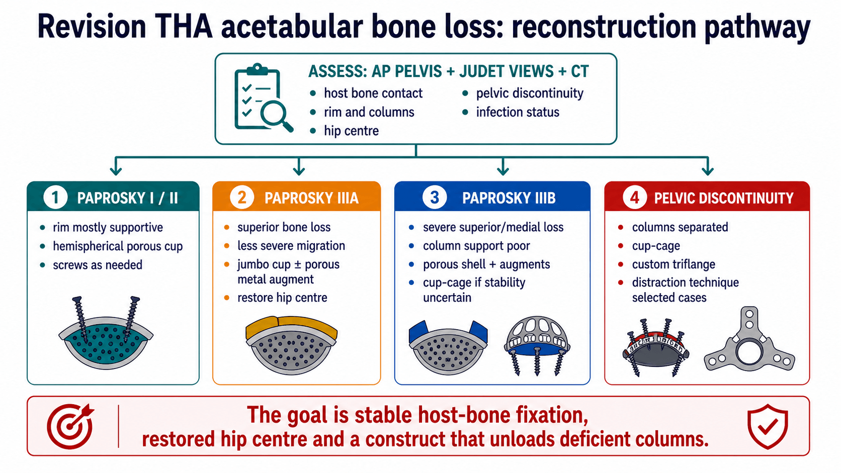
| Pelvic discontinuity | Superior ilium separated from inferior ischiopubic segment through the acetabulum. | Construct must stabilise or unitise the pelvis; standard cup fixation is often inadequate. |
| Femoral metaphyseal loss | Proximal femur cannot support the stem. | Shift fixation distally with a tapered fluted stem if diaphysis allows. |
| Non-reconstructable femur | No reliable proximal or diaphyseal host fixation. | Consider proximal femoral replacement or allograft-prosthetic composite in selected cases. |
Pathophysiology
Bone loss in revision THA usually develops from osteolysis, loosening, stress shielding, infection, periprosthetic fracture, adverse local tissue reaction or repeated previous surgery. Each mechanism leaves a different reconstruction problem.
Failure Mechanism to Bone-Loss Pattern
| Mechanism | Typical Bone Problem | Planning Consequence |
|---|---|---|
| Polyethylene wear and osteolysis | Cavitary acetabular or proximal femoral defects, sometimes with intact rim. | Assess whether fixation remains possible with shell/stem exchange and grafting. |
| Aseptic loosening | Progressive migration, rim deficiency, femoral cortical thinning or pedestal formation. | Plan extraction and reconstruction; classify both sides. |
| Infection | Bone destruction plus compromised soft tissues. | Treat infection pathway first; reconstruction may need staged strategy. |
| Periprosthetic fracture | Femoral cortical tube disrupted; implant may be loose. | Use Vancouver plus Paprosky femoral planning; fixation must bypass fracture and deficient bone. |
| Repeated revision | Combined acetabular/femoral deficiency, abductor damage and instability risk. | Expect higher constraint, complex exposure and restricted rehabilitation. |
| Pelvic discontinuity | Columns no longer form a continuous ring. | Cup-cage, triflange or distraction technique rather than simple hemispherical cup. |
Clinical Presentation and Assessment
History and examination
Ask what failed and what has already been done. Important history includes original diagnosis, approach, implant type, fixation method, bearing surface, prior infection, wound issues, instability episodes, fractures, metal-on-metal exposure, antibiotics, anticoagulation, neurological symptoms and functional goals.
Examination must document:
- Gait, Trendelenburg sign, abductor function and walking aids.
- Limb length, fixed deformity, flexion contracture and rotational profile.
- Scar position and soft-tissue envelope.
- Neurovascular status, especially sciatic/peroneal symptoms.
- Pain with rotation, trochanteric pain and signs of loosening or instability.
- Spine and pelvic obliquity when leg length or instability is part of the problem.
First-line imaging and tests
Revision THA Imaging and Tests
| Investigation | How To Order It | What It Answers |
|---|---|---|
| AP pelvis and lateral hip | Standing or standardised AP pelvis plus lateral of affected hip. | Migration, hip centre, loosening, osteolysis, offset, leg length and component position. |
| Full femur radiographs | Include hip to knee with entire implant and distal femur. | Stem length, cement, cortical defects, distal hardware and bypass planning. |
| Judet views | Oblique pelvic views when columns/discontinuity are uncertain. | Anterior and posterior column support. |
| CT with metal artefact reduction | Pelvis and/or femur depending defect. | Column integrity, discontinuity, version, bone stock, osteolysis and occult fracture. |
| ESR, CRP and aspiration | Screen for infection; aspirate when markers, symptoms or history are suspicious. | Defines whether revision is aseptic or infection pathway. |
| Implant records | Obtain stickers, op notes and bearing/taper details. | Determines extraction tools, compatibility and backup components. |
Investigations
The investigation plan must answer four questions before theatre: is the joint infected, is the acetabular column support intact, where can the femur obtain fixation, and what implant/extraction equipment is required.
How Each Test Changes The Operation
| Test | Decision It Supports | Unsafe Shortcut |
|---|---|---|
| ESR, CRP and aspiration when indicated | Aseptic revision versus infection pathway, culture strategy and staging. | Calling a loose implant aseptic without infection workup. |
| AP pelvis and lateral hip | Migration, hip centre, offset, leg length, loosening and gross osteolysis. | Using AP pelvis alone to choose augments or cages. |
| Full-length femur radiographs | Stem length, cement mantle, cortical tube, distal hardware, fracture and bypass length. | Planning femoral revision without seeing the whole stem. |
| Judet views | Anterior and posterior column integrity when discontinuity is possible. | Missing posterior column deficiency. |
| CT with metal artefact reduction | Column support, discontinuity, component version, osteolysis, cortical defects and occult fracture. | Assuming a cup can grip host bone without cross-sectional mapping. |
Bone-Loss Mapping
Paprosky Acetabular Classification
| Type | Bone-Loss Pattern | Reconstruction Meaning |
|---|---|---|
| I | Minimal bone loss; hemispherical shape and rim supportive. | Porous hemispherical cup usually sufficient. |
| IIA/IIB/IIC | Distorted hemisphere with superior, lateral or medial bone loss. | Cup with screws, graft or limited augment depending host contact. |
| IIIA | Severe superior bone loss but some column/rim support remains. | Jumbo cup, porous shell and augments; restore hip centre if possible. |
| IIIB | Severe superior/medial migration with poor column support; discontinuity risk. | Cup-cage, custom triflange, distraction or complex augment strategy. |
Management
Management is selected by fixation biology and mechanical stability. The surgeon should not choose an implant because it is familiar; the implant must solve the defect pattern.
Acetabular Decision Pathway
| Defect Situation | Preferred Direction | Why |
|---|---|---|
| Paprosky I/II with supportive rim | Porous hemispherical shell with screws; graft contained defects as required. | Enough host bone exists for initial stability and ingrowth. |
| Superior segmental loss but columns partly supportive | Jumbo cup or porous shell plus modular augment. | Augment converts an unsupported segment into a stable platform. |
| Severe medial/superior loss or suspected discontinuity | Plan cup-cage, custom triflange or distraction rather than shell alone. | The construct must bridge or unitise deficient columns. |
| Contained bone loss in younger patient | Consider impaction grafting when stable containment and surgeon experience allow. | May restore bone stock but fails if initial stability is poor. |
Acetabular Reconstruction
Acetabular Reconstruction Options
| Option | Best Use | Limitations and Pitfalls |
|---|---|---|
| Porous hemispherical shell | Paprosky I/II and selected IIIA with enough host bone contact. | Fails if rim/columns cannot support initial stability. |
| Jumbo cup | Superior bone loss where large shell restores contact. | Can raise hip centre or over-ream if used indiscriminately. |
| Porous metal augment | Segmental superior, posterior or medial defects with shell contact possible. | Augment must support shell and be mechanically stable; cement only at augment-shell interface when used. |
| Cup-cage | Severe bone loss or discontinuity when a shell alone may not be stable. | Cage protects shell while ingrowth occurs; risk of dislocation, infection and cage fatigue remains. |
| Custom triflange | Massive defects, pelvic discontinuity, failed cages or unusual anatomy. | Requires CT-based manufacture, longer lead time and counselling about high complication risk. |
| Acetabular distraction | Selected chronic pelvic discontinuity with porous shell and augments. | Technique-sensitive; requires careful patient and defect selection. |
| Impaction grafting | Contained or reconstructable defects, especially where bone-stock restoration matters. | Use caution in severe uncontained defects or when primary stability is weak. |
Femoral Reconstruction
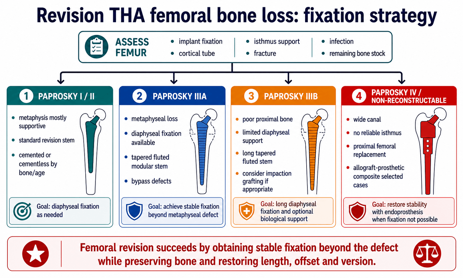
Core Femoral Principle
Bypass the defect, obtain axial and rotational stability, restore length/offset/version, and avoid creating a fracture during extraction.
When Host Fixation Is Not Enough
Paprosky IV, severe Vancouver B3 fractures, tumour-like bone loss or failed multiple revisions may require proximal femoral replacement or allograft-prosthetic composite.
Femoral Reconstruction Options
| Option | When To Choose | Key Technical Point |
|---|---|---|
| Cemented revision stem | Older patient, poor bone, intact cement mantle strategy or selected low-demand cases. | Cement technique and version control are critical. |
| Tapered fluted modular stem | Paprosky IIIA/selected IIIB with diaphyseal fixation available. | Achieve axial/rotational stability beyond defect and restore version with modularity. |
| Extensively porous-coated stem | Selected femora with adequate diaphyseal engagement. | Mismatch, thigh pain and stress shielding must be considered. |
| Impaction grafting | Younger patient or contained femoral deficiency where bone stock restoration is realistic. | Technique-sensitive; needs intact cortical tube and stable cemented stem construct. |
| Proximal femoral replacement | Non-reconstructable proximal femur, massive bone loss, severe B3 fracture or salvage setting. | Higher dislocation/infection risk; restore abductors and soft-tissue tension where possible. |
Operative Technique
Revision THA bone loss operation: PIPADRAW sequence
Theatre Sequence
| Step | What The Surgeon Does | Pitfall |
|---|---|---|
| Position | Usually lateral decubitus for posterior/lateral revision; ensure full femur access and ability to extend incision. | Positioning that prevents distal femoral exposure makes ETO or fracture control harder. |
| Imaging/equipment | Have AP pelvis/full femur templates, extraction systems, burrs, cables, augments, cages, stems, bearings and backup constraint. | Starting without backup implants converts a planned reconstruction into improvisation. |
| Preparation | Antibiotics/cultures per infection plan, blood availability, cell salvage if used, previous incision strategy. | Giving antibiotics before cultures may compromise microbiology if infection is suspected. |
| Approach | Use prior approach where safe; extensile posterior, lateral or anterolateral exposure by implant, scar and surgeon familiarity. | Poor soft-tissue handling increases instability and wound complications. |
| Dissection | Identify abductors, sciatic nerve risk zone, pseudocapsule, implants, cables/screws and osteolytic membrane. | Aggressive membrane removal can damage remaining host bone. |
| Removal | Remove liner/head first; assess fixation; use curved blades/extraction tools for cup; use ETO for difficult stem/cement removal when appropriate. | Uncontrolled extraction causes iatrogenic fracture and worsens bone loss. |
| Reconstruction | Rebuild acetabulum to host bone/columns and femur to reliable fixation zone; restore hip centre, offset, length and version. | Stable-looking components can still be biomechanically wrong if hip centre or version is poor. |
| At-risk structures | Sciatic nerve, superior gluteal neurovascular bundle, femoral vessels medially, abductors, greater trochanter and peroneal nerve stretch. | Lengthening and scar dissection increase nerve risk. |
| Aftercare | Weight bearing by construct, bone loss, ETO fixation and fracture risk; dislocation precautions and abductor rehabilitation. | Allowing full weight bearing after tenuous fixation can fail the reconstruction. |
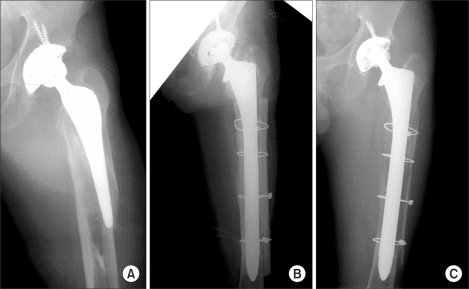
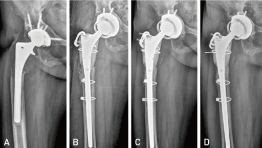
Extended trochanteric osteotomy
Use ETO when a well-fixed stem, long cement mantle, distal ingrowth, cement restrictor, femoral deformity or high fracture risk makes direct extraction unsafe. The aim is controlled access while preserving the vascularised osteotomy fragment.
Key steps:
- Plan osteotomy length from implant/cement extent; commonly about 12 to 16 cm from the greater trochanter in described techniques.
- Preserve vastus lateralis and abductor attachments to maintain biology.
- Round osteotomy corners with burr to reduce stress risers.
- Open the osteotomy in a controlled fashion, remove stem/cement, reconstruct the canal, then close with cables or wires.
- Protect against trochanteric migration, nonunion, fracture and abductor dysfunction.
Complications and Failure Management
Complications
| Complication | Why It Happens | Prevention or Management |
|---|---|---|
| Dislocation | Abductor deficiency, altered hip centre, constrained reconstruction, soft-tissue damage. | Restore offset/length/version, choose bearing strategy, consider dual mobility or constrained liner in selected cases. |
| Infection | Long surgery, multiple revisions, dead space and compromised host. | Optimise, culture, debride, antibiotic plan and staged reconstruction when indicated. |
| Aseptic loosening | Poor host fixation, inadequate column support, failed ingrowth or overloaded cage. | Use appropriate host bone fixation, augments, cage/triflange and protected rehabilitation. |
| Nerve injury | Limb lengthening, traction, scar dissection or screw/cage placement. | Document pre-op status, limit acute lengthening, protect sciatic nerve and use safe screw corridors. |
| Periprosthetic fracture | Extraction, weak cortex, stress risers or inadequate bypass. | ETO, cables/struts, long stems and bypass defects. |
| ETO nonunion or migration | Poor biology, inadequate fixation or excessive stripping. | Preserve attachments, cable fixation, protected weight bearing and revision fixation if symptomatic failure. |
Guidelines, Registries and Global Practice
Revision burden is rising worldwide as the primary THA population grows and ages. National joint registries are the best source of global epidemiology because they capture whole populations rather than single-centre series.
Global Epidemiology and Registry Signals
| Source | Signal | Planning Relevance |
|---|---|---|
| Aseptic loosening and instability | Across major registries (AOANJRR, NJR, AJRR, SHAR) aseptic loosening, dislocation and infection are consistently the leading reasons for revision THA. | These mechanisms generate most acetabular and femoral bone loss; anticipate them in workup. |
| Rising revision volume | Registries report a steady absolute rise in revision procedures as the primary arthroplasty cohort expands and survives longer. | Demand for augments, cup-cages, triflanges and tapered fluted stems is increasing globally. |
| Re-revision risk | Registry data show re-revision risk is higher than first-time revision, particularly after instability and infection. | Counsel patients that complex bone-loss reconstruction is not always a single definitive operation. |
| Highly porous metal trend | Outcome literature and registry-linked series show a shift away from cemented reconstruction toward highly porous shells and modular porous augments. | Mirror contemporary practice; cemented sockets are now selective. |
Society Guidance and Consensus, Side by Side
| Body | Emphasis Relevant to Bone Loss | Practical Takeaway |
|---|---|---|
| AAOS (US) | Systematic infection workup before revision (serology then aspiration) and structured pre-operative planning. | Exclude periprosthetic joint infection before any aseptic bone-loss reconstruction. |
| BOA / British Hip Society (UK) | Complex revision and pelvic discontinuity concentrated in higher-volume revision units with multidisciplinary input. | Refer massive bone loss and discontinuity to experienced revision teams. |
| AO Foundation | Mechanical principles: bypass the defect, obtain stable fixation in host bone and protect biology during exposure. | Fixation strategy is defined by where reliable host bone remains. |
| EFORT / European consensus | Standardised classification (Paprosky), infection exclusion and registry-informed implant selection. | Use a shared classification language and registry-supported implants. |
High-Resource versus Limited-Resource Practice
| Dimension | Well-Resourced Setting | Limited-Resource Setting |
|---|---|---|
| Imaging | CT with metal artefact reduction and Judet views routinely available. | May rely on plain films and Judet views; CT access can be limited, raising the value of careful radiographic assessment. |
| Implants | Full range of porous augments, cup-cages, custom triflanges and modular tapered stems. | Custom and modular options may be unavailable; structural allograft, cages and standard revision implants are used more. |
| Custom triflange | CT-based manufacture feasible with adequate lead time. | Manufacturing lead time, cost and supply chain often make custom triflange impractical. |
| Follow-up | Registry capture and structured surveillance. | Surveillance may be opportunistic; emphasise durable, forgiving constructs. |
Controversies and Areas of Uncertainty
Cup-cage versus custom triflange
Both manage severe defects and discontinuity. Off-the-shelf cup-cage avoids manufacturing lead time and cost, while custom triflange is matched to unique anatomy. High-quality comparative data are limited and choice remains largely surgeon- and resource-dependent.
Acetabular distraction
Distraction with a porous shell and augments is a newer technique for chronic pelvic discontinuity that aims to use elastic recoil for stability. Evidence is mostly single-centre cohorts; its place relative to cup-cage and triflange is still being defined.
Dual mobility versus constrained liners
Dual mobility reduces dislocation while preserving motion, but late intraprosthetic dissociation is a concern. Constrained liners can salvage severe abductor deficiency but transmit higher loads to fixation. The threshold between them is not standardised.
Modularity in tapered fluted stems
Modular fluted tapered stems aid version and length control but introduce a modular junction at risk of corrosion or fracture. Whether modular or monoblock stems are preferable in a given defect remains debated.
Evidence Signals
Acetabular bone-loss update
- Paprosky remains the most commonly used acetabular bone-loss classification.
- Careful radiological assessment can diagnose bone-loss pattern and chronic pelvic discontinuity before surgery.
- Contemporary practice increasingly uses highly porous shells with modular porous metal augments.
Acetabular reconstruction review
- Pre-operative evaluation includes history, examination, infection workup and detailed radiographic planning.
- Paprosky classification is based on column integrity and guides treatment strategy.
- Uncemented biological fixation techniques are preferred in many contemporary reconstructions.
Clinical Scenarios
Use these scenarios to practise clinical reasoning and management decisions
"A patient presents with a painful loose acetabular component. AP pelvis shows superior migration and medial wall deficiency. CT suggests poor posterior column support but no clear acute infection."
"A well-fixed cementless femoral stem must be removed during revision THA. The proximal femur is thin and there is concern that extraction will fracture the femur."
"Intra-operatively, during a revision THA for a loose cup, you find motion between the superior and inferior hemipelvis when you stress the acetabulum. CT had shown medial migration and a broken Kohler line."
References
- Sanghavi SA, Paprosky WG, Sheth NP. Evaluation and Management of Acetabular Bone Loss in Revision Total Hip Arthroplasty: A 10-year Update. J Am Acad Orthop Surg. 2024;32(10):e466-e475. doi:10.5435/JAAOS-D-23-00645.
- Fryhofer GW, Ramesh S, Sheth NP. Acetabular reconstruction in revision total hip arthroplasty. J Clin Orthop Trauma. 2020;11(1):22-28. doi:10.1016/j.jcot.2019.11.004.
- Hasenauer MD, Paprosky WG, Sheth NP. Treatment options for chronic pelvic discontinuity. J Clin Orthop Trauma. 2018;9(1):58-62. doi:10.1016/j.jcot.2017.09.009.
- Abdel MP, Trousdale RT, Berry DJ. Pelvic Discontinuity Associated With Total Hip Arthroplasty: Evaluation and Management. J Am Acad Orthop Surg. 2017;25(5):330-338. doi:10.5435/JAAOS-D-15-00260.
- Taunton MJ, Fehring TK, Edwards P, Bernasek T, Holt GE, Christie MJ. Pelvic discontinuity treated with custom triflange component: a reliable option. Clin Orthop Relat Res. 2012;470(2):428-434. doi:10.1007/s11999-011-2126-1.
- De Martino I, Strigelli V, Cacciola G, et al. Survivorship and Clinical Outcomes of Custom Triflange Acetabular Components in Revision Total Hip Arthroplasty: A Systematic Review. J Arthroplasty. 2019;34(10):2511-2518. doi:10.1016/j.arth.2019.05.032.
- Sershon RA, McDonald JF 3rd, Nagda S, Hamilton WG, Engh CA Jr. Custom Triflange Cups: 20-Year Experience. J Arthroplasty. 2021;36(9):3264-3268. doi:10.1016/j.arth.2021.05.005.
- Brown JM, Mistry JB, Cherian JJ, et al. Femoral Component Revision of Total Hip Arthroplasty. Orthopedics. 2016;39(6):e1129-e1139. doi:10.3928/01477447-20160819-06.
- Wyles CC, Hannon CP, Viste A, et al. Extended Trochanteric Osteotomy in Revision Total Hip Arthroplasty. JBJS Essent Surg Tech. 2023;13(3):e21.00003. doi:10.2106/JBJS.ST.21.00003.
- Jones SA. Impaction Grafting Made Easy. J Arthroplasty. 2017;32(9S):S54-S58. doi:10.1016/j.arth.2017.02.045.
- Lee JM, Kim TH. Acetabular Cup Revision Arthroplasty Using Morselized Impaction Allograft. Hip Pelvis. 2018;30(2):65-77. doi:10.5371/hp.2018.30.2.65.
- Fink B, Ahmadian A, Sax FH, Schuster P. Revision total hip arthroplasty using a modular fluted, tapered revision femoral component and interlocking screws in Vancouver B3 periprosthetic fractures with insufficient bone at the isthmus. Bone Joint J. 2024;106-B(4):344-351. doi:10.1302/0301-620X.106B4.BJJ-2023-0899.R1.
- Wyles CC, Hannon CP, Viste A, Perry KI, Trousdale RT, Berry DJ, Abdel MP. Extended Trochanteric Osteotomy in Revision Total Hip Arthroplasty. JBJS Essent Surg Tech. 2023;13(3):e21.00003. doi:10.2106/JBJS.ST.21.00003.
Revision THA Bone Loss Cheat Sheet
Clinical summary
Workup
- •History, op notes and implant details
- •ESR/CRP ± aspiration
- •AP pelvis, lateral and full femur
- •Judet views for columns
- •CT for discontinuity/version/bone stock
Acetabulum
- •Paprosky I/II: porous shell
- •IIIA: shell plus augment/jumbo cup
- •IIIB: poor columns, plan backup
- •Discontinuity: cup-cage/triflange/distraction
- •Restore hip centre and host fixation
Femur
- •Classify Paprosky femur
- •Plan extraction and ETO
- •Fix beyond deficient bone
- •Restore length, offset and version
- •Prepare fracture and salvage options
"Exclude infection, map bone loss, classify acetabulum and femur, then choose a construct that obtains stable fixation."