Sanders Classification of Calcaneal Fractures
- Reading the wrong slice. Sanders is read on the CORONAL CT at the widest part of the posterior facet — not the sagittal or axial view. Wrong slice = wrong type.
- Counting displacement instead of lines. The type is the number of fracture LINES crossing the posterior facet (I none, II one, III two, IV three+) — a displaced two-part fracture is still a Type II, not a Type III.
- Forgetting Sanders only describes the posterior facet. It says nothing about Böhler angle, heel varus, calcaneocuboid involvement or the soft-tissue envelope — all of which drive the actual decision.
- Offering primary arthrodesis for the wrong type. Primary subtalar arthrodesis is discussed for the unreconstructable Type IV, NOT for Type II/III.
- Overstating surgery / operating too early. RCTs (Buckley; UK Heel/Griffin) show similar 2-year outcomes vs non-operative for typical fractures with more complications (OR ~7.5) — select patients, and never operate before the soft tissues settle (positive wrinkle test).
The Sanders classification system
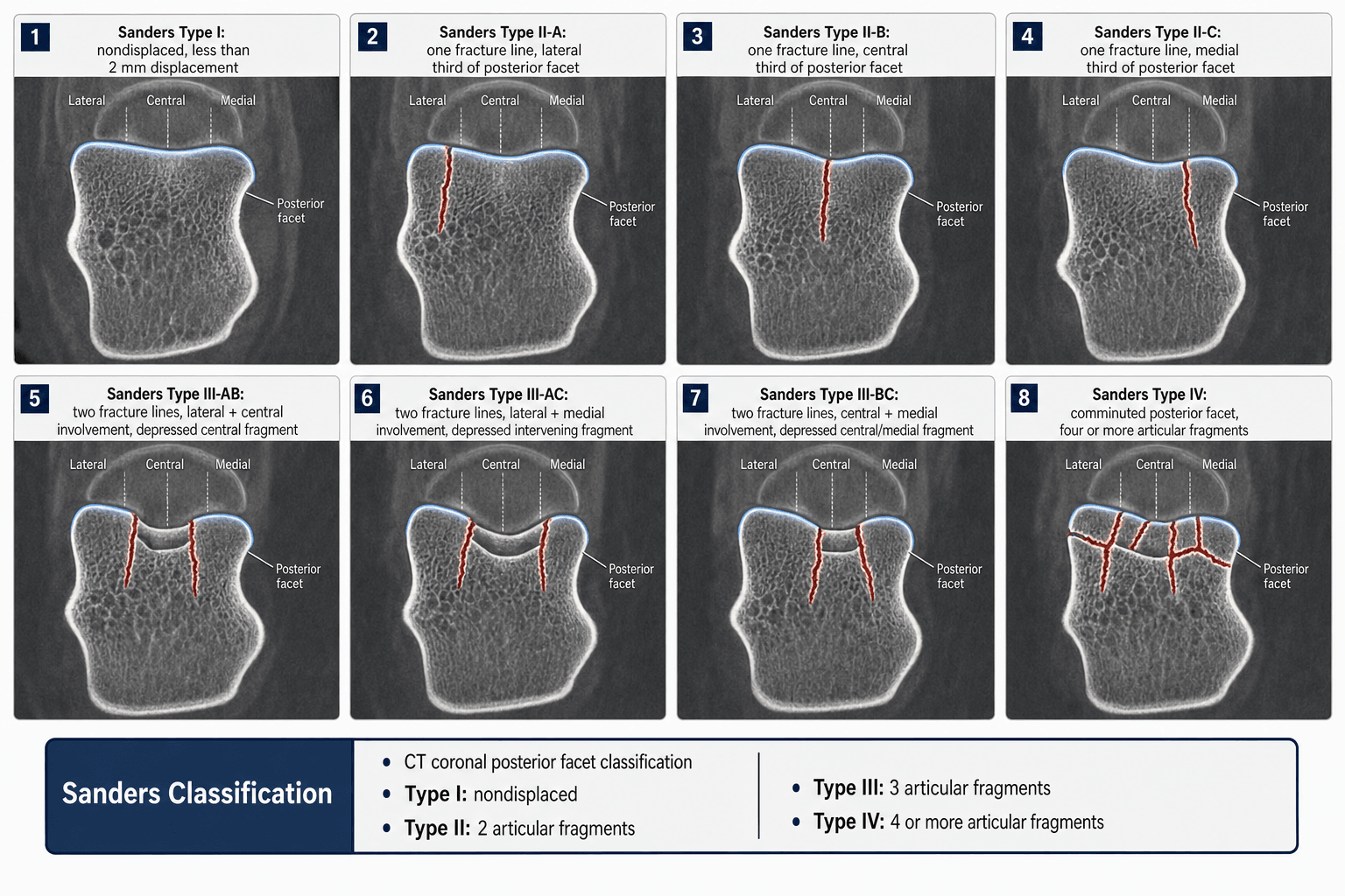
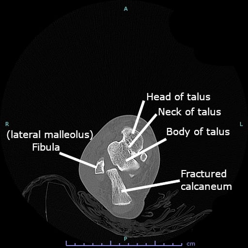
The Sanders classification is applied to the coronal CT image at the widest part of the posterior facet of the calcaneus (where the sustentaculum tali and posterior facet are broadest). Vertical reference lines divide the facet into columns:
- Line A — most lateral, through the lateral border of the posterior facet.
- Line B — central.
- Line C — most medial, near the sustentaculum (medial border).
The number of fracture lines crossing the posterior facet determines the type.
- Lines through posterior facet
- None (non-displaced articular fracture)
- Fragment pattern
- Single — no articular displacement
- Displacement
- None — congruent joint (under 2 mm)
- Lines through posterior facet
- One line (subtypes IIA, IIB, IIC)
- Fragment pattern
- Two fragments of the posterior facet
- Displacement
- Displaced single split
- Lines through posterior facet
- Two lines (subtypes IIIAB, IIIAC, IIIBC)
- Fragment pattern
- Three fragments with a depressed central piece
- Displacement
- Displaced with central depression
- Lines through posterior facet
- Three or more lines
- Fragment pattern
- Four or more fragments — highly comminuted
- Displacement
- Comminuted and incongruent
Subscripts give the line location: IIA/IIB/IIC for a single line in the lateral/central/medial position; IIIAB/IIIAC/IIIBC for two lines in those combinations. Medial fractures (IIC, IIIBC) involve the sustentaculum and are harder to fix. The depressed central fragment in Type III is the key to the reduction.
0–1–2–3 linesThe four types
Hook:Count the fracture lines crossing the posterior facet; the number of articular fragments is one more than the line count (up to the comminuted Type IV).
Sanders is read on a single CORONAL CT slice at the widest part of the posterior facet — using the wrong slice, or trying to read it on the sagittal or axial view, is the commonest error. Identify the slice, count the lines crossing the posterior facet, then name the type and subscripts.
Sanders grades the joint, but the examiner wants the patient: a calcaneal fracture is a high-energy axial-load injury (a fall from height or a road traffic collision driving the talus into the calcaneus), so screen for what comes with it:
- The "lover's" / "Don Juan" fracture pattern — the same axial load transmits up the skeleton, so around 10% have an associated thoracolumbar spine (burst) fracture, and others have the contralateral calcaneus, tibial plateau, pilon, or hip/acetabulum injured. Examine and image the spine and the opposite limb, and complete an ATLS survey in a fall-from-height.
- Foot compartment syndrome — occurs in roughly 10% of calcaneal fractures (the deep/calcaneal compartment); a missed compartment syndrome contracts the intrinsics into a fixed claw-toe deformity. Assess for a tense, swollen foot and disproportionate pain; measure compartment pressures if suspected (foot fasciotomy is considered, though its role is debated against the wound-healing risk).
- The soft-tissue envelope and open fractures — fracture blisters and severe swelling are the rule; an open calcaneal fracture (often a transverse medial wound) is a high-infection emergency needing urgent debridement. Operate on closed fractures only once swelling settles (a positive wrinkle test).
Type interpretation & operative decision
The type drives prognosis and the operative decision: the higher the type, the harder the reconstruction, the higher the complication rate, and the greater the chance of post-traumatic subtalar arthritis.
- 1Classify on the coronal CTCount the posterior-facet lines (I non-displaced, II one line, III two lines, IV ≥3). Note subscripts and the medial (sustentacular) involvement.
- 2Wait for the soft tissuesOperate once swelling settles (positive wrinkle test, ~1–3 weeks). Smoking, diabetes and vascular disease sharply raise wound-complication risk.
- 3Type I → non-operative; II/III → ORIFType I: early mobilisation. Type II/III: anatomical reduction of the posterior facet, restore height/length/width and Böhler angle, bone-graft the metaphyseal defect, lateral/sinus-tarsi plate — but counsel that RCTs show similar 2-year outcomes vs non-operative for typical fractures.
- 4Type IV → ORIF vs primary arthrodesisThe comminuted facet is often not reconstructable (0% anatomical in Sanders' series); discuss ORIF ± primary subtalar arthrodesis (RCT: comparable outcomes, faster definitive treatment, fewer late fusions) pre-operatively.
- Recommended treatment
- Non-operative; early mobilisation; percutaneous fixation if Böhler angle severely collapsed
- Expected outcome
- Generally good; low arthritis risk
- Key operative point
- Confirm the surface is truly non-displaced on CT
- Recommended treatment
- ORIF (extensile lateral or sinus tarsi) with plate/screws
- Expected outcome
- Best chance of anatomical reduction (86% in Sanders' series) and a good outcome
- Key operative point
- Anatomical reduction of the posterior facet is the single most important step
- Recommended treatment
- ORIF; technically demanding; sinus tarsi/percutaneous as a less-invasive option
- Expected outcome
- Intermediate; the central depressed fragment is the key challenge
- Key operative point
- Elevate the central depressed fragment and bone-graft the metaphyseal defect
- Recommended treatment
- ORIF often not feasible; primary subtalar arthrodesis a valid option (especially older/low-demand)
- Expected outcome
- Worst prognosis; high subtalar arthritis rate
- Key operative point
- Decide ORIF vs primary fusion pre-operatively if the facet is not reconstructable
A Sanders Type IV fracture with a comminuted posterior facet is the principal indication to discuss primary subtalar arthrodesis with the patient. Attempting to reconstruct a non-reconstructable joint risks a second, more difficult operation and a worse outcome — make this decision pre-operatively, not after a failed reconstruction. Equally, in a smoker/diabetic/vasculopath with a high-risk soft-tissue envelope, weigh non-operative care or a limited approach against the substantial wound-complication risk.
Limitations & modern context
- Reliability is good for Types I, II and IV but only moderate for Type III (distinguishing IIIAB from IIIBC, and IIB from IIIAB, on the coronal slice).
- Sanders describes only the posterior facet — not calcaneocuboid involvement, heel varus, Böhler angle, or the soft-tissue state, all of which influence outcome.
- It was developed in the era of extensile lateral ORIF; it is less directly applicable when a sinus tarsi or percutaneous approach is planned (reduction judged fluoroscopically).
- Slice selection matters — multidetector CT may show comminution on adjacent slices not captured by the single Sanders slice; 3D and weight-bearing cone-beam CT give a fuller picture.
- It is purely morphological — it does not incorporate patient factors (smoking, diabetes, vascular disease, work demands) that strongly drive wound healing, infection and function.
- High-level RCTs temper the surgical default: Buckley 2002 and the UK Heel (Griffin) 2014 trials found operative and non-operative care give similar 2-year outcomes for typical displaced fractures, with more complications after surgery — so the Sanders type informs, but does not by itself dictate, the operate-or-not decision.
Sanders is the CT/prognostic system, but the examiner pairs it with the plain-film classification and parameters — and one true emergency:
- Essex-Lopresti classification — based on the secondary fracture line: a tongue-type fracture has the secondary line exit posteriorly through the tuberosity, so the posterior facet stays attached to the tuberosity as one "tongue"; a joint-depression type has the secondary line exit just behind the posterior facet, leaving the facet fragment separate from the tuberosity. Both share a primary line (from the angle of Gissane) that creates a constant (sustentacular/anteromedial) fragment — held to the talus by the interosseous and deltoid ligaments and the key reduction reference.
- The tongue-type emergency — a displaced tongue fragment can tent and threaten the posterior heel skin, causing pressure necrosis. This is a surgical emergency: urgent reduction (often a percutaneous Essex-Lopresti/Westhues maneuver) to take tension off the skin, regardless of the eventual definitive plan.
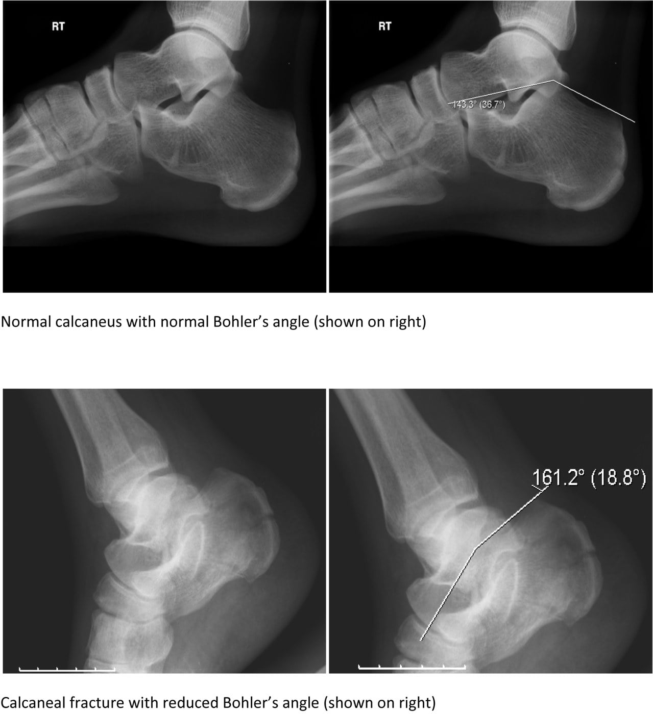
- The radiographic parameters — Böhler's tuber-joint angle (normal ~20–40°) falls with loss of calcaneal height; the crucial angle of Gissane (normal ~120–145°) at the lateral calcaneus rises with posterior-facet collapse. A reduced Böhler angle correlates with a worse outcome and is part of operative planning. Sanders grades only the posterior facet, so always read these on the lateral film.
Viva practice
- Read on the CORONAL CT at the widest posterior facet (not sagittal/axial); lines A (lateral), B (central), C (medial).
- I non-displaced, II one line/two-part, III two lines/three-part (depressed centre), IV ≥3 lines/comminuted.
- Anatomical posterior-facet reduction is the key to ORIF; in Sanders' series 86% anatomical in II vs 0% in IV.
- Type IV is where primary subtalar arthrodesis is discussed, not Type II.
- RCTs (Buckley, UK Heel): similar 2-year outcomes operative vs non-operative for typical fractures, more complications with surgery → select patients.
- Sanders describes only the posterior facet — also assess heel varus, Böhler angle, calcaneocuboid joint and soft tissues.
Mobilise–Fix–Fix-harder–FuseTreatment by Sanders type
Hook:I mobilise, II fix, III fix harder, IV consider fuse — and select the patient (fit, non-compensated, good soft tissues) before operating.
Exam viva
Practise clinical reasoning and management decisions out loud
“A 35-year-old roofer falls from a height and lands on his heels. CT shows a depressed intra-articular calcaneal fracture. The coronal CT at the widest part of the posterior facet shows one fracture line through the central part of the posterior facet, with the lateral fragment displaced. How would you classify the injury and outline your operative plan?”
“A 52-year-old manual worker sustains a comminuted intra-articular calcaneal fracture. CT shows three fracture lines through the posterior facet with at least four articular fragments. Outline the classification, prognosis and the operative options you would discuss.”
Exam cheat sheet
How to read the CT
- Coronal CT slice at the widest part of the posterior facet (not sagittal/axial)
- Lines A (lateral), B (central), C (medial) divide the facet into columns
- Count the fracture lines crossing the posterior facet — that defines the type
The four types
- Type I: non-displaced (under 2 mm) — no line crossing the articular surface
- Type II: one line — two fragments (IIA, IIB, IIC)
- Type III: two lines — three fragments with a depressed central piece (IIIAB, IIIAC, IIIBC)
- Type IV: three or more lines — four or more fragments, comminuted
Treatment & prognosis
- Type I: non-operative; early mobilisation
- Type II: ORIF; best prognosis after anatomical reduction (86% anatomical in Sanders' series)
- Type III: ORIF; elevate the central fragment, bone-graft the defect
- Type IV: ORIF often not feasible; primary subtalar arthrodesis is a valid option
- RCTs show operative ≈ non-operative at 2 years for typical fractures, with more surgical complications
Key pitfalls
- Read on the single coronal slice — not the sagittal/axial view
- Describes only the posterior facet — not heel varus, Böhler angle, calcaneocuboid joint or soft tissues
- Type IV (not II) is where primary arthrodesis is discussed
- Inter-observer agreement is moderate for Type III (IIIAB vs IIIBC)
Evidence
Operative treatment in 120 displaced intraarticular calcaneal fractures. Results using a prognostic computed tomography scan classification
- Introduced the CT-based four-type classification (with IIA/B/C and IIIAB/AC/BC subscripts) from 120 operatively treated displaced intra-articular calcaneal fractures.
- Anatomical articular reduction was achieved in 86% of Type II, 60% of Type III and 0% of Type IV fractures.
- Good/excellent clinical results occurred in 73% (Type II), 70% (Type III) and only 9% (Type IV) — outcome tracked the type, and Type IV did not improve even with surgical experience.
Displaced intra-articular fractures of the calcaneus
- Comprehensive instructional review of displaced intra-articular calcaneal fractures and their ORIF through an extensile lateral approach.
- Outcome correlates strongly with Sanders type; the technical priorities are restoring height, length and width, anatomical reduction of the posterior facet, bone grafting the defect and lateral plate fixation.
- Details the soft-tissue, blister and compartment-syndrome considerations around the lateral approach.
According to PubMed, the classification and its type-specific reduction/outcome figures (86% anatomical in II, 0% in IV; 73%/70%/9% good results) come from Sanders et al. 1993 (PMID 8472475); the ORIF technique from Sanders 2000 (DOI 10.2106/00004623-200002000-00009); the operative-vs-non-operative equivalence (with selected surgical benefit) from Buckley et al. 2002 (DOI 10.2106/00004623-200210000-00001) and the UK Heel Trial, Griffin et al. 2014 (DOI 10.1136/bmj.g4483); and the Sanders-IV ORIF-vs-primary-arthrodesis comparison from Buckley et al. 2014 (DOI 10.1097/BOT.0000000000000191).