Seinsheimer Classification of Subtrochanteric Fractures
Examiners expect you to name all five types (and the sub-patterns) and explain what drives each — fragment count and geometry in types I–IV, and intertrochanteric extension in type V. Be precise about Type III: IIIA is when the lesser trochanter is a separate third fragment, and IIIB is when the third fragment is a butterfly of the shaft. Then explain why medial cortex (calcar) integrity is the critical factor for stability, and justify cephalomedullary nailing over plate fixation for unstable patterns.
The Seinsheimer classification
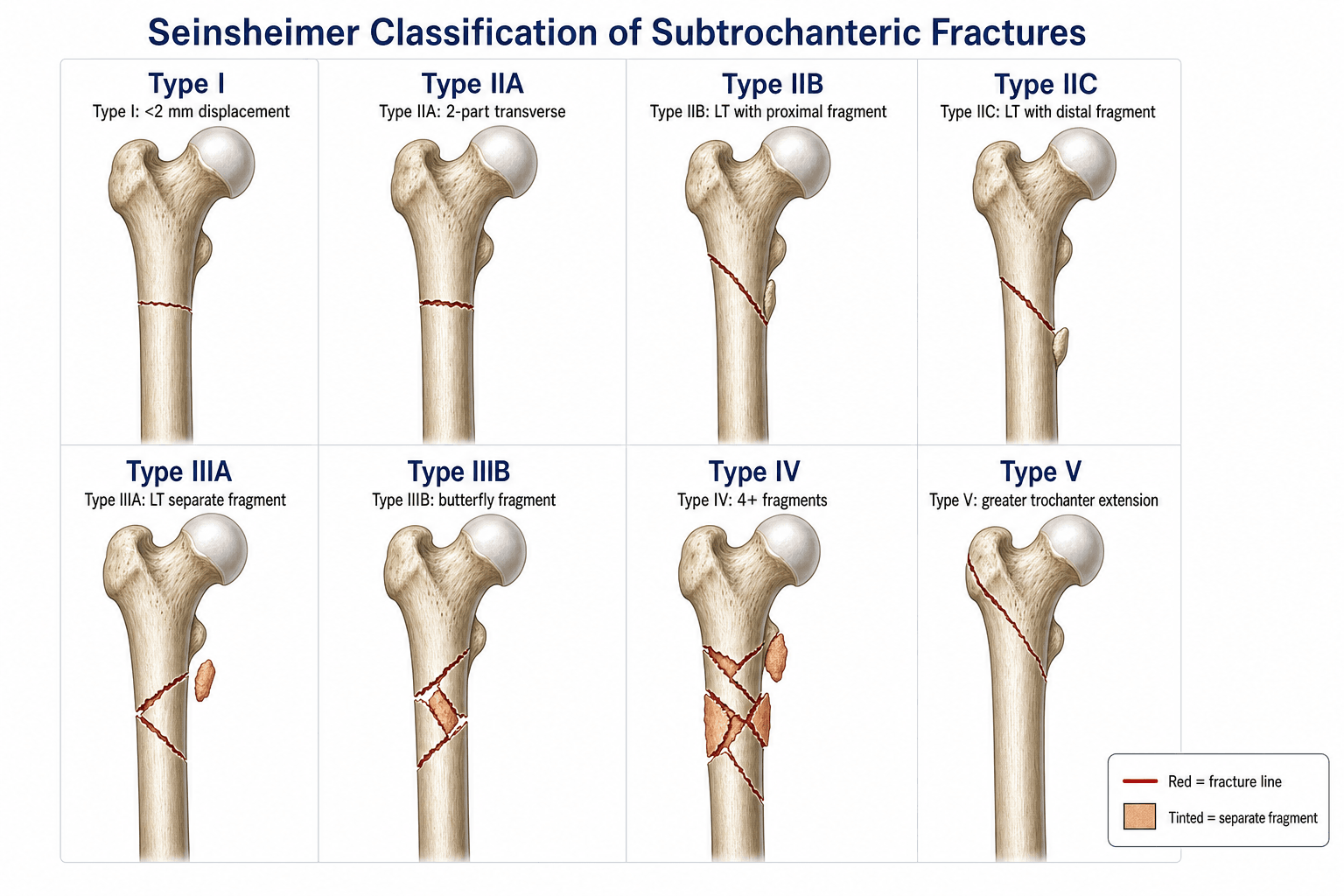
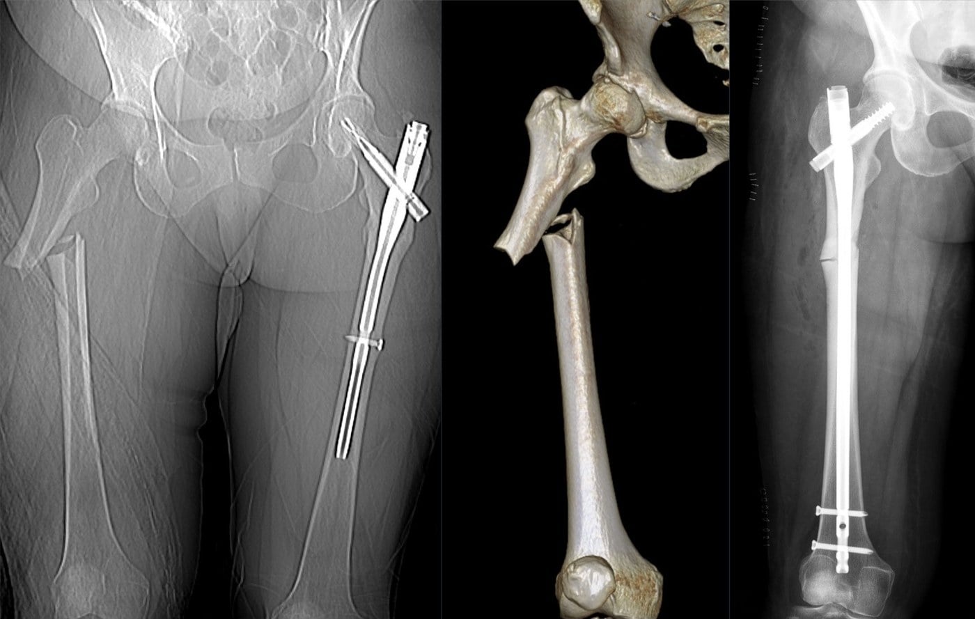
The Seinsheimer system classifies subtrochanteric fractures by the number of fragments, the geometry of the fracture line, and whether the fracture extends into the intertrochanteric region.
- Fragments
- Non-displaced (under 2 mm)
- Geometry
- Any pattern, minimally displaced
- Stability
- Stable
- Fragments
- Two-part
- Geometry
- Transverse fracture line
- Stability
- Moderate
- Fragments
- Two-part
- Geometry
- Spiral; lesser trochanter on the PROXIMAL fragment
- Stability
- Moderate
- Fragments
- Two-part
- Geometry
- Spiral; lesser trochanter on the DISTAL fragment
- Stability
- Moderate
- Fragments
- Three-part
- Geometry
- Spiral; lesser trochanter is a SEPARATE third fragment
- Stability
- Unstable
- Fragments
- Three-part
- Geometry
- Spiral; the third part is a BUTTERFLY fragment of the shaft
- Stability
- Unstable
- Fragments
- Four or more
- Geometry
- Comminuted, medial cortex destroyed
- Stability
- Very unstable
- Fragments
- Variable
- Geometry
- Subtrochanteric with intertrochanteric (greater-trochanter) extension
- Stability
- Unstable
I · II (2-part) · III (3-part) · IV (4+) · V (eXtends)Count the fragments: types I to V
Hook:Count the fragments to get the type; the LETTER then tells you where the lesser trochanter sits (II) or whether it is the third fragment (III).
The most error-prone distinctions are within types II and III. In IIB the lesser trochanter stays with the PROXIMAL fragment; in IIC it goes with the DISTAL fragment. In IIIA the lesser trochanter is a separate third fragment (the posteromedial calcar buttress is lost); in IIIB the third fragment is a butterfly of the shaft. Loss of the posteromedial buttress (IIIA, IV) is what makes reduction and fixation hardest.
Subtrochanteric biomechanics and deforming forces
The subtrochanteric region is the most highly stressed zone of the femur. Understanding the biomechanics explains why these fractures are difficult to treat and why implant selection matters.
- Location
- Medial cortex (calcar)
- Effect
- Highest in the body (classically ~1200 psi, Koch 1917)
- Clinical Relevance
- Medial cortex fracture = loss of the load-bearing column
- Location
- Lateral cortex
- Effect
- High but lower than medial compression
- Clinical Relevance
- Lateral wall integrity needed for the nail entry point
- Location
- Lesser trochanter
- Effect
- Flexion and external rotation of the proximal fragment
- Clinical Relevance
- Proximal fragment flexes and externally rotates
- Location
- Greater trochanter
- Effect
- Abduction of the proximal fragment
- Clinical Relevance
- Contributes to the varus, abducted proximal fragment
- Location
- Distal shaft
- Effect
- Medial displacement and shortening of the shaft
- Clinical Relevance
- Shaft adducts and shortens
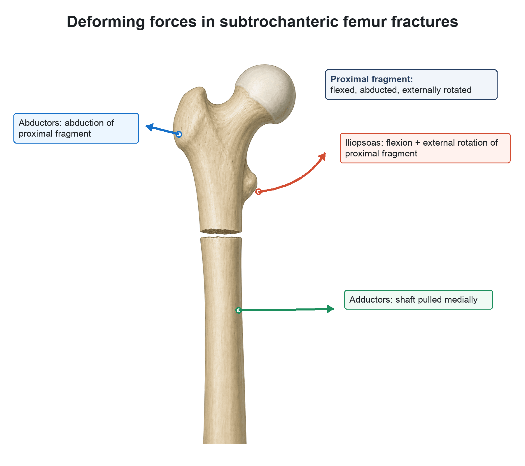
Flexed · Abducted · Externally rotatedDeforming forces on the proximal fragment
Hook:The classic deformity is a flexed, abducted, externally rotated proximal fragment with a medially-displaced shaft — overcome these BEFORE nailing; the nail cannot reduce the fracture alone.
The subtrochanteric region is predominantly cortical bone with a tenuous blood supply. Unlike intertrochanteric fractures (cancellous bone with good healing potential), subtrochanteric fractures carry a higher rate of nonunion and implant failure. Preserve the biological environment by minimising soft-tissue stripping — this is the rationale for indirect reduction and intramedullary fixation over open plating.
Implant selection
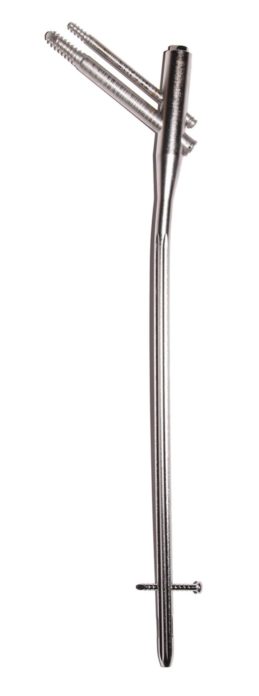
The choice of implant is driven by fracture stability, medial cortex integrity, and the need to protect the entire femur. Cephalomedullary nailing is the mainstay of treatment for displaced patterns.
- 1Classify + imagePlain films ± CT to define fragment count, geometry, medial-cortex status and any intertrochanteric extension (type V).
- 2Reduce FIRSTOvercome the deforming forces (flexed/abducted/ER proximal fragment) by positioning, traction and percutaneous tools — the nail will not reduce the fracture.
- 3Long cephalomedullary nailTrochanteric-entry long nail protecting the whole femur is the workhorse for types II–V; reduce before reaming to avoid varus/malrotation.
- 4Adjuncts / alternativesCerclage wire to hold a medial/butterfly fragment (IIIA/B, IV); a proximal femoral locking plate or blade plate when the lateral wall is destroyed or in revision.
Nail · Achieve reduction · Include whole femur · Lateral wallImplant principles: NAIL
Hook:Why nail over plate: shorter lever arm, load-sharing, early weight-bearing, and preserved biology (no soft-tissue stripping).
In the viva, always justify why a nail over a plate: the intramedullary position gives a shorter lever arm (less bending stress on the implant), the nail is load-sharing rather than load-bearing, it permits early weight-bearing, and the biological environment is preserved. Plates (blade plate, DCS, locking plates) are reserved for revision, malunion correction, or when the lateral wall prevents nail entry.
Because this is high-stress cortical bone, the complication rate is higher than for intertrochanteric fractures — and a common viva is "your nailed subtrochanteric fracture hasn't united; what now?":
- The common complications — nonunion (often from a varus or medial-gap malreduction), varus malunion (the commonest malalignment, typically from a too-medial nail start point or an unreduced flexed proximal fragment), implant failure / nail or screw breakage (the implant fatigues across a nonunion), cephalic screw cut-out, and malrotation.
- Managing an established nonunion — first rule out and treat infection and optimise the patient (vitamin D, calcium, stop smoking, review bisphosphonates). Then revise the fixation: either an exchange reamed nail (for a viable canal / hypertrophic nonunion) or conversion to a fixed-angle device (a 95° blade plate, dynamic condylar screw, or proximal femoral locking plate) with compression and autogenous bone graft — always correcting the varus and restoring the medial buttress, the loss of which usually caused the failure.
Limitations and modern context
- The Seinsheimer system is less widely used than the AO/Müller classification in contemporary practice. The AO system provides a universal fracture language across all bones, whereas Seinsheimer is specific to the subtrochanteric femur — but it remains a focused, biomechanically logical framework and still appears in exams.
- The Russell-Taylor classification (based on piriformis-fossa involvement and lesser-trochanter integrity) is an alternative that more directly guided nail entry-point selection, but is also less used now with the shift to trochanteric-entry nails.
- Modern cephalomedullary nails with trochanteric entry points (long Gamma 3, PFNA-II, InterTAN) have largely overcome older entry-point limitations — a trochanteric-entry nail can be used even when the piriformis fossa is fractured.
- Bisphosphonate-related atypical subtrochanteric fractures are a distinct entity — transverse, non-comminuted, with a medial spike and lateral cortical beaking — and often fit Seinsheimer IIA, but carry a high risk of propagation and nonunion from suppressed bone turnover.
- Registry data show cephalomedullary nails for subtrochanteric fractures carry a re-operation rate of roughly 5–10%, with varus malunion and screw cut-out the dominant failure modes.
The examiner expects you to define the region and to place Seinsheimer among the other systems it names:
- The subtrochanteric region — the zone from the lesser trochanter to about 5 cm distal (the proximal femoral diaphysis just below the lesser trochanter), a high-stress, predominantly cortical area.
- Fielding classification — by the level of the fracture relative to the lesser trochanter: Type I at the level of the lesser trochanter, Type II within 2.5 cm below it, Type III between 2.5 and 5 cm below.
- Russell-Taylor — by piriformis-fossa involvement (Type I spares the fossa, Type II extends into it) and lesser-trochanter/medial-cortex integrity (A intact, B involved); it historically guided the nail entry point but is less pivotal now with trochanteric-entry nails.
- AO/OTA — the universal alphanumeric system grading morphology as simple (A), wedge (B) or complex/comminuted (C); it gives a common language across bones but does not encode piriformis-fossa involvement.
- Bottom line — Seinsheimer (fragment geometry) and Russell-Taylor (entry point/medial cortex) are the focused subtrochanteric systems, Fielding describes the level, and AO/OTA is the universal descriptor.
Viva practice
- I non-displaced; II two-part (IIA transverse, IIB spiral LT-proximal, IIC spiral LT-distal); III three-part (IIIA LT separate fragment, IIIB butterfly); IV comminuted; V intertrochanteric extension.
- The three-part spiral was Seinsheimer's failure-prone pattern.
- Medial cortex (calcar) integrity = stability; its loss (IIIA, IV) is the key problem.
- Reduce before you ream — the nail cannot reduce the fracture.
- Long trochanteric-entry cephalomedullary nail is the workhorse; cerclage for medial/butterfly fragments; plate for lateral-wall loss/revision.
- Atypical (bisphosphonate) fractures are a distinct biology — transverse, medial spike, high nonunion risk.
Exam viva
Practise clinical reasoning and management decisions out loud
“A 72-year-old woman sustains a fall at home. Radiographs show a spiral subtrochanteric fracture with the lesser trochanter as a separate third fragment. How would you classify this fracture and what is your management plan?”
“A 58-year-old man in a high-energy motorbike collision has a highly comminuted subtrochanteric fracture with more than four fragments and destruction of the medial cortex, extending proximally into the intertrochanteric region. Discuss the classification, the biomechanical challenge, and your operative strategy.”
Exam cheat sheet
Types I to V (count the fragments)
- Type I: non-displaced or under 2 mm — stable
- Type II (two-part): IIA transverse · IIB spiral, LT on the proximal fragment · IIC spiral, LT on the distal fragment
- Type III (three-part): IIIA lesser trochanter is a separate fragment · IIIB the third part is a butterfly fragment
- Type IV: four or more fragments — comminuted, medial cortex destroyed
- Type V: subtrochanteric with intertrochanteric (greater-trochanter) extension
Biomechanics
- Subtrochanteric region = within ~5 cm distal to the lesser trochanter
- Medial cortex (calcar) carries the highest compressive load — its loss = instability
- Proximal fragment: flexed, abducted, externally rotated; shaft adducted medially
- Predominantly cortical bone, poorer blood supply — higher nonunion risk than intertrochanteric fractures
Implant selection
- Long trochanteric-entry cephalomedullary nail is the workhorse for displaced types II–V
- Reduce before reaming — the nail cannot reduce a subtrochanteric fracture alone
- Cerclage wires assist medial/butterfly-fragment containment (IIIA, IIIB, IV)
- Plates (blade plate, locking plate) reserved for revision, malunion, or lateral-wall loss
- Biological fixation: indirect reduction, preserve soft tissues, bridge comminution
Evidence
Subtrochanteric fractures of the femur
- Reported 56 patients with subtrochanteric fractures and proposed the five-type (eight-pattern) classification by fragment number and geometry.
- Of 47 fractures treated with internal fixation there were 9 fixation failures.
- The three-part spiral subtrochanteric fracture occurred 18 times and accounted for 8 of the 9 fixation failures — the failure-prone pattern.
Cephalomedullary nails in high-energy proximal femur fractures in young patients: prospective randomized comparison of trochanteric versus piriformis fossa entry portal
- Prospective RCT of 34 patients (17 per group), aged 18–50, with high-energy proximal femur fractures (subtrochanteric, intertrochanteric or ipsilateral neck/shaft) — trochanteric (Long Gamma) versus piriformis (Russell-Taylor Recon) entry nails.
- No difference in blood loss, incision length, operative duration, reduction quality, ease of use, union rate, varus malunion or outcome.
- Both devices gave predictably good results in these difficult fractures.
Gamma and other cephalocondylic intramedullary nails versus extramedullary implants for extracapsular hip fractures in adults
- Systematic review of 43 RCTs, predominantly TROCHANTERIC fractures; the Gamma nail had increased operative/later femoral fracture and reoperation rates versus the sliding hip screw (SHS).
- For trochanteric fractures the SHS appeared superior; mortality and medical complications did not differ between implants.
- For SUBTROCHANTERIC fractures (and some unstable patterns), intramedullary nails tended to have fewer healing complications than fixed-angle plates.
According to PubMed, the classification (and the failure-prone three-part spiral — 8 of 9 fixation failures in 56 patients) is from Seinsheimer 1978 (J Bone Joint Surg Am 1978;60(3):300-6; PMID 649632). Trochanteric-entry nailing is validated by Starr et al. 2006 (DOI 10.1097/00005131-200604000-00002); the nail-versus-plate evidence (with the subtrochanteric nuance) is the Parker & Handoll Cochrane review (DOI 10.1002/14651858.CD000093.pub5); cerclage-assisted reduction by Codesido et al. 2017 (DOI 10.1007/s00402-017-2722-y); and atypical-fracture criteria by Shane et al. 2010 (DOI 10.1002/jbmr.253).