Coxa Saltans | Internal vs External | Iliopsoas vs ITB
- External Snapping (ITB): Visible snap over the lateral hip. 'I saw it pop out'.
- Internal Snapping (Iliopsoas): Deep audible snap/clunk with hip extension from flexion/abduction.
- Intra-articular: True mechanical symptoms (locking/catching) - usually painful.
- Voluntary: Many patients can reproduce the snap voluntarily.
- Ober's Test: Assessing ITB tightness is key for external type.
- “Ask the patient to reproduce the snap!
- “Psoas snap: Flexion + Abduction + External Rotation to Extension
- “ITB snap: Flexion/Extension in lateral decubitus
- “Painful vs Painless: Painless snapping requires NO treatment.
Overview and Epidemiology
Snapping hip syndrome, or coxa saltans, is a palpable or audible snap around the hip during movement. It is classified by the structure that causes the snap: one outside the joint laterally (external) or anteriorly (internal), or a lesion inside it (intra-articular).
Who. It is most common in females, in whom the wider pelvis increases the ITB angle, and in adolescents and young adults. Among athletes it is seen in dancers (ballet), gymnasts and runners. In adolescents, habitual snapping can be associated with anxiety or compulsion.
Anatomy. Coxa vara makes the greater trochanter prominent and increases ITB friction, and in a leg-length discrepancy the longer leg has the tighter ITB. Increased femoral anteversion and a narrow bi-iliac width, common in dancers, are the other anatomical predispositions.
Activity. Sports involving repetitive hip flexion and extension, such as cycling and running, predispose to it. So do dance, especially ballet with its extreme hip positions, martial arts with high kicks, and kicking sports such as soccer.
Control. The biomechanical factors are gluteal weakness with poor hip control, core instability, tight hip flexors, and ITB tightness from overuse.
Pathophysiology and Mechanisms
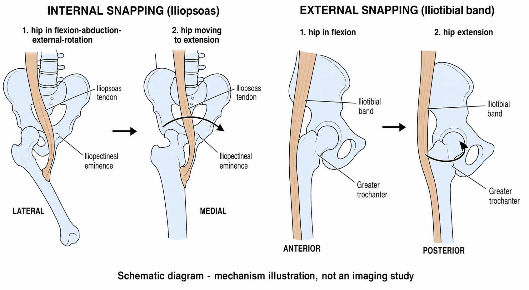
Internal snapping. The iliopsoas tendon snaps over the iliopectineal eminence or the femoral head. It happens as the hip moves from flexion, abduction and external rotation (FABER) into extension, adduction and internal rotation, when the tendon flips from lateral to medial across the eminence. It is often associated with a painful iliopsoas bursitis.
External snapping. The thickened posterior fibres of the iliotibial band, or the anterior border of gluteus maximus, snap over the greater trochanter. It occurs during flexion and extension, classically in single-leg stance or running, and is associated with trochanteric bursitis (greater trochanteric pain syndrome).
Intra-articular snapping. Mechanical interposition inside the joint causes locking or catching, and is not a true snap over bone. It must be distinguished from the vacuum phenomenon, the noise of the suction seal. The structures responsible are:
- Labral tear (the most common)
- Loose bodies (chondromatosis)
- Ligamentum teres tears
- Chondral flaps
Classification Systems
Anatomical. The anatomical classification places the snap relative to the joint, which helps guide the anatomical target: extra-articular lateral (external: iliotibial band or gluteus maximus), extra-articular anterior (internal: iliopsoas tendon), and intra-articular (labrum, loose bodies, cartilage flaps). The three types sound, look and provoke differently.
- External (ITB)
- Sometimes
- Internal (Iliopsoas)
- Often Loud 'Clunk'
- Intra-articular
- Click/Catch
- External (ITB)
- Visible 'Jump' of ITB
- Internal (Iliopsoas)
- Not visible
- Intra-articular
- Not visible
- External (ITB)
- Lateral (Greater Troch)
- Internal (Iliopsoas)
- Anterior (Groin)
- Intra-articular
- Deep Groin/C-sign
- External (ITB)
- Walking/Running
- Internal (Iliopsoas)
- Extension from Frog-leg
- Intra-articular
- Rotation/Pivoting
Functional. The functional classification decides whether to treat at all, and treatment is only for the first type:
- Painful: pathological, associated with bursitis or tissue damage, and needs treatment
- Painless: physiological, a noisy hip, and needs no treatment
- Voluntary: the patient demonstrates it on demand, often with a psychological component
- Involuntary: occurs during gait or sport
Some patients, often adolescents, habitually snap the hip as a tic. Surgery in painless voluntary snappers has unpredictable, often poor, outcomes. Avoid operating on the 'party trick'.
Clinical Assessment
History. Ask the patient to describe the sound (a pop, click or clunk) and to show where it is. A lateral snap, with the patient pointing to the trochanter, is external; a groin snap is internal or intra-articular. Ask whether it hurts, because that decides whether it needs treating, and then ask the patient to do it now: demonstration is diagnostic.
Is it intra-articular? Rule out a labral tear. Intra-articular pathology presents with sharp groin pain and mechanical locking or catching.
Examining for external snapping. Ober's test assesses ITB tightness. To reproduce the snap, lie the patient on the side and passively flex and extend the hip while compressing the ITB against the greater trochanter; the band is felt to jump.
Examining for internal snapping. The Thomas test assesses psoas tightness (fixed flexion deformity). For the dynamic test the patient lies supine and actively extends the hip from flexion, abduction and external rotation, and a clunk is palpable anteriorly.
Examining the joint. FADIR is the impingement and labral-loading test, and the scour test grinds the femoral head.
Investigations
Radiographs are usually normal. Their job is to look for the intra-articular associations: cam or pincer FAI and hip dysplasia (DDH).
Dynamic ultrasound is the diagnostic test of choice for snapping. It shows the tendon snapping over bone in real time, can show bursitis, and can be used to perform a diagnostic injection.
MRI and MR arthrography are for ruling out intra-articular pathology, above all a labral tear. MR arthrography is the gold standard for labral tears and the discriminator between intra- and extra-articular causes. MRI may also show iliopsoas bursitis in internal snapping, and gluteal tendinopathy or ITB thickening in external snapping.
Differential Diagnosis
The central exam skill is separating benign extra-articular snapping from intra-articular and other mimics of the painful, noisy hip.
- Location & Character
- Anterior groin, audible deep clunk
- Key Discriminator
- Reproduced extending from FABER; tendon flips over eminence
- Confirm With
- Dynamic ultrasound
- Location & Character
- Lateral, visible jump over trochanter
- Key Discriminator
- Visible/palpable jump; positive Ober
- Confirm With
- Clinical + dynamic US
- Location & Character
- Deep groin, C-sign, locking/catching
- Key Discriminator
- Mechanical locking, positive FADIR, cam/pincer on XR
- Confirm With
- MR arthrogram
- Location & Character
- Lateral, point tenderness, night pain
- Key Discriminator
- Tenderness over trochanter, abductor weakness, no true snap
- Confirm With
- MRI (gluteal tendinopathy)
- Location & Character
- Intermittent true locking
- Key Discriminator
- Episodic mechanical block, normal between episodes
- Confirm With
- CT / MRI / arthroscopy
- Location & Character
- Groin, activity-related, no snap
- Key Discriminator
- Pain with hop test, risk factors (RED-S)
- Confirm With
- MRI
- Location & Character
- Lower groin, no snap
- Key Discriminator
- Pain on resisted adduction / sit-up
- Confirm With
- MRI / clinical
Management Algorithm
Treatment is a ladder, and only painful snapping that has failed the lower rungs reaches surgery.
Treatment Ladder
If painless, explain that the hip is noisy but normal. No treatment beyond education.
Stretching of the ITB and psoas, core and gluteal strengthening, and activity modification.
Ultrasound-guided steroid injection into the iliopsoas bursa or the trochanteric bursa, which is both diagnostic and therapeutic.
Only for refractory painful snapping, which is rare. The technique depends on the type.
Surgical Technique
Internal snapping: arthroscopic psoas fractional lengthening. The goal is to lengthen the tendon without releasing it completely, preserving power. Only the tendinous portion is cut, at the musculotendinous junction, leaving the muscular sleeve intact; this reduces tension and stops the snapping while aiming to retain flexion power. Whether it truly preserves more strength than complete tenotomy has not been shown (see Controversies).
Access to the psoas. The release is made by hip arthroscopy or endoscopy, at one of three levels: transcapsular from the central compartment at the level of the labrum, transcapsular from the peripheral compartment, or endoscopically at the lesser trochanter.
External snapping: ITB release or Z-plasty. The goal is to reduce ITB tension over the greater trochanter. The options are:
- Open Z-plasty, a formal Z-lengthening of the ITB
- Cruciate or transverse incision, a cross-cut or transverse release of the ITB over the trochanter
- Endoscopic release, a diamond-shaped defect created over the greater trochanter, with lower morbidity than open Z-plasty for suitable patients
- Bursectomy, excision of the inflamed trochanteric bursa
Complications
- Risk
- Common (Internal)
- Note
- Radiological atrophy near-universal after tenotomy; early weakness usually compensates
- Risk
- ~5% arthroscopic / over 20% open
- Note
- Insufficient release, re-scarring; lower with arthroscopic technique
- Risk
- Rare
- Note
- Usually asymptomatic
- Risk
- Rare
- Note
- LFCN (external/anterior portals); genital paraesthesia with central-compartment psoas tenotomy
Iliopsoas-Impingement Labral Lesion (Not the Same as FAI)
The joint is usually involved. At arthroscopy most internal snappers have coexisting intra-articular pathology: synovitis, chondropathy or labral lesions. One lesion is worth knowing by name, the labral tear caused by iliopsoas impingement. It is the one anterior labral tear caused by the tendon, so its treatment differs from FAI, and that makes it a high-yield discriminator.
Mechanism. A tight or inflamed iliopsoas tendon lies directly in front of the anterior capsulolabral complex. With repetitive flexion and extension it abuts and inflames the labrum where it crosses it, the anterior labrum at around 3 o'clock (right hip), producing a focal "kissing" tear with overlying synovitis. In an FAI tear a bony cam or pincer lesion drives the labral damage instead.
- Iliopsoas-impingement lesion
- Direct tendon abutment on the labrum
- FAI labral tear
- Bony cam/pincer abnormal contact
- Iliopsoas-impingement lesion
- Focal, directly anterior (~3 o'clock), isolated
- FAI labral tear
- Anterosuperior, often with chondral damage
- Iliopsoas-impingement lesion
- Normal alpha angle, no pincer over-coverage
- FAI labral tear
- Cam (raised alpha angle) and/or pincer present
- Iliopsoas-impingement lesion
- Localised synovitis/bruising deep to the psoas tendon
- FAI labral tear
- Labrochondral separation at the cam/pincer zone
- Iliopsoas-impingement lesion
- Address the iliopsoas (release at labral level) +/- labral debridement
- FAI labral tear
- Correct the bone (osteoplasty/rim trim) + repair labrum
Why it matters. When the tear is genuinely caused by iliopsoas impingement (a focal anterior tear, normal bony morphology, an inflamed tendon), addressing the iliopsoas at the labral level is a legitimate part of treatment, alongside conservative care of the labral lesion. It is the exception to the rule, set out under Outcomes, against isolated psoas release for intra-articular pathology. The dangerous error runs the other way: ascribing a tear to the psoas when an unrecognised cam or pincer lies behind it, releasing the tendon, and leaving the FAI undertreated. General labral-tear classification and repair technique belong to the hip labral tear topic.
There are three reasons to release an iliopsoas tendon: internal snapping, iliopsoas tendinopathy after THA, and a labral tear caused by iliopsoas impingement.
Iliopsoas Tendinopathy After Total Hip Arthroplasty
Persistent groin pain after a well-fixed, non-infected THA is a classic exam scenario, and iliopsoas impingement or tendinopathy is a recognised, treatable, component-related cause of it.
Mechanism. The iliopsoas tendon runs immediately in front of the anterior acetabular rim. It catches on the hard metal edge during active hip flexion if the acetabular component is oversized, prominent, under-recessed (sitting proud of the anterior wall), or excessively retroverted or uncovered anteriorly, or if a screw is prominent or cement extrudes. The result is anterior groin pain, often with a clunk, on tasks that load active flexion: rising from a low chair, climbing stairs, getting out of a car, the straight-leg raise.
Work-up. The dangerous causes must be excluded first, and the diagnosis is then confirmed by injection:
- Infection and loosening are always excluded before pain is labelled iliopsoas, with inflammatory markers, plain films for lucency or migration, and aspiration if there is any doubt
- Examination finds pain on resisted active hip flexion or a resisted straight-leg raise, reproduced and localised anteriorly
- CT measures anterior overhang or prominence of the acetabular component relative to the bony rim and assesses version; MRI or ultrasound shows the inflamed tendon
- An image-guided injection of local anaesthetic into the iliopsoas tendon sheath gives temporary relief that confirms the diagnosis, and is the key discriminator
- Indication
- First line, all cases
- Action
- Activity modification, physiotherapy, NSAIDs
- Indication
- Diagnostic + therapeutic
- Action
- Local anaesthetic +/- steroid into the tendon sheath
- Indication
- Refractory pain, cup only mildly prominent and otherwise well-positioned
- Action
- Arthroscopic/endoscopic or open release
- Indication
- Cup grossly oversized, malpositioned, or retroverted
- Action
- Revise the component - treat the underlying cause
The decision turns on whether the cup is the problem. A well-positioned cup that is only slightly prominent can be managed with tendon release, but a grossly oversized or malpositioned component should be revised rather than chasing the tendon. Detailed acetabular revision technique belongs to the revision-THA topic.
Detailed Rehabilitation
Weeks 0-4: mobilisation and activation. Glute bridges, 3 sets of 15 with the focus on the squeeze, and banded clamshells, 3 sets of 15. Add a kneeling-lunge psoas stretch held for 30 seconds and patient-guided foam rolling of the ITB for self-myofascial release.
Weeks 4-8: strengthening. Single-leg deadlifts (RDL) for the posterior chain, lateral band walks to recruit gluteus medius, Bulgarian split squats for eccentric psoas control, and forward and backward monster walks against a band.
Weeks 8 onwards: return to sport. Plyometrics such as box jumps for landing mechanics, 45-degree cutting drills, and sport-specific work: kicking for soccer, pointe work for ballet.
After a psoas release, avoid active hip flexion against resistance for the first 4 weeks.
Postoperative Care
After psoas release the patient bears weight as tolerated with crutches for 2 weeks. Range of motion is unlimited from the start, using a stationary bike, to prevent scarring. Strengthening moves from isometrics in weeks 1-4 to concentric loading in weeks 4-8 and sport-specific work from week 8.
Return to sport takes 6-8 weeks after surgery for external snapping and 12-16 weeks after surgery for internal snapping, because of the weakness.
Outcomes and Prognosis
Selection decides the result. A painful snap that fails conservative care is the key surgical indication, and outcomes are good in properly selected patients: those with refractory pain who have failed a structured trial of physiotherapy. Painless snapping, a large proportion of cases on ultrasound, is benign and does poorly with surgery.
External snapping. The majority resolve with conservative management and surgery is rarely required. For refractory cases, ITB Z-plasty resolves snapping in close to 100% of carefully selected hips, with durable results reported out to about 7 years.
Internal snapping. Arthroscopic tenotomy resolves snapping in roughly 93% of hips. The cost in atrophy and weakness, and the lower recurrence of arthroscopic over open release, are set out under Complications and Controversies.
Intra-articular snapping. Outcomes depend on the underlying lesion: labral tear, loose body or FAI. Treat the primary pathology, for example with labral repair or cam and pincer correction; an isolated psoas release is inappropriate and may worsen instability.
Guidelines, Registries & Global Practice
Global Epidemiology
- Snapping hip is reported in 5-10% of the general population in some series; many are painless and physiologic.
- Markedly over-represented in classical ballet (cohorts report internal snapping in up to ~90% of dancers, the large majority painless), gymnasts, runners, and football/soccer players.
- Female predominance is consistent across populations (over 80% female in surgical series), partly reflecting pelvic geometry and the dance/gymnastics population.
Society Guidance, Side by Side
No condition-specific international guideline exists for snapping hip; practice is consensus- and registry-poor. Relevant overlapping guidance:
- Relevant Position
- Conservative first-line; surgery only for refractory painful snapping
- Practical Implication
- Physiotherapy + activity modification before any release
- Relevant Position
- Image and treat the painful hip; rule out FAI/labral pathology before extra-articular surgery
- Practical Implication
- MR arthrogram for suspected intra-articular cause
- Relevant Position
- Painless snapping in dancers needs reassurance, not intervention
- Practical Implication
- Avoid operating on the 'party trick'
- Relevant Position
- Where surgery indicated, arthroscopic/endoscopic over open
- Practical Implication
- Lower recurrence and complication rates
Registry Note
Snapping hip itself is not tracked in arthroplasty registries (NJR, AJRR, AOANJRR, SHAR), but iliopsoas impingement/tendinopathy after THA is a recognised cause of post-replacement groin pain - check anterior acetabular component prominence and consider release or cup revision in refractory cases.
High- vs Limited-Resource Practice
- Well-resourced settings: Dynamic ultrasound and MR arthrogram readily available; image-guided injection and hip arthroscopy/endoscopy offered for refractory cases.
- Limited-resource settings: Diagnosis is clinical (history of reproducible snap + Ober/Thomas tests); management is reassurance, structured physiotherapy, and selective landmark-guided injection. Open Z-plasty remains a valid, low-technology option where endoscopy is unavailable.
Related pages: Psoas Major for the anatomy that explains why the tendon snaps over the iliopectineal eminence and why releasing it costs flexion strength; Labral Tears of the Hip for the anterior labral lesion that iliopsoas impingement produces - distinct from an FAI tear in position and mechanism, as this page's own section sets out; Femoroacetabular Impingement for the bony impingement that must be excluded before a snap is blamed for a patient's groin pain, and which is frequently found at the same arthroscopy; Hip Arthroscopy for the access, traction and portal considerations behind the peripheral-versus-central release comparison carded above - the genital paraesthesia in that trial was a traction complication, not a tenotomy one; Greater Trochanteric Pain Syndrome, Trochanteric Bursitis and Athletic Pubalgia for the lateral and groin mimics that produce pain without a true snap; and Total Hip Arthroplasty Indications for the prosthetic context of post-arthroplasty iliopsoas tendinopathy, where an overhanging component is the cause and release alone may not be the answer.
Controversies & Areas of Uncertainty
Where to release. Tenotomy at the level of the labrum (central compartment) is the most-used site, over 90% in pooled series. In one RCT, transcapsular release from the peripheral compartment was associated with fewer cases of genital paraesthesia and better short-term scores. The optimal location and technique remain undefined.
Tenotomy or fractional lengthening. Both abolish snapping, and iliopsoas atrophy is near-universal regardless of technique. High-quality comparative data on whether partial or fractional release truly preserves more strength than complete tenotomy are lacking.
Does the weakness matter? Radiological atrophy is common, yet most patients recover functional strength and return to sport. Whether residual weakness is clinically meaningful for elite athletes and dancers (grand battement power) is debated and under-studied.
Snapping with FAI: what to fix? Because intra-articular pathology is found in most internal snappers, the question is whether to add iliopsoas release when correcting FAI or labral pathology or to treat the joint alone. It is unresolved, and over-release risks anterior microinstability.
MCQ Practice Points
Q: What structure does the psoas tendon snap over? A: Iliopectineal Eminence (pelvic brim) or the Femoral Head.
Q: What structure does the ITB snap over? A: Greater Trochanter. Specifically the posterior third of the ITB.
Q: What is the most common significant complication of psoas release? A: Hip Flexion Weakness. Can be permanent and disabling for athletes.
Q: What is the investigation of choice? A: Dynamic Ultrasound. Allows real-time visualization of the snapping event.
Q: Which demographic is most affected? A: Young Females. Particularly dancers and gymnasts (flexibility + anatomy).
Exam Viva Scenarios
Practise clinical reasoning and management decisions out loud
“A 16-year-old female dancer complains of a loud 'clunk' in her groin when she extends her leg from a high kick. It is occasionally painful. She can reproduce it on demand.”
“A 22-year-old cyclist has lateral hip pain and a visible snap over the trochanter when walking. He has failed foam rolling and stretching. Ober's test is positive.”
“A 30-year-old footballer presents with groin pain and locking. He describes a deep 'clunk'. He has a positive FADIR test. Plain films show a Cam lesion.”
Classification
- Internal: Iliopsoas (Groin clunk)
- External: ITB (Lateral pop)
- Intra-articular: Labrum (Click/Catch)
- Voluntary vs Involuntary
Diagnosis
- Clinical Reproduction is key
- Dynamic Ultrasound = Gold Standard
- MRI to exclude labral tear
- Ober Test for ITB tightness
Management
- Painless = No treatment
- Painful = Physio + Injection
- Surgery = Last resort (Release/Lengthening)
- Risk: Flexion Weakness (Psoas)
Anatomy
- Psoas to Iliopectineal Eminence
- ITB to Greater Trochanter
- Labrum to Acetabular Rim
- Bursa involved in both
Evidence Base
Arthroscopic Iliopsoas Tenotomy - Outcomes & Safety
- Snapping resolved in 93% of hips
- Radiological psoas atrophy is near-universal (92%) but usually clinically tolerated
- Early flexion weakness common; most recover
- High-quality comparative data still lacking
Open vs Arthroscopic Iliopsoas Release
- Arthroscopic release has ~4x lower recurrence than open
- Fewer overall complications than open release
- Three distinct surgical indications recognised
- Effective regardless of indication
Dynamic Ultrasound Diagnosis of Snapping Hip
- Dynamic US localises the cause in over 90% of cases
- Provides real-time tendon-to-symptom correlation
- Iliopsoas was the dominant cause in this series
- A large proportion of snapping hips are painless
ITB Z-Plasty for Refractory External Snapping
- Z-plasty resolved snapping in 100% of hips
- Surgery for external snapping is rarely needed
- Patient selection is critical to results
- Predictable, durable outcomes when indicated
Peripheral vs Central Compartment Tenotomy (RCT)
- Peripheral-compartment release reduced genital paraesthesia
- Better 1-year WOMAC than central approach
- Intra-articular pathology coexists in most internal snappers
- Approach selection affects both outcome and complications
Modified Z-Plasty for Gluteus Maximus Tightness
- Gluteus maximus tightness is a distinct external-snap cause
- Modified Z-plasty gave durable 7-year results
- No abductor weakness or recurrence
- Functional impairment, not just noise, drove surgery