AITFL + PITFL + IOL + ITL | External Rotation Mechanism | Subtle on Plain Films | Fix if Unstable
- External rotation is the primary mechanism - AITFL tears first, then IOL, then PITFL
- Clear space greater than 6mm on mortise view indicates instability
- Tibiofibular overlap less than 6mm (AP) or less than 1mm (mortise) suggests diastasis
- Clinical tests: squeeze test, external rotation stress test, Cotton test (fibular translation)
- Surgical fixation: position ankle in dorsiflexion and neutral rotation to avoid malreduction
- Screw vs suture button: both effective, suture button allows physiologic motion
- “High ankle sprain = syndesmotic injury requiring 2-3x longer recovery than lateral sprain
- “Always rule out syndesmotic injury in ankle fractures - Maisonneuve pattern has proximal fibula fracture
- “Squeeze test compresses tibia/fibula at mid-calf - reproduces pain at syndesmosis
- “External rotation stress view shows diastasis - compare to contralateral side
Overview and Epidemiology
Syndesmotic instability, commonly called a high ankle sprain, is disruption of the ligaments that bind the distal tibia and fibula together, allowing abnormal motion and widening (diastasis) of the ankle mortise. The name distinguishes it from the much more common lateral ankle sprain of the ATFL and CFL: the syndesmotic ligaments lie above the ankle joint proper, between tibia and fibula, rather than connecting the talus to the fibula.
Who. Syndesmotic injury accounts for 1-11% of all ankle sprains in the general population, with a much higher incidence in contact sports such as American football, rugby and ice hockey. It is present in 23% of operatively treated ankle fractures, and it is associated with the Maisonneuve fracture, a proximal fibula fracture with syndesmotic disruption.
Mechanism. External rotation of the foot relative to the leg is the most common mechanism: the foot is planted and the body rotates over it, in cutting sports, in a tackle, or when the foot is caught in a hole. Hyperdorsiflexion and eversion are less common causes. Internal rotation injuries are rare and produce different patterns.
Why it matters. The injury is slow to recover from and easy to miss:
- Return to sport takes 8-12 weeks, against 2-4 weeks for a lateral ankle sprain
- Up to 20% are missed on the initial clinical examination
- Even a millimetre of malreduction raises the risk of post-traumatic arthritis (the 1mm rule, under Pathophysiology)
- Undertreated injuries leave chronic pain and instability
Anatomy and Biomechanics of the Syndesmosis
The distal tibiofibular joint is a fibrous joint held by four ligaments. Their shares of syndesmotic stability come from Ogilvie-Harris's cadaver study (see the Evidence Base), and the posterior figure has to be quoted with its composition.
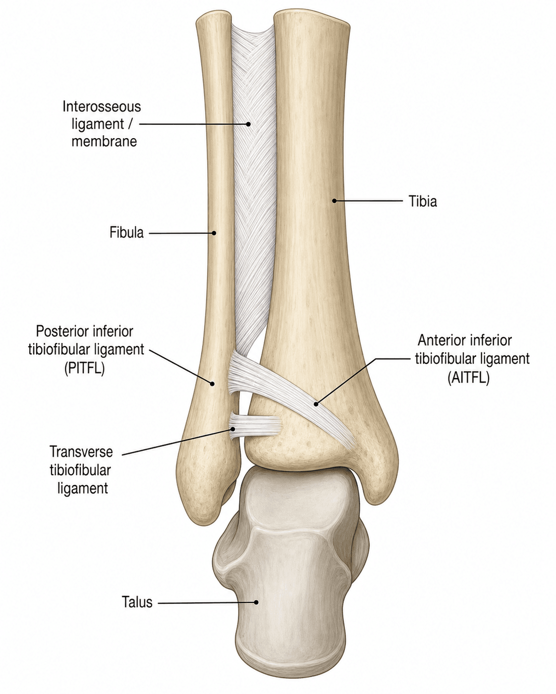
- Attachments and form
- Anterior tubercle of the tibia (Chaput) to the anterior fibula, running obliquely downward from tibia to fibula
- Share of stability
- 35%, the largest single ligament
- In injury
- Tears first; the most commonly injured
- Attachments and form
- Distal continuation of the interosseous membrane
- Share of stability
- 22%
- In injury
- Fails after the AITFL
- Attachments and form
- Posterior tubercle of the tibia (Volkmann) to the posterior fibula
- Share of stability
- 9%
- In injury
- Tears last
- Attachments and form
- Deep portion of the PITFL: a labrum-like extension posteriorly that contacts the posterior talus
- Share of stability
- 33%
- In injury
- Part of the posterior complex
The posterior complex. Behind the joint the intermalleolar ligament lies with the posterior inferior tibiofibular ligament. The superficial PITFL and the deep transverse ligament together contribute 42%, which is where the teaching that the PITFL is the strongest component comes from. That is true of the complex, not of the superficial ligament, which contributes least of the four; ligament by ligament, the AITFL is the largest single contributor. In tensile testing the PITFL was stronger than the AITFL, but the difference was not statistically significant (Beumer). The clinical point stands: the posterior complex is the key stabiliser, and if it is intact the syndesmosis is usually stable.
The mortise is meant to move. The talar dome is wider anteriorly by 2-3mm. In dorsiflexion the wider anterior talus pushes the fibula laterally, a physiological widening of up to 1-2mm, and the syndesmotic ligaments allow this normal motion while preventing excessive separation. They also contribute to tibiotalar contact mechanics.
Restoring the fibula. A reduction has to put back the fibula's length, its rotation and the tibiofibular width:
- Shortening of more than 2mm alters ankle biomechanics
- External rotation malreduction narrows the mortise posteriorly
- The anatomic tibiofibular relationship must be restored
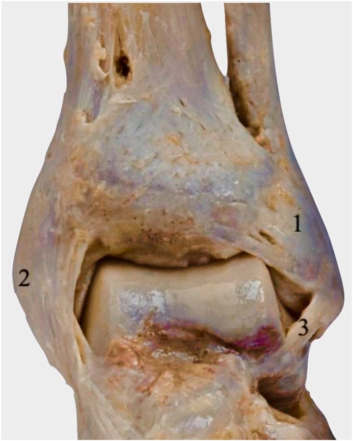
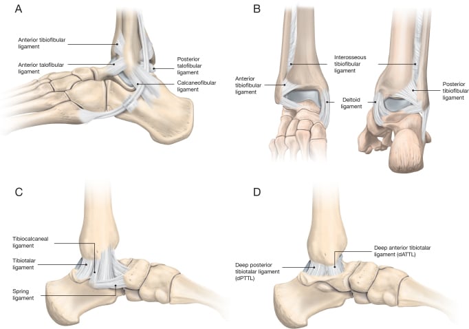
Pathophysiology
The sequence of failure. External rotation applied to the foot while the leg is fixed fails the ligaments in a predictable order, and complete syndesmotic disruption requires failure of all three. The oblique orientation of the tibiofibular ligaments is what converts external rotation of the talus into fibular displacement.
- AITFL. Tensioned first, it tears partially or completely depending on the magnitude of the force, and allows the anterior syndesmosis to begin widening. It tested weaker than the PITFL, although not significantly so.
- Interosseous ligament. Continued external rotation propagates the injury proximally; the ligament tears from distal to proximal, may extend several centimetres up the interosseous membrane, and creates a potential space for fibular displacement.
- PITFL. Last to fail. It may avulse with a posterior malleolus fracture (the Volkmann fragment), and its complete disruption allows frank diastasis.
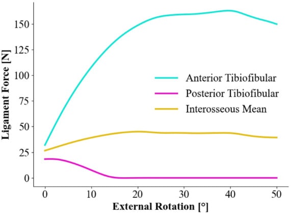
What diastasis does to the joint. The normal tibiofibular clear space is 2-5mm. When the fibula displaces laterally the tibiotalar contact area falls, contact pressure rises, and the tibial plafond is loaded asymmetrically.
Cadaver studies show that 1mm of lateral fibular displacement reduces tibiotalar contact area by 42% and increases peak contact pressure by 40%. This is why anatomic reduction is essential: even small degrees of malreduction lead to accelerated arthritis, and "close enough" is not acceptable for syndesmotic injuries.
Altered kinematics. The syndesmosis loses its normal motion during dorsiflexion, and the fibula cannot translate laterally as the talus widens anteriorly. The talus sits abnormally in the mortise through the gait cycle, and shear stress on the articular cartilage increases.
From overload to arthritis. Chronic overload of the medial or lateral tibial plafond produces subchondral bone stress and microfracture. The cartilage breaks down over months to years, and post-traumatic osteoarthritis develops.
Why it heals slowly. A high ankle sprain takes 2-3 times longer to recover than a lateral sprain, for reasons that lie in the ligaments and the load they carry:
- The syndesmotic ligaments have a lower blood supply than the lateral ankle ligaments
- Weight bearing continuously stresses the healing syndesmosis, and the mortise must stay stable during dynamic activity
- Without fixation the ligaments may heal in an elongated position, and interposed scar tissue prevents anatomic healing
- Incomplete healing leads to chronic instability and pain
Classification Systems
Each system describes a different injury. West Point grades an isolated syndesmotic injury by its stability and drives treatment; Lauge-Hansen places the syndesmotic injury inside an ankle fracture pattern and predicts what else is injured; Edwards and DeLee focuses on isolated syndesmotic injuries and the medial or lateral injury that accompanies them.
The West Point Ankle Grading System is the most commonly used classification for syndesmotic instability.
- Description
- Ligament disruption but no diastasis on static or stress films
- Stability
- Stable
- Treatment
- Conservative
- Description
- Diastasis only with stress testing, reduces spontaneously
- Stability
- Unstable
- Treatment
- Consider fixation (depends on activity level)
- Description
- Frank diastasis on static radiographs
- Stability
- Unstable
- Treatment
- Operative fixation mandatory
Latent injuries can be treated conservatively with protected weight bearing. Subluxation may be managed conservatively in low-demand patients but often requires fixation in athletes, and dislocation always requires operative fixation.
Clinical Presentation and Examination
History. The mechanism is an external rotation injury, a twist on a planted foot, or a direct blow. The pain is anterior, just above the joint line, where a lateral ankle sprain hurts more distally. Inability to bear weight immediately after the injury, and pain lasting weeks rather than the days of a typical lateral sprain, are part of the picture.
Red flags. These suggest that an "ankle sprain" is a syndesmotic injury:
- A high-energy mechanism
- Persistent pain despite treatment for "ankle sprain"
- Proximal fibular tenderness (Maisonneuve)
- Pain on walking more than 2 weeks after the injury
Clinical tests. No single clinical test is perfectly sensitive, so the tests are combined, and if the mechanism and presentation suggest a syndesmotic injury, imaging proceeds even when they are negative. Palpation of the syndesmotic ligaments is the sensitive sign (about 92%). The squeeze test is the most specific test (about 85-88%), not the most sensitive, so a negative squeeze test does not exclude injury; the external rotation stress test is also highly specific.
- Technique
- Compress tibia and fibula together at mid-calf
- Positive
- Pain at the ankle (distal syndesmosis), not at the compression site
- Accuracy and use
- Sensitivity 30-92%, specificity 88-95%
- Technique
- Stabilise the leg and externally rotate the foot, knee flexed to 90 degrees
- Positive
- Pain at the anterior or posterior syndesmosis
- Accuracy and use
- Sensitivity 20-71%, specificity 85-97%
- Technique
- Grasp the calcaneus and talus and translate laterally, comparing with the other side
- Positive
- Lateral shift of the fibula relative to the tibia of more than 2mm
- Accuracy and use
- More commonly performed intraoperatively, under anaesthesia
- Technique
- Dorsiflex the ankle and apply an external rotation force
- Positive
- Pain at the syndesmosis
- Accuracy and use
- May be difficult to perform acutely because of pain
Palpation. Work through the ankle in sequence so that associated injuries are not missed, comparing with the other side, which is essential for detecting subtle abnormality:
- Anterior syndesmosis, just above the joint between tibia and fibula: tenderness suggests AITFL injury
- Posterolateral ankle: tenderness suggests PITFL injury
- Proximal fibula, head and neck, always: tenderness indicates a possible Maisonneuve fracture
- Medial malleolus and deltoid ligament, often injured in combination with syndesmotic disruption
- Along the interosseous membrane: proximal tenderness may indicate a high interosseous membrane injury
Function. Functional assessment gauges severity and, later, recovery. Inability to bear weight suggests a significant injury, and the gait is antalgic, the foot held externally rotated to avoid pain and push-off weak because dorsiflexion hurts. End-range dorsiflexion is painful as the talus widens the mortise, and so are plantarflexion and external rotation; compare with the uninjured side.
Strength and hopping. The heel raise (gastrocnemius) is often painful; test tibialis anterior with resisted dorsiflexion, and eversion and inversion strength. In the subacute phase, inability to perform a single-leg hop suggests persistent instability, and the hop test is useful for return-to-sport decisions.
- Distinguishing features
- Pain ABOVE the joint line over the anterior syndesmosis; positive squeeze and dorsiflexion-external rotation tests; prolonged recovery
- Key investigation
- Mortise/stress radiographs (clear space greater than 6mm), MRI
- Distinguishing features
- Pain and swelling distal/anterolateral over the lateral ligaments; anterior drawer positive; rapid recovery
- Key investigation
- Clinical; radiograph if Ottawa rules positive
- Distinguishing features
- Proximal fibular tenderness with medial-side injury; distal fibula may look normal
- Key investigation
- Full-length tibia-fibula radiographs; mortise view
- Distinguishing features
- Focal lateral malleolar bony tenderness and deformity
- Key investigation
- AP, mortise, lateral radiographs
- Distinguishing features
- Medial tenderness and widened medial clear space; often accompanies syndesmotic injury
- Key investigation
- Mortise view (medial clear space), stress views, MRI
- Distinguishing features
- Deep ankle pain, mechanical catching or effusion, often after an inversion injury
- Key investigation
- MRI or CT
- Distinguishing features
- Posterolateral pain behind the fibula, painful resisted eversion, snapping
- Key investigation
- Dynamic ultrasound or MRI
Investigations and Imaging
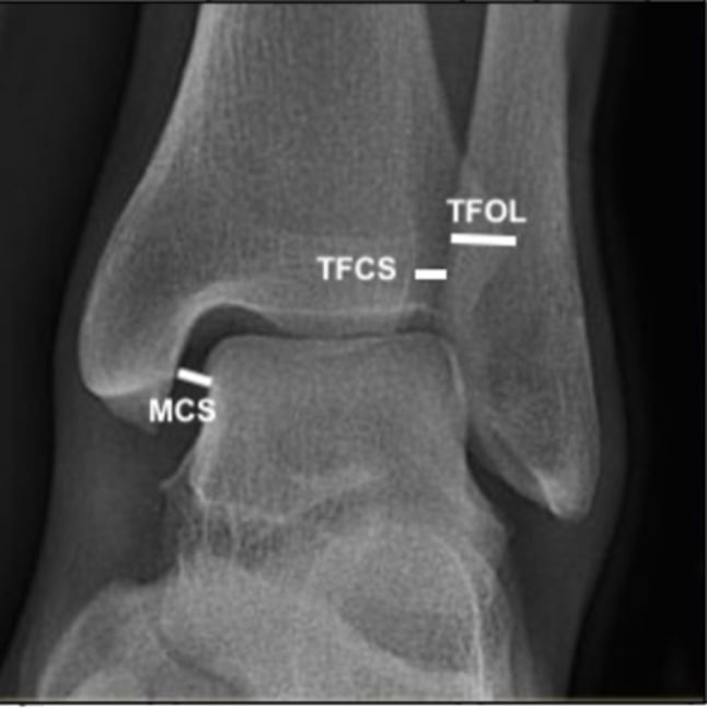
Plain radiographs. The standard AP, mortise and lateral series is the initial imaging and is often diagnostic. The mortise view is the most important, and the tibiofibular clear space is the most reliable plain-film sign of diastasis:
- Tibiofibular clear space, measured 1cm above the plafond: over 6mm on the AP or mortise suggests instability
- Tibiofibular overlap: under 6mm on the AP or under 1mm on the mortise is abnormal
- Medial clear space, between medial malleolus and talus: should equal the superior clear space; widening of more than 1mm relative to it indicates deltoid injury
- Talocrural angle: should be symmetric
- Lateral view: assess for a posterior malleolus fracture, which is associated with PITFL injury (its fixation is under Surgical Technique)
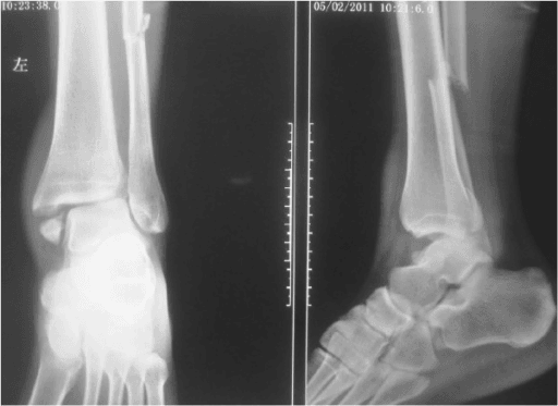
Weight-bearing radiographs may reveal diastasis that is not apparent on non-weight-bearing films, and are useful in chronic or subtle cases. A standing mortise view, ideally with the contralateral side for comparison, is the cheapest test for dynamic instability.
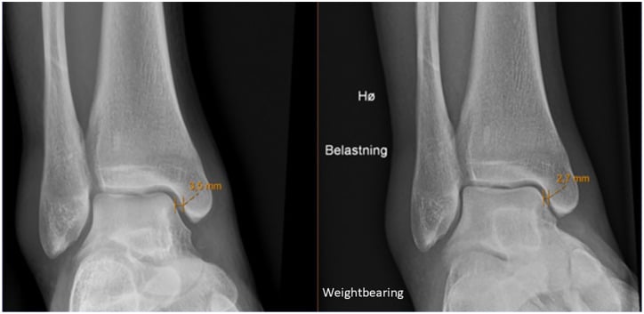
Stress radiographs are the gold standard for diagnosing latent syndesmotic instability. They are indicated when static films are equivocal, when clinical suspicion is high despite normal static films, and to separate West Point latent from subluxation in an isolated syndesmotic injury.
- External rotation stress view: an external rotation force on the foot while a mortise film is taken, manually or with a device; widening of the medial or tibiofibular clear space is positive
- Gravity stress view: the patient lies lateral decubitus with the foot hanging off the table, and gravity provides the stress; less painful than manual stress
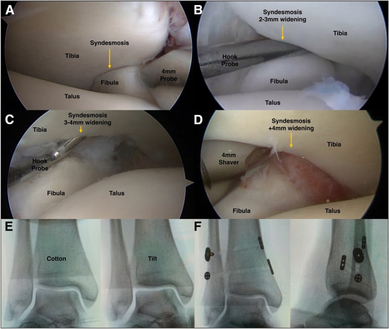
- Radiographic Cotton test: a lateral translation force on the foot, measuring fibular displacement; more than 2mm compared with the contralateral side indicates instability
Always obtain bilateral stress views for comparison. Normal syndesmotic width varies between individuals, and a 1mm side-to-side difference is significant.
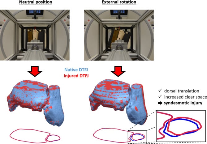
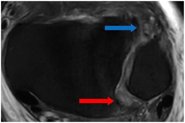
MRI confirms the extent of the ligament injury, assesses associated injuries (osteochondral lesions, deltoid, lateral ligaments), and is indicated for chronic syndesmotic pain with normal radiographs. Findings include fluid signal and discontinuity in a torn AITFL or PITFL, bone bruising in the distal tibia or fibula, and associated deltoid or lateral ligament injuries. MRI names which ligaments are torn but does not measure instability, which remains a loaded or stress assessment.
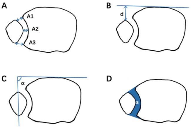
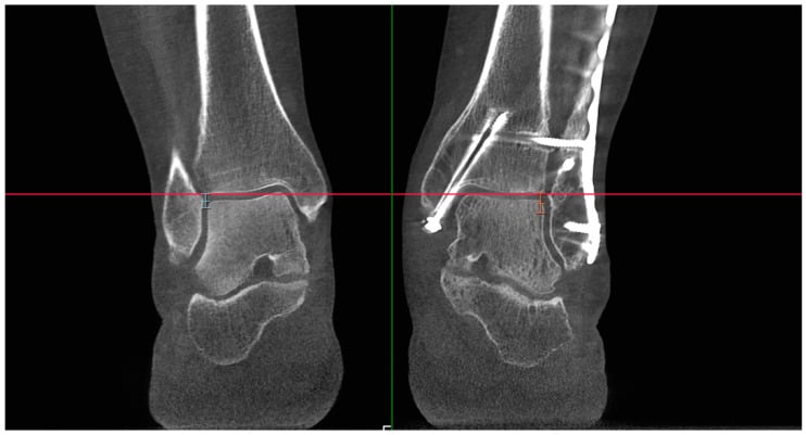
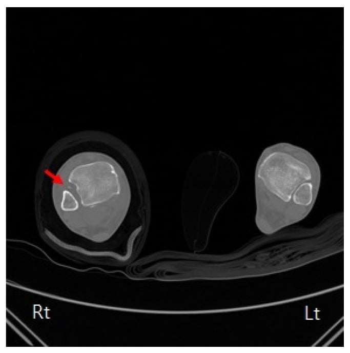
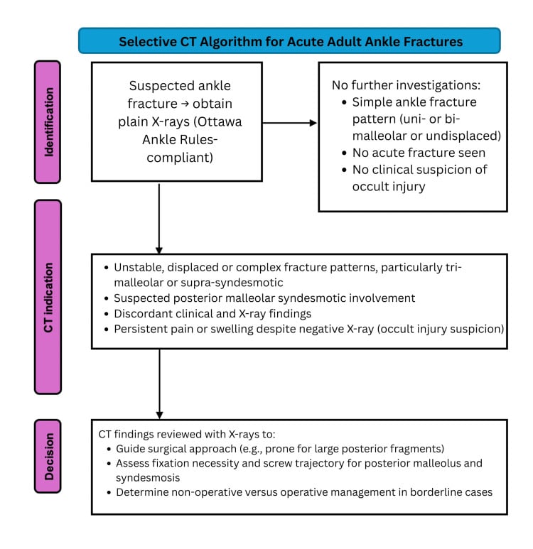
CT assesses the size of a posterior malleolus fracture, evaluates syndesmotic reduction after fixation, and detects malreduction (fibular malposition). Axial images at the level of the tibial plafond measure the fibula's position in the incisura and show rotational malreduction. Incisura shape varies widely between individuals, so the uninjured side is the only reliable normal, and side-to-side comparison is how malreduction is actually detected.
Ultrasound gives a dynamic assessment of syndesmotic stability, but it is less commonly used and operator-dependent.
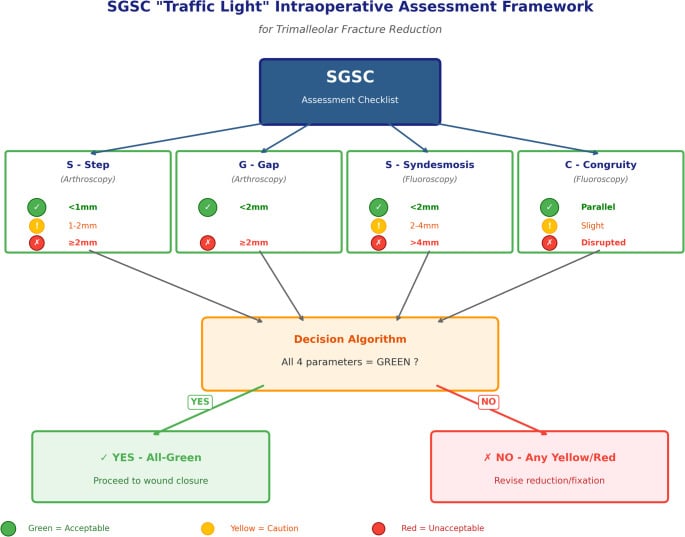
Arthroscopy visualises syndesmotic widening directly, and widening of more than 2mm is instability. Widening is graded through the anteromedial portal as normal, 2 to 3mm, 3 to 4mm or over 4mm. Direct visualisation is the most sensitive assessment of instability; it is done before and after fixation and is useful intraoperatively to assess the reduction.
- Advantages
- Fast, low cost, readily available. Clear space and overlap measurements
- Limitations
- May miss latent instability
- Best Use
- Initial screening, frank diastasis
- Advantages
- Gold standard for latent instability. Bilateral comparison
- Limitations
- Requires contralateral views, can be painful
- Best Use
- Equivocal cases, isolated syndesmotic injury
- Advantages
- Visualises ligaments, assesses associated injuries
- Limitations
- Expensive, longer acquisition time
- Best Use
- Chronic pain, pre-operative planning
- Advantages
- Excellent for bony detail, post-operative reduction assessment
- Limitations
- Radiation, poor soft tissue detail
- Best Use
- Posterior malleolus, assess reduction quality
6126-1-2 Rule for Radiographic Assessment
Hook:Remember the critical measurements: 6-1-2 for clear space, overlap, and translation
Management Algorithm
The decision. Surgical decision-making is based on stability assessment and fracture pattern. A West Point latent injury, stable on stress, is treated conservatively. A subluxation that opens only on stress is considered for fixation when athletic demands are high, with a conservative trial otherwise. Frank diastasis, an unstable ankle fracture and a Maisonneuve injury are fixed, and in an ankle fracture the decision is made on the table with a Cotton test after the fracture is fixed.
- Stability
- Stable on stress views
- Treatment
- Conservative: boot 4-6 weeks, progressive weight bearing
- Key Points
- Return to sport 8-12 weeks (longer than lateral sprain)
- Stability
- Unstable on stress only (West Point subluxation)
- Treatment
- Consider fixation if athletic demands high, otherwise trial conservative
- Key Points
- MRI can confirm extent of ligament injury
- Stability
- Unstable (West Point dislocation)
- Treatment
- Operative fixation mandatory
- Key Points
- 1-2 screws or suture button construct
- Stability
- Unstable if disrupted
- Treatment
- Fix fracture, then assess syndesmosis with Cotton test intraoperatively
- Key Points
- Fix if more than 2mm fibular translation or diastasis on stress
Who. West Point latent injuries, stable on stress testing, and isolated AITFL sprains without diastasis, in a patient with no associated fracture who can bear weight without significant pain.
The protocol. Protection comes first, then load, then proprioception and sport, with the boot worn for 4-6 weeks in total:
- 0-2 weeks: RICE (rest, ice, compression, elevation); a controlled ankle motion (CAM) boot or below-knee cast; non-weight bearing initially, progressing as tolerated; a short course of NSAIDs for pain, kept short to avoid delayed healing; crutches until single-leg standing is comfortable
- 2-6 weeks: protected weight bearing in the boot; gentle range of motion (alphabet exercises, plantarflexion and dorsiflexion, avoiding inversion and eversion initially); progressive resistance exercises
- 6-12 weeks: wean from the boot to a lace-up ankle brace; proprioceptive training (balance exercises, single-leg stance); strengthening of gastrocnemius, tibialis anterior and peroneals; sport-specific drills for athletes; gradual return to activity
Follow-up radiographs at 2-4 weeks make sure that delayed diastasis has not developed. Return to sport follows the criteria under Prevention and Return to Sport.
Surgical Technique
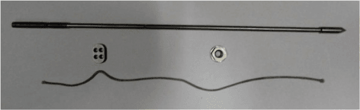
Syndesmotic screw fixation is the traditional technique, with proven long-term results.
Set-up. The patient is supine with a bump under the ipsilateral hip, with an optional thigh tourniquet and fluoroscopy for AP, mortise and lateral views.
Reduction. Restore fibular length first if there is a fracture. Hold the ankle in dorsiflexion and neutral rotation: the talus is wider anteriorly, so dorsiflexion locks it in the mortise and prevents over-compression. Apply the reduction clamp from fibula to tibia 2cm above the joint line with gentle compression, then confirm the reduction on fluoroscopy:
- AP: tibiofibular overlap over 6mm
- Mortise: clear space under 6mm and overlap over 1mm
- Lateral: fibula centred in the incisura
The screw. It is placed to a standard recipe:
- 2-3cm above the tibial plafond
- Directed 30 degrees anterior to posterior, parallel to the joint line, with the drill perpendicular to the long axis of the fibula
- A 3.5mm or 4.5mm cortical screw, inserted with a non-lag technique
- Some surgeons add a second screw 1-2cm proximal to the first, on the same trajectory, for increased stability
Three cortices or four. Tricortical purchase (both fibular cortices and the near tibial cortex) provides adequate stability while allowing some physiological motion and the screw is less likely to break, and it is increasingly favoured. Quadricortical purchase through all four cortices is more rigid, with a higher breakage rate, and requires screw removal before full weight bearing.
Finish. Remove the clamp and take final AP, mortise and lateral images, confirming that the clear space, overlap and fibular position are anatomic.
Removal. A non-bioabsorbable screw is removed at 8-12 weeks to prevent it breaking with weight bearing and to allow full activity, and weight bearing is protected until the screw is removed or a bioabsorbable screw is incorporated. Practice varies: some centres remove routinely and others retain the screw until it is symptomatic, and the evidence does not mandate routine removal.
Complications
Malreduction, as over-compression or as fibular malposition in rotation or translation, is the intraoperative complication that matters. It is recognised on intraoperative fluoroscopy on multiple views and prevented with the ankle in dorsiflexion and neutral rotation and a gentle clamp; the detail is under Surgical Technique.
Intra-articular screw. A screw placed too distal or on the wrong trajectory can enter the joint, presenting with pain, limited motion and radiographic signs. Stay 2cm above the plafond and confirm on the lateral view.
Neurovascular injury. The superficial peroneal nerve is at risk at the fibular incision site, and the deep peroneal nerve or anterior tibial vessels, rarely, with an anterior approach. Careful dissection and knowledge of the anatomy prevent it.
Wound complications occur in 5-10% of ankle fracture surgery, with diabetes, smoking and soft-tissue injury as the risk factors. Delay surgery if blisters are present, and handle the soft tissues carefully.
Early screw breakage follows weight bearing too early on rigid fixation. It is usually asymptomatic if the syndesmosis has healed, and is prevented by protected weight bearing until screw removal or union.
Loss of reduction. The syndesmosis can re-widen despite fixation, which may indicate inadequate fixation or continued instability; revision fixation is the answer if it is detected early.
Postoperative Care and Rehabilitation
The implant sets the pace. A rigid screw is protected from full load until it comes out, while a suture button allows physiological motion and weight bearing as tolerated from the start.
- Screw fixation
- Splint or boot, non-weight bearing; elevation above heart level; ice; DVT prophylaxis if indicated; sutures out at 10-14 days
- Suture button fixation
- Splint or boot, weight bearing as tolerated; ice and elevation; DVT prophylaxis if indicated; sutures out at 10-14 days
- Screw fixation
- CAM boot; non-weight bearing continues with a quadricortical screw, partial weight bearing with a tricortical screw in some protocols; gentle plantarflexion and dorsiflexion; avoid inversion and eversion
- Suture button fixation
- Boot continues; full weight bearing progressed rapidly; ankle pumps immediately and range of motion in all planes by 2 weeks; resistance band exercises begin
- Screw fixation
- Screw removal at 8-12 weeks if not bioabsorbable, then full weight bearing over 2 weeks; wean from boot to brace; range of motion extended to inversion and eversion; strengthening begins
- Suture button fixation
- Wean from boot to brace by 6 weeks; progressive strengthening (heel raises bilateral then unilateral, resisted dorsiflexion, band work for peroneals and invertors); balance exercises
- Screw fixation
- Progressive strengthening; proprioception (balance board, single-leg exercises); sport-specific drills; return to sport 12-16 weeks after screw removal
- Suture button fixation
- Sport-specific training and agility drills; gradual return to sport at 12-16 weeks
- Screw fixation
- 20-28 weeks (5-7 months)
- Suture button fixation
- 12-16 weeks (3-4 months)
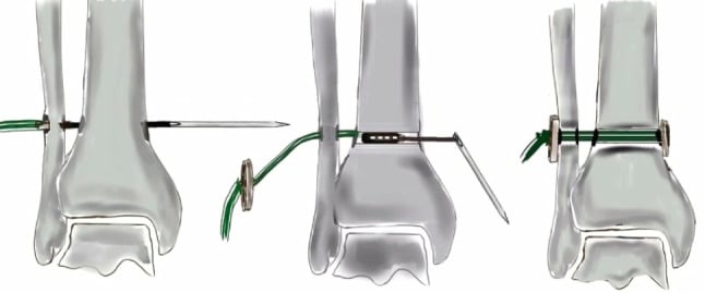
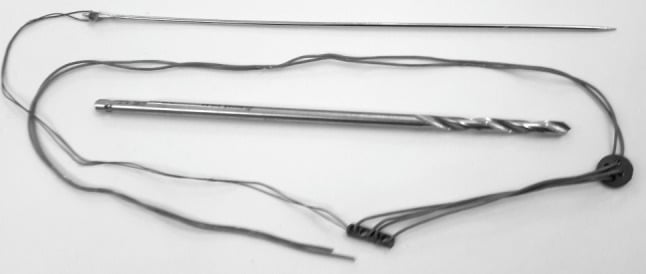
Why the button is faster. Physiological motion and earlier weight bearing allow faster rehabilitation, early motion reduces stiffness, and there is no re-operation for implant removal and no wait for it. The result is a return to sport approximately 2-3 months earlier than after screw fixation, a major benefit for athletes.
Exercises. The programme moves from motion to strength to sport:
- 0-6 weeks: ankle pumps (plantarflexion and dorsiflexion in the boot), alphabet tracing for range of motion, towel scrunches for intrinsic strength, and isometric contractions against resistance without motion
- 6-12 weeks: resistance-band dorsiflexion, plantarflexion, inversion and eversion; heel raises, bilateral progressing to unilateral; toe raises for tibialis anterior; single-leg stance on a firm surface, progressing to a foam pad or balance board
- 12+ weeks: plyometrics (box jumps, single-leg hops), agility work (ladder drills, cone drills, cutting), sport-specific exercises, and the single-leg hop test to assess readiness for return to sport
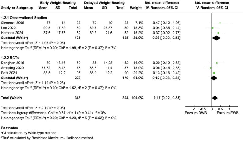
Timing of weightbearing. Pooled evidence across observational studies and trials shows that early loading gives slightly better function without a clear increase in failure.
Prevention and Return to Sport
Prevention. Four strategies are used:
- Neuromuscular training: proprioceptive exercises in pre-season training, balance and agility drills, and core and hip strengthening, which improves lower-limb control
- Bracing or taping: prophylactic bracing in high-risk sports (football, basketball) reduces the incidence of ankle injuries by 30-50%; lace-up braces or high-top shoes provide lateral support
- Field maintenance: avoid holes and uneven surfaces, and use footwear suited to the surface
- Conditioning: an adequate warm-up, gradual increases in training intensity, and avoiding fatigue, since injury risk increases when fatigued
Return to sport. The athlete must meet all of the objective criteria:
- Pain-free weight bearing, running and cutting
- Full range of motion equal to the contralateral side, especially dorsiflexion, which is critical for push-off
- Strength 90% of the contralateral side on single-leg heel raise and hop test
- Functional testing passed: single-leg hop over 90% of the contralateral side, and agility
- Radiographic stability, with no diastasis on stress views
The athlete should also be confident with cutting and jumping, with no apprehension or fear of re-injury.
Graduated return. Load is rebuilt over six weeks:
- Week 1: straight-line jogging at 50% effort
- Week 2: increased pace and longer distances at 75% effort
- Week 3: change of direction, cutting at 50% speed
- Week 4: sport-specific drills at increasing intensity
- Week 5: non-contact practice
- Week 6 and beyond: full-contact practice, then return to competition
After return. Continue proprioceptive training indefinitely and maintain strength and conditioning. A lace-up ankle brace is worn for 6-12 months after return, with taping as an alternative, and weaned as confidence improves.
Athletes who return to sport before meeting objective criteria have a significantly higher re-injury rate. The prolonged recovery time for syndesmotic injuries is frustrating for athletes but essential for proper healing. Premature return leads to chronic instability.
Guidelines, Registries & Global Practice
Global epidemiology:
Syndesmotic injuries account for roughly 1-11% of all ankle sprains but are markedly over-represented in collision and cutting sports played worldwide (American football, rugby league and union, soccer, ice hockey and Australian Rules football). In the United States Military Academy cohort the syndesmosis was involved in a substantial minority of ankle sprains, and elite-football injury surveillance confirms a prolonged return-to-play burden relative to lateral sprains. The JAAOS review by Hunt and colleagues emphasises that the historic paucity of high-level data has produced genuine practice variation in diagnosis and fixation choice across regions (DOI, PMID 26498585).
Side-by-side guidance:
- Diagnosis emphasis
- Stress testing and arthroscopy to grade isolated injuries; unstable injuries need stabilisation
- Fixation guidance
- Anatomic reduction is paramount; screw or dynamic fixation both acceptable
- Evidence level
- Expert consensus / Level V
- Diagnosis emphasis
- Mortise clear space and stress views; CT for reduction assessment
- Fixation guidance
- Trend toward dynamic (suture button) fixation; screw still standard, removal optional
- Evidence level
- Level I-II RCT/meta-analysis
- Diagnosis emphasis
- Restore fibular length and rotation first, then assess syndesmosis (Cotton/hook test)
- Fixation guidance
- Anatomic reduction under direct or CT control; avoid over-compression and malrotation
- Evidence level
- Principle-based / Level V
- Diagnosis emphasis
- Early senior assessment; CT where reduction is uncertain
- Fixation guidance
- Stabilise unstable syndesmosis; implant choice surgeon-led
- Evidence level
- Standard of care / Level V
- No national joint registry tracks syndesmotic implants specifically; the best comparative data come from RCTs and meta-analyses rather than registries
- Level I meta-analysis (Grassi et al., AJSM 2020) shows dynamic fixation reduces malreduction and reoperation versus screws (DOI, PMID 31188642)
- CT-based RCT evidence (Kortekangas et al., Injury 2015) shows dynamic constructs maintain reduction better at 2 years (DOI, PMID 25769201)
- Implant choice: screw fixation remains common globally for cost reasons; suture button uptake is higher in athletic and well-resourced centres
- Screw removal: routine removal practised in some centres, retain-until-symptomatic in others; evidence does not mandate routine removal
- Reduction assessment: increasing use of intraoperative or postoperative CT (vs plain films alone) where available
MCQ Practice Points
Answer: AITFL (Anterior Inferior Tibiofibular Ligament)
The sequential failure pattern is: AITFL → IOL → PITFL. The AITFL is relatively weaker and positioned anteriorly where it receives maximal tension during external rotation. The PITFL complex tears last and accounts for 42 per cent of resistance to diastasis - though note what that number contains: superficial PITFL 9 per cent plus deep transverse ligament 33 per cent. Taken ligament by ligament, the AITFL is the single largest contributor at 35 per cent, so "the PITFL is the strongest" is true of the complex, not of the superficial ligament an examiner may be pointing at.
Answer: Greater than 6mm
Harper and Keller (1989) established that a clear space greater than 6mm on mortise view has 100% sensitivity and 94% specificity for syndesmotic diastasis. This is measured 1cm above the tibial plafond on the mortise radiograph.
Answer: Maisonneuve fracture - YES, syndesmosis requires fixation
This is a Maisonneuve fracture pattern (Lauge-Hansen PER IV). The entire syndesmosis is disrupted from the proximal fibula fracture down to the ankle. The distal fibula appears intact but the syndesmotic ligaments are completely torn. Fix the medial malleolus, then perform Cotton test to confirm syndesmotic instability, then fix syndesmosis. Do NOT fix the proximal fibula fracture.
Answer: Dorsiflexion
The talus is wider anteriorly by 2-3mm. Positioning the ankle in dorsiflexion locks the wider anterior talus in the mortise, preventing over-compression of the syndesmosis. If the ankle is in plantarflexion during fixation, the narrower posterior talus allows over-compression, and when the patient dorsiflexes post-operatively, the ankle will be too tight.
Answer: Lateral fibular translation test - greater than 2mm requires fixation
The Cotton test assesses lateral translation of the fibula relative to the tibia. The examiner grasps the calcaneus and talus and translates laterally while palpating the fibula. Greater than 2mm of translation (compared to contralateral side) indicates syndesmotic instability requiring fixation. This is often performed intraoperatively under anesthesia after fibula or medial malleolus fixation.
Answer: Allows physiologic motion and faster return to sport (no removal needed)
Suture button (TightRope) allows 1-2mm of physiologic syndesmotic motion, permits earlier weight bearing and range of motion, and does not require removal. This translates to faster return to sport: 12-16 weeks vs 20-28 weeks with screw fixation. RCTs show equivalent functional outcomes but lower re-operation rate with suture button.
Answer: 40% increase in contact pressure
Biomechanical cadaver studies show that even 1mm of lateral fibular displacement reduces tibiotalar contact area by 42% and increases peak contact pressure by 40%. This explains the high rate of post-traumatic arthritis with malreduction and why anatomic reduction is essential.
Answer: NOT the squeeze test — palpation/ligament tenderness is the most sensitive; the squeeze test is the most SPECIFIC.
This is the commonest inversion in the topic. A systematic review with meta-analysis of 6 studies, 512 participants and 13 clinical tests (DOI) found the highest sensitivity in palpation (92%, 95% CI 79-98) and the dorsiflexion lunge (75%, 64-84), while the highest specificity was the squeeze test (85%, 81-89) followed by external rotation (78%, 73-82). A separate cohort against MRI (DOI) agrees: syndesmosis ligament tenderness was most sensitive (92%) along with inability to single-leg hop (89%), while the squeeze test had the highest specificity (88%).
Why the inversion is dangerous: treating the squeeze test as the sensitive test invites using a negative one to exclude injury. It cannot — its sensitivity is poor, so the error direction is under-diagnosis of exactly the injury that causes late diastasis and arthritis.
What to do instead. Both papers recommend the same strategy: cluster the SENSITIVE findings to screen — syndesmotic ligament tenderness, inability to hop, dorsiflexion lunge, dorsiflexion-external-rotation stress — then apply a SPECIFIC test (squeeze) to rule in. And note the ceiling the meta-analysis is explicit about: no clinical test, alone or clustered, stratifies a stable from an unstable syndesmosis. That decision needs imaging, stress examination or arthroscopy — which is why intra-operative assessment carries the fixation decision.
- AITFL tears first in external rotation injuries
- PITFL complex contributes 42 per cent of resistance to diastasis (superficial 9 per cent plus deep transverse 33 per cent); the AITFL alone contributes 35 per cent
- Inferior transverse ligament is the deep portion of PITFL, and is the larger contributor of the two
- Sequential failure: AITFL → IOL → PITFL
- Squeeze test: most specific, not most sensitive (compress at mid-calf, pain at ankle); palpation of ligament tenderness is the most sensitive
- External rotation stress test: also highly specific
- Cotton test: assess lateral fibular translation (greater than 2mm = unstable)
- All tests should be compared to contralateral side
- Clear space greater than 6mm on mortise view = diastasis
- Tibiofibular overlap less than 6mm on AP or less than 1mm on mortise = abnormal
- Medial clear space should equal superior clear space
- Stress views needed if static films equivocal
- West Point: latent (stable), subluxation (unstable with stress), dislocation (frank diastasis)
- Maisonneuve: PER IV injury - proximal fibula fracture + complete syndesmotic disruption
- Position ankle in dorsiflexion and neutral rotation during fixation
- Screw placed 2-3cm above plafond, 30 degrees anterior to posterior
- Tricortical vs quadricortical: both effective, tricortical lower breakage rate
- Suture button allows physiologic motion and faster return to sport
- 1mm malreduction increases contact pressure by 40%
- Screw vs suture button: equivalent outcomes, suture button faster return to sport
- Return to sport: 8-12 weeks conservative, 12-16 weeks suture button, 20-28 weeks screw
- Arthritis risk 10-30% at 5-10 years, higher with malreduction
Exam Viva Scenarios
Practise clinical reasoning and management decisions out loud
“A 22-year-old male rugby player presents 3 days after sustaining an external rotation injury to his ankle during a tackle. He describes immediate pain above the ankle joint and difficulty bearing weight. Ottawa ankle rules applied - radiographs show no fracture. Squeeze test is positive. Mortise view shows clear space of 5.5mm. External rotation stress views show clear space widening to 7mm, with 2mm difference compared to contralateral side. What is your diagnosis and management?”
“A 35-year-old female presents with an ankle injury after a fall. She has swelling and tenderness over the medial malleolus and proximal fibula. Ankle radiographs show a medial malleolus fracture and widened medial clear space, but the fibula appears intact distally. Proximal tibia-fibula radiographs show a proximal fibula fracture. What is your diagnosis and surgical plan?”
“You are asked to review a patient in clinic 2 weeks after syndesmotic screw fixation for an ankle fracture. The patient reports persistent pain and difficulty mobilizing. You obtain radiographs and notice the tibiofibular clear space is 7mm on the mortise view. What is your assessment and management?”
Key Anatomy
- AITFL: tears first, 35% of stability
- IOL: interosseous ligament, 22% stability
- PITFL complex: 42% stability (superficial 9% + deep transverse 33%), tears last
- ITL: deep PITFL, labrum-like function
- Sequential failure: AITFL → IOL → PITFL
Mechanism and Epidemiology
- External rotation of foot on planted ankle
- 1-11% of all ankle sprains, 23% of ankle fractures
- Common in contact sports: rugby, AFL, football
- Maisonneuve: proximal fibula fracture + complete syndesmotic disruption
Clinical Tests (Compare to Contralateral)
- Squeeze test: most specific, not most sensitive (compress mid-calf, pain at ankle); palpation most sensitive
- External rotation stress: also highly specific
- Cotton test: greater than 2mm fibular translation = unstable
- Palpate entire fibula (rule out Maisonneuve)
Radiographic Assessment
- Mortise view: clear space greater than 6mm = diastasis (MOST IMPORTANT)
- AP: tibiofibular overlap less than 6mm abnormal
- Mortise: overlap less than 1mm abnormal
- Stress views if equivocal (bilateral for comparison)
- Medial clear space = superior clear space (if wider = deltoid injury)
West Point Classification
- Latent: stable on stress → conservative
- Subluxation: unstable on stress → consider fixation (athlete)
- Dislocation: frank diastasis → operative mandatory
Surgical Technique Principles
- Ankle in DORSIFLEXION and NEUTRAL rotation (critical)
- Screw 2-3cm above plafond, 30° anterior-posterior
- Tricortical (3 cortices) vs quadricortical (4 cortices) - both work, tricortical less breakage
- Cotton test intraop: greater than 2mm translation = fix
- Check reduction: AP, mortise, lateral fluoroscopy
Screw vs Suture Button
- Screw: rigid, remove at 8-12 weeks, RTP 20-28 weeks
- Button: allows motion, no removal, RTP 12-16 weeks
- Both equivalent outcomes, button faster rehab
- Button higher cost but lower re-operation rate
Complications
- Malreduction (20-50%): even 1mm → 40% increased contact pressure
- Screw breakage: 25-50% if not removed by 12 weeks
- Post-traumatic arthritis: 10-30% at 5-10 years (higher with malreduction)
- Chronic instability: inadequate initial fixation, premature removal
Return to Sport
- Conservative: 8-12 weeks (latent injuries)
- Suture button: 12-16 weeks
- Screw: 20-28 weeks (includes removal time)
- Criteria: pain-free, full ROM, 90% strength, functional testing
- Use ankle brace for 6-12 months after return
Critical Numbers to Remember
- 6mm: clear space threshold on mortise view
- 1mm: malreduction increases pressure by 40%
- 2mm: Cotton test translation threshold
- 2-3cm: screw distance above plafond
- 30°: screw trajectory (anterior-posterior)
- 8-12 weeks: screw removal timing
Evidence Base
Dynamic vs Static Fixation: Level I Meta-analysis of RCTs
- Meta-analysis of randomised controlled trials comparing dynamic (suture button) versus static (screw) syndesmotic fixation
- Dynamic fixation significantly reduced overall complications (RR 0.55) and inadequate reduction at final follow-up (RR 0.36)
- Lower risk of recurrent diastasis or clinical instability with dynamic fixation (RR 0.10)
- REOPERATION OVERALL WAS NOT REDUCED: RR 0.64, P=0.07, not significant. The benefit appears only in the subgroup compared with PERMANENT screws (RR 0.24, P=0.007) - because a retained screw generates its own removal operations
- AOFAS was higher with dynamic fixation (+6.06 at 3 months, +5.21 at 12, +8.60 at 24), but the Olerud-Molander score showed NO DIFFERENCE - the two functional instruments disagree
- VAS pain lower at 6 months (-0.73) and 12 months (-0.52), and range of motion 4.36 degrees greater
- Overall quality of evidence graded MODERATE TO VERY LOW, owing to risk of bias, heterogeneity, indirectness and small numbers
Clear Space Measurement: Radiographic Criteria
- Cadaveric radiographic study (12 lower limbs) defining the normal distal tibiofibular relationship on AP and mortise views
- Tibiofibular clear space on AP and mortise views should be less than approximately 6mm
- Tibiofibular overlap on AP view should be greater than approximately 6mm (or 42% of fibular width)
- Tibiofibular overlap on mortise view should be greater than approximately 1mm
- Tibiofibular clear space was the single most reliable parameter for detecting early syndesmotic widening
Relative Contribution of Each Syndesmotic Ligament to Stability
- Eight fresh-frozen cadaver specimens on a hydraulic test system, with sequential cutting of the four component ligaments and measurement of the percentage resistance to 2 mm of diastasis
- ANTERIOR INFERIOR TIBIOFIBULAR LIGAMENT 35 per cent - the largest single contributor
- DEEP posterior inferior tibiofibular (transverse) ligament 33 per cent
- Interosseous ligament 22 per cent
- SUPERFICIAL posterior inferior tibiofibular ligament only 9 per cent
- The authors conclude that the syndesmosis must be assessed both anteriorly AND posteriorly at examination, and that the interosseous ligament can be seen arthroscopically
Syndesmotic Ligament Biomechanics
- Ex vivo tensile testing of bone-ligament-bone complexes from 10 fresh-frozen cadaveric lower limbs, average age at death 72 years
- THE HEADLINE IS NOT SIGNIFICANT: the posterior tibiofibular ligament exhibited greater strength than the anterior tibiofibular ligament and the posterior tibiotalar deltoid, but the paper states explicitly that the difference was NOT statistically significant, and there were no significant differences in stiffness between the three
- The FAILURE MODES are the robust part of the study: the AITFL failed predominantly by substance rupture near its fibular insertion, PITFL failures were split evenly between substance rupture and fibular avulsion, and the posterior tibiotalar deltoid ruptured most often near its talar insertion
- The comparison with the lateral collateral and deltoid ligaments is taken from the LITERATURE, not measured in this study - the authors cite it as context for their inference about ankle constraint
Reduction Quality Predicts Functional Outcome
- Multicentre retrospective review of 51 ankle fractures treated with syndesmotic screw fixation across three university hospitals
- On postoperative radiographs, 16% of syndesmoses were not anatomically reduced
- Quality of syndesmotic reduction was the ONLY significant predictor of functional outcome (p = 0.04)
- Reduction alone accounted for 15-18% of the variance in validated functional scores (SMFA, Olerud-Molander)
- 8 patients were judged to have had inappropriate or absent indications for the screw, and the authors conclude that 16 per cent of syndesmotic screws may have been unnecessary
- THE OUTCOME DENOMINATOR IS SMALLER THAN THE SERIES: only 39 of the 51 patients were available at final follow-up, at a mean of 18.1 months
- Overall results were good (mean SMFA functional index 11.4, Olerud-Molander 74.1, VAS pain 1.7), and 70 per cent of the injuries were pronation-external-rotation
Suture Button vs Single Screw: Randomised Trial
- 97 patients randomised to suture button (n=48) or a single quadricortical syndesmotic screw (n=49), with 90% followed to 2 years
- Higher median AOFAS (96 vs 86) and Olerud-Molander (100 vs 90) scores in the suture button group at 2 years
- Less pain on walking and at rest, and higher EQ-5D index, with suture button
- Tibiofibular side-to-side difference of 2mm or more on CT at 2 years in 20/40 screw versus 8/40 suture button patients (p = 0.009)
- 7 screw patients developed symptomatic recurrent diastasis versus none with suture button
Reduction Accuracy on Bilateral CT: TightRope vs Screw
- Prospective RCT (43 patients) comparing TightRope versus a single 3.5mm tricortical screw in PER/Weber-C fractures, assessed with bilateral CT
- Postoperative malreduction rates were similar between groups
- Intraoperative CT of dynamically fixed ankles was misleading unless the ankle was supported at 90 degrees, mimicking malreduction
- At follow-up CT beyond 2 years, three patients in the screw group and one in the TightRope group had a malreduced syndesmosis - a difference that was NOT statistically significant (P=0.33)
- No significant difference in Olerud-Molander, VAS, Foot and Ankle Outcome Score, RAND-36 or the incidence of osteoarthritis on cone-beam CT
- On blinded retrospective radiologist review of the intra- and post-operative CT, exactly ONE patient in each group had an incongruent syndesmosis
Diagnostic Accuracy of Clinical Tests (MRI Reference)
- Cross-sectional diagnostic accuracy study of 87 acute ankle sprains across 9 Australian clinics, with MRI as the reference standard
- Squeeze test was the most specific clinical TEST (specificity 88 per cent) - but its positive likelihood ratio of 2.15 has a 95% CI of 0.86 to 5.39, which CROSSES 1, so a positive squeeze test does not by itself significantly raise the probability of injury
- Dorsiflexion-external rotation stress test: sensitivity 71 per cent, negative LR 0.46 (CI 0.27-0.79) - this one does exclude, and injury was nearly four times more likely when it was positive (OR 3.9, p=0.004)
- Syndesmosis ligament tenderness was the most sensitive sign at 92 per cent, negative LR 0.28 (CI 0.09-0.89), with OR 4.04 (p=0.048) for injury when positive
- Inability to perform a single-leg hop had 89 per cent sensitivity, but its negative LR of 0.37 has a CI of 0.13 to 1.03 that crosses 1
- The most specific finding overall was not a test at all but PAIN OUT OF PROPORTION to the apparent injury (specificity 79 per cent, positive LR 3.05, CI 1.68-5.55) - the only positive likelihood ratio on the page whose interval excludes 1
- No single test was sufficiently accurate alone; combining sensitive and specific tests improved diagnosis
References
-
Hermans JJ, Beumer A, de Jong TA, Kleinrensink GJ. Anatomy of the distal tibiofibular syndesmosis in adults: a pictorial essay with a multimodality approach. J Anat. 2010;217(6):633-645. doi:10.1111/j.1469-7580.2010.01302.x
-
Waterman BR, Belmont PJ, Cameron KL, Deberardino TM, Owens BD. Epidemiology of ankle sprain at the United States Military Academy. Am J Sports Med. 2010;38(4):797-803. doi:10.1177/0363546509350757
-
Nussbaum ED, Hosea TM, Sieler SD, Incremona BR, Kessler DE. Prospective evaluation of syndesmotic ankle sprains without diastasis. Am J Sports Med. 2001;29(1):31-35. doi:10.1177/03635465010290010301
-
Hunt KJ, Phisitkul P, Pirolo J, Amendola A. High ankle sprains and syndesmotic injuries in athletes. J Am Acad Orthop Surg. 2015;23(11):661-673. doi:10.5435/JAAOS-D-13-00135
-
van Dijk CN, Longo UG, Loppini M, Florio P, Maltese L, Ciuffreda M, Denaro V. Classification and diagnosis of acute isolated syndesmotic injuries: ESSKA-AFAS consensus and guidelines. Knee Surg Sports Traumatol Arthrosc. 2016;24(4):1200-1216. doi:10.1007/s00167-015-3942-8
-
Harper MC, Keller TS. A radiographic evaluation of the tibiofibular syndesmosis. Foot Ankle. 1989;10(3):156-160. doi:10.1177/107110078901000308
-
Beumer A, van Hemert WL, Swierstra BA, Jasper LE, Belkoff SM. A biomechanical evaluation of the tibiofibular and tibiotalar ligaments of the ankle. Foot Ankle Int. 2003;24(5):426-429. doi:10.1177/107110070302400509
-
Alonso A, Khoury L, Adams R. Clinical tests for ankle syndesmosis injury: reliability and prediction of return to function. J Orthop Sports Phys Ther. 1998;27(4):276-284. doi:10.2519/jospt.1998.27.4.276
-
Sman AD, Hiller CE, Rae K, Linklater J, Black DA, Nicholson LL, Refshauge KM. Diagnostic accuracy of clinical tests for ankle syndesmosis injury. Br J Sports Med. 2015;49(5):323-329. doi:10.1136/bjsports-2013-092787
-
Sikka RS, Fetzer GB, Sugarman E, Wright RW, Fritts H, Boyd JL, Fischer DA. Correlating MRI findings with disability in syndesmotic sprains of NFL players. Foot Ankle Int. 2012;33(5):371-378. doi:10.3113/FAI.2012.0371
-
Lubberts B, Verhaven E, D'Hooghe P, Vereecke E, van Dijk CN. Epidemiology and return to play following isolated syndesmotic injuries of the ankle: a prospective cohort study of 3677 male professional footballers in the UEFA Elite Club Injury Study. Br J Sports Med. 2019;53(15):959-964. doi:10.1136/bjsports-2017-097710
-
Weening B, Bhandari M. Predictors of functional outcome following transsyndesmotic screw fixation of ankle fractures. J Orthop Trauma. 2005;19(2):102-108. doi:10.1097/00005131-200502000-00006
-
Kukreti S, Faraj A, Miles JN. Does position of syndesmotic screw affect functional and radiological outcome in ankle fractures? Injury. 2005;36(9):1121-1124. doi:10.1016/j.injury.2005.02.015
-
Lubberts B, Vopat BG, Wolf JC, Linn MS, Guss D, Nahed R, DiGiovanni CW. Arthroscopically assisted syndesmotic reduction: a systematic review of biomechanical and clinical outcomes. Arthroscopy. 2017;33(8):1585-1594. doi:10.1016/j.arthro.2017.03.005
-
Grass R, Rammelt S, Biewener A, Zwipp H. Peroneus longus ligamentoplasty for chronic instability of the distal tibiofibular syndesmosis. Foot Ankle Int. 2003;24(5):392-397. doi:10.1177/107110070302400504
-
Schepers T, Van Lieshout EM, de Vries MR, Van der Elst M. Increased rates of wound complications with locking plates in distal fibular fractures. Injury. 2011;42(10):1125-1129. doi:10.1016/j.injury.2011.01.009
-
Manjoo A, Sanders DW, Tieszer C, MacLeod MD. Functional and radiographic results of patients with syndesmotic screw fixation: implications for screw removal. J Orthop Trauma. 2010;24(1):2-6. doi:10.1097/BOT.0b013e3181a9f7a5
-
Laflamme M, Belzile EL, Bédard L, van den Bekerom MP, Glazebrook M, Pelet S. A prospective randomized multicenter trial comparing clinical outcomes of patients treated surgically with a static or dynamic implant for acute ankle syndesmosis rupture. J Orthop Trauma. 2015;29(5):216-223. doi:10.1097/BOT.0000000000000245
-
Andersen MR, Frihagen F, Hellund JC, Madsen JE, Figved W. Randomized trial comparing suture button with single syndesmotic screw for syndesmosis injury. J Bone Joint Surg Am. 2018;100(1):2-12. doi:10.2106/JBJS.16.01011
-
Kortekangas TH, Flinkkilä T, Niinimäki J, Leskelä HV, Savolainen V, Pakarinen H, Ohtonen P, Ristiniemi J. A prospective randomised study comparing TightRope and syndesmotic screw fixation for accuracy and maintenance of syndesmotic reduction assessed with bilateral computed tomography. Injury. 2015;46(6):1119-1126. doi:10.1016/j.injury.2015.02.004