The Nerve Under Your Arthroscopy Portal
- It is the nerve of the LATERAL compartment - peroneus longus and brevis - so eversion is its motor signature.
- It pierces the crural fascia in the distal third of the leg, classically 10 to 12 cm proximal to the tip of the lateral malleolus, though the cadaveric range is 3 to 18 cm.
- It runs in the lateral compartment throughout in 73 per cent of legs, but crosses into or branches within the ANTERIOR compartment in more than a quarter.
- It divides into the medial and intermediate dorsal cutaneous nerves, which supply the entire dorsum of the foot except the first web space and the lateral border.
- It is the structure most commonly injured in anterior ankle arthroscopy, and the injury is preventable by marking the nerve before the foot is insufflated.
- “Plantarflex and invert the foot before you prepare the skin - the nerve becomes a visible subcutaneous band in most people.
- “The fourth web space is superficial peroneal; the lateral border of the foot and the lateral side of the little toe are sural — but hold that map loosely. In 260 dissected feet the textbook pattern was present in only 35 per cent, with six patterns described and the sural nerve frequently supplying more than its diagram allows. Use the four-point sensory map to raise a hypothesis, never to exclude a nerve.
- “In a cadaveric ankle arthroscopy course, a single lecture on portal complications cut the nerve injury rate from 25 per cent to 3.6 per cent.
- “A fascial defect over the lateral compartment was present in almost half the legs operated for entrapment - look for a muscle hernia.
The commonest iatrogenic injury in ankle arthroscopy.
- The nerve, usually the intermediate dorsal cutaneous branch, crosses the anterolateral ankle in subcutaneous fat.
- Joint distension and a tourniquet make it invisible.
- Mark it FIRST, before prepping, before distension, with the foot plantarflexed and inverted.
- Then incise skin only and spread bluntly to capsule.
The nerve lies in the compartment you are releasing.
- It sits just posterior to the anterior intermuscular septum.
- Blind scissors driven distally toward the lateral malleolus divide it.
- Identify the septum, release the anterior compartment in front of it and the lateral compartment behind it, and see the nerve distally.
- Where the Nerve Lies
- Dividing from the common peroneal nerve within peroneus longus
- What It Is Doing
- Entering the lateral compartment
- What Puts It At Risk
- Fibular neck fracture, positioning, proximal fibular surgery
- Where the Nerve Lies
- Lateral compartment, between the peronei and the anterior intermuscular septum
- What It Is Doing
- Supplying peroneus longus then brevis
- What Puts It At Risk
- Lateral compartment fasciotomy, fibular shaft plating
- Where the Nerve Lies
- Pierces the crural fascia and becomes subcutaneous
- What It Is Doing
- Dividing into medial and intermediate dorsal cutaneous nerves
- What Puts It At Risk
- Entrapment, distal fasciotomy, distal fibular plate incision
- Where the Nerve Lies
- Subcutaneous, crossing the anterolateral ankle and extensor retinaculum
- What It Is Doing
- Purely sensory
- What Puts It At Risk
- Anterolateral arthroscopy portal, lateral ligament surgery
- Where the Nerve Lies
- Subcutaneous over the tarsus and metatarsals
- What It Is Doing
- Dorsal digital branches
- What Puts It At Risk
- Hallux valgus, lesser toe and midfoot surgery, tight dressings
MEDIAL then INTERMEDIATEThe Two Terminal Branches
Hook:Everything on the dorsum EXCEPT the first web, the lateral border and the medial border.
TWO MUSCLES, ONE NERVE, NO ARTERYLateral Compartment
Hook:The only compartment in the leg without its own named artery.
PITFinding the Nerve Before You Cut
Hook:Plantarflex, Invert, Toe-flex - then mark it, because after distension you will never see it again.
Overview
The superficial peroneal nerve - the superficial fibular nerve in current terminology - is the larger of the two terminal branches of the common peroneal nerve. Its root value is L4, L5 and S1, predominantly L5 and S1. It is a mixed nerve: motor to the two muscles of the lateral compartment, and sensory to almost the whole dorsum of the foot.
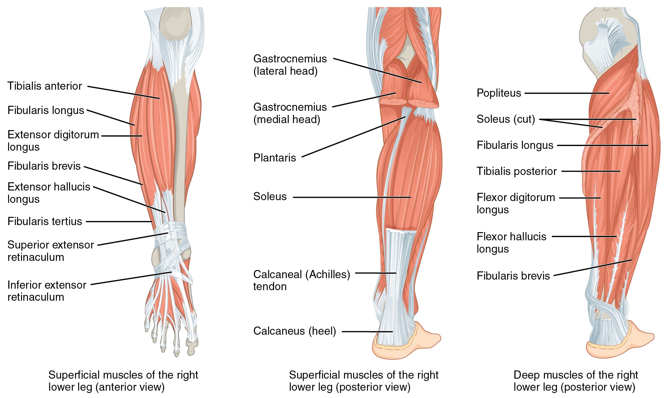
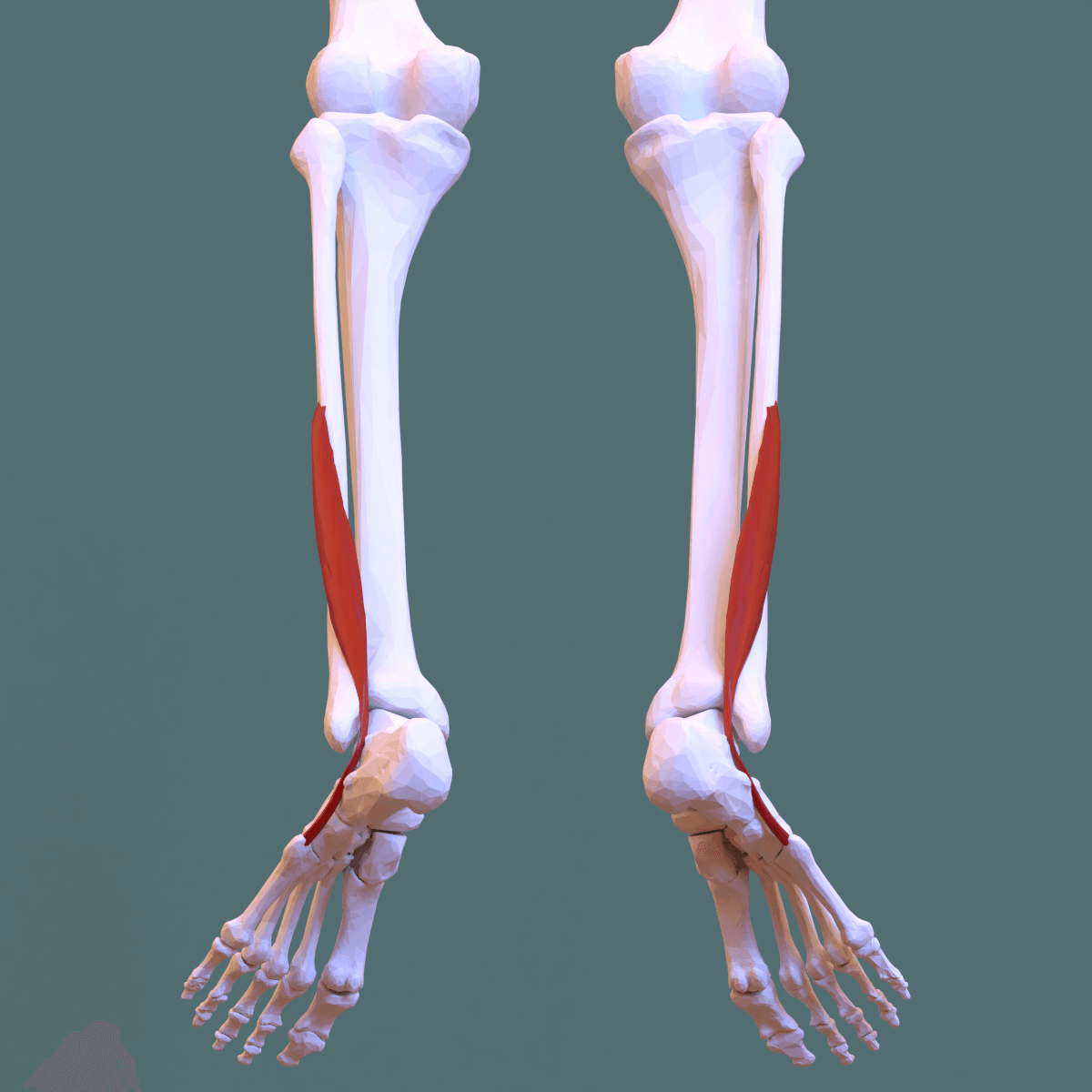
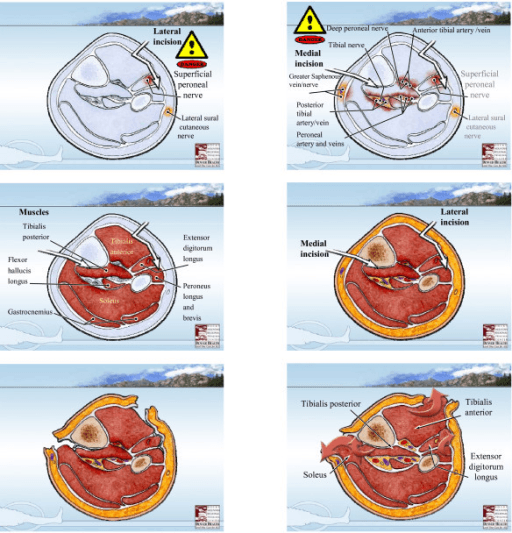
It arises at the fibular neck, within the substance of peroneus longus, at the same point the deep peroneal nerve is given off. From there it descends in the lateral compartment, between the peronei behind and the anterior intermuscular septum in front, supplying peroneus longus and then peroneus brevis. In the distal third of the leg it does the thing that defines its surgical relevance: it pierces the deep crural fascia and becomes subcutaneous, crossing the anterolateral ankle in a layer of fat about as thick as a fingernail.
Three features combine badly:
- It is subcutaneous for its last 10 cm and crosses exactly where anterolateral ankle incisions and arthroscopy portals are placed.
- Its exit point is wildly variable. In the largest cadaveric study, of 85 legs, the nerve or its branches pierced the crural fascia anywhere between 3 and 18 cm proximal to the lateral malleolus. The teaching figure of 10 to 12 cm is a mean, not a boundary.
- The operative environment hides it. A tourniquet empties the veins that mark it, joint distension for arthroscopy balloons the soft tissues and effaces it, and prone or lateral positioning changes its apparent line.
The remedy is procedural rather than anatomical: look for the nerve, mark it, and do so before anything is inflated or exsanguinated. In a cadaveric ankle arthroscopy course, adding a single lecture on portal-related complications reduced the superficial peroneal nerve injury rate from 25 per cent to 3.6 per cent - which tells you the injury is a knowledge and technique problem, not bad luck.
The nerve also matters as the motor supply to eversion. Loss of eversion is the finding that separates a common peroneal nerve lesion from an isolated deep peroneal lesion, and peroneus longus weakness is a component of the cavovarus foot in hereditary neuropathy.
Course, Motor Supply and Relations
Origin
- Terminal branch of the common peroneal nerve, arising at the fibular neck within the substance of peroneus longus, beneath the arcade of that muscle.
- Root value L4, L5 and S1, predominantly L5 and S1.
In the lateral compartment
- Descends between the peroneus longus and brevis posteriorly and the anterior intermuscular septum anteriorly.
- Gives motor branches to peroneus longus proximally and to peroneus brevis more distally.
- Compartment variation is the examinable point. In 85 cadaveric legs, the nerve:
- ran within the lateral compartment from origin to fascial exit in 73 per cent,
- took a variable course in the lateral compartment then crossed into the anterior compartment in 14 per cent,
- divided in two, with branches in both the anterior and lateral compartments, in 12 per cent,
- and in one leg never lay deep to peroneus longus at all, running along the deep surface of the fascia throughout.
- Practically: more than a quarter of legs have superficial peroneal nerve tissue in the anterior compartment. That is why a surgeon releasing the anterior compartment must not assume the nerve is safely elsewhere.
The fascial exit
- The nerve pierces the deep crural fascia in the distal third of the leg to become subcutaneous.
- The classic teaching figure is 10 to 12 cm proximal to the tip of the lateral malleolus.
- The measured cadaveric range is 3 to 18 cm - a range that makes any single number unsafe to rely on.
- The exit point is a fixed tethering point and is the site of superficial peroneal nerve entrapment.
Terminal branches
- Medial dorsal cutaneous nerve: the more medial branch. Crosses the anterior ankle and extensor retinaculum, and supplies the medial side of the hallux and the second and third web spaces - the adjacent sides of the second, third and fourth toes.
- Intermediate dorsal cutaneous nerve: the more lateral branch. Crosses the anterolateral ankle and supplies the dorsolateral foot, and the third and fourth web spaces - the adjacent sides of the third, fourth and fifth toes.
- The division frequently occurs before the nerve pierces the fascia, which is why two separate subcutaneous branches are often found at the ankle rather than a single trunk.
- Articular branches supply the ankle and subtalar joints.
The parent nerve is developed in common peroneal nerve anatomy and its motor twin in deep peroneal nerve anatomy. The procedures that threaten it most are covered in ankle impingement syndromes and compartment syndrome.
Surface Anatomy and Examination
The technique to identify the nerve clinically before you incise
This is the single most useful practical skill on this page, and it should be performed before every anterolateral ankle incision.
- Position the patient sitting or supine with the leg free, before prep, before tourniquet, before any joint distension.
- Fully plantarflex the ankle and invert the foot. This tents the nerve over the anterolateral ankle.
- Actively flex the fourth toe - or ask the patient to curl the lesser toes. This tightens the intermediate dorsal cutaneous branch specifically and brings it into relief.
- Look tangentially across the skin with the light behind you. In most people one or two fine subcutaneous bands become visible crossing the anterolateral ankle obliquely.
- Palpate along the band by rolling a fingertip across it, and confirm with a Tinel sign if the nerve is irritable.
- Mark it with a skin pen and plan the portal or incision away from the mark, or to cross it at right angles with the branch under direct vision.
- If nothing is visible - in an oedematous, obese or previously operated ankle - assume the nerve is exactly where you want to cut and dissect accordingly: skin only, then blunt spread.
Named tests and manoeuvres
- How to Perform
- Fully plantarflex and invert the foot with the fourth toe flexed
- Positive Finding
- A visible or palpable subcutaneous band over the anterolateral ankle
- What It Means / False Positives
- Identifies the nerve pre-operatively; fails in oedematous or obese ankles
- How to Perform
- Percuss 10-12 cm proximal to the lateral malleolus, anterior to the fibula
- Positive Finding
- Paraesthesia radiating into the dorsum of the foot
- What It Means / False Positives
- Localises entrapment at the fascial exit; compare with the other leg
- How to Perform
- Sustained thumb pressure over the tender exit point
- Positive Finding
- Reproduction of the exertional pain and dysaesthesia
- What It Means / False Positives
- First of the three provocative manoeuvres for entrapment
- How to Perform
- Maintain pressure over the exit point and resist active dorsiflexion-eversion
- Positive Finding
- Reproduction of symptoms
- What It Means / False Positives
- Second provocative manoeuvre; tensions the nerve against the fascial edge
- How to Perform
- Passively plantarflex and invert while palpating the nerve
- Positive Finding
- Reproduction of symptoms
- What It Means / False Positives
- Third provocative manoeuvre; stretches the nerve at the exit
- How to Perform
- Ankle plantarflexed, resist eversion
- Positive Finding
- Weakness of peroneus longus and brevis
- What It Means / False Positives
- Test in plantarflexion; in dorsiflexion, EDL and peroneus tertius mask peroneal weakness
- How to Perform
- Test first web, fourth web, dorsum of second toe, lateral border of the foot
- Positive Finding
- A pattern rather than a single deficit
- What It Means / False Positives
- Separates deep peroneal, superficial peroneal and sural territories in seconds
- How to Perform
- Palpate the distal lateral leg with the patient standing or after exercise
- Positive Finding
- A soft, reducible bulge - a muscle hernia through a fascial defect
- What It Means / False Positives
- Present in nearly half of legs operated for entrapment
In the largest prospective series of superficial peroneal nerve entrapment, plain radiographs, superficial peroneal nerve conduction studies and intramuscular pressure measurements at rest after exercise were normal in every patient, while the provocative clinical tests were positive in all of them.
The lesson is the same one that applies to radial tunnel syndrome in the upper limb: this is a clinical diagnosis. A normal study does not exclude it, and the decision to operate rests on a consistent history, reproducible provocative signs and a positive response to a diagnostic block. Conversely, an unexpectedly abnormal study should prompt a search for a more proximal or a generalised cause.
Complications
- Mechanism
- Blade through subcutaneous fat after joint distension
- How to Avoid It
- Mark the nerve first, needle-localise, transilluminate, incise skin only
- What to Do
- Repair if recognised; otherwise block, observe, then explore if a neuroma forms
- Mechanism
- Blind scissors aimed at the lateral malleolus
- How to Avoid It
- Extend the incision or use a distal counter-incision and see the nerve
- What to Do
- Repair or resect and bury the stump in the peroneal muscle
- Mechanism
- Assuming the nerve is always in the lateral compartment
- How to Avoid It
- Remember that 27 per cent of legs have nerve tissue anteriorly
- What to Do
- Identify the intermuscular septum before releasing either compartment
- Mechanism
- Medial dorsal cutaneous branch in the dorsomedial flap
- How to Avoid It
- Identify and retract the branch with the flap; avoid dorsomedial retraction pressure
- What to Do
- Usually recovers; treat a neuroma by excision and proximal transposition
- Mechanism
- Divided branch left in subcutaneous fat under the shoe
- How to Avoid It
- Repair, or resect and bury proximally in muscle
- What to Do
- Desensitise, confirm with a block, then excise and transpose
- Mechanism
- Nerve stretched over the anterolateral ankle at the time of injury
- How to Avoid It
- Not preventable - recognise it as a cause of persistent lateral ankle pain
- What to Do
- Reassure, desensitise, proprioceptive rehabilitation
- Mechanism
- Wrong diagnosis, or incomplete release proximal and distal to the exit
- How to Avoid It
- Confirm with provocative tests and a diagnostic block first
- What to Do
- Re-examine for chronic exertional compartment syndrome and a proximal cause
- Mechanism
- Loss of eversion added to loss of dorsiflexion, unsplinted
- How to Avoid It
- Ankle-foot orthosis at neutral from day one
- What to Do
- Serial casting, tendo-Achilles lengthening, posterior tibial tendon transfer
Consequences of permanent loss
- Sensory: numbness over most of the dorsum of the foot. Well tolerated in itself, but a neuroma on the dorsum sits directly under the shoe and can be genuinely disabling.
- Motor: loss of eversion. In isolation this produces a subtle instability with ankle inversion sprains; combined with a foot drop it produces an equinovarus foot.
- Protective sensation of the dorsum is not critical in the way plantar sensation is, so the risk of neuropathic ulceration is low compared with a tibial nerve lesion.
Clinical Relevance
The commonest iatrogenic injury in the operation
Injury to the superficial peroneal nerve at the anterolateral portal is the most frequent complication of anterior ankle arthroscopy, and the branch usually involved is the intermediate dorsal cutaneous nerve.
The evidence that it is preventable
- In a controlled cadaveric course study of 60 novice surgeons, one group received standard teaching on portal placement and the other received an additional lecture specifically on portal-related complications and the structures at risk.
- The rate of superficial peroneal nerve injury fell from 25 per cent in the standard group to 3.6 per cent in the group taught about complications.
- The same study found hindfoot endoscopy significantly safer than anterior arthroscopy overall - 5 per cent of structures injured versus 13.9 per cent.
Portal technique that protects the nerve
- Mark the nerve before anything else - plantarflexion, inversion, fourth toe flexion, skin pen.
- Establish the anteromedial portal first, medial to the tibialis anterior tendon, where the saphenous nerve and vein are the structures at risk.
- Localise the anterolateral portal with a needle under direct arthroscopic vision, and use transillumination through the skin to identify the nerve as a shadow.
- Place the portal lateral to the peroneus tertius tendon, at or just above the joint line.
- Incise skin only with the blade, then spread bluntly with a mosquito haemostat down to capsule, and introduce a blunt trocar.
- Never use an anterocentral portal - the deep peroneal nerve and anterior tibial artery lie directly beneath it.
- Use non-invasive distraction rather than an invasive distractor where possible, and avoid excessive traction time.
- Examine and document superficial peroneal territory sensation postoperatively.
If it is injured
- A new area of numbness on the dorsum of the foot after arthroscopy is common and often a neuropraxia from portal stretch; it usually recovers over weeks to a few months.
- A painful, Tinel-positive spot at the portal that persists beyond three to six months indicates a divided branch or a branch tethered in scar, and warrants a diagnostic block and consideration of exploration.
- Excision of a symptomatic neuroma with proximal transposition into muscle is the salvage - excision alone recreates the problem in the same subcutaneous plane.
Surgical Relevance
- 10 to 12 cm proximal to the tip of the lateral malleolus - the classic fascial exit point, and the site of entrapment.
- 3 to 18 cm - the actual measured cadaveric range of the exit. Treat 10 to 12 cm as the place to start looking, not as a safe boundary.
- 73 per cent - proportion of legs in which the nerve stays in the lateral compartment throughout. 27 per cent have nerve tissue in the anterior compartment.
- 25 per cent falling to 3.6 per cent - the arthroscopy portal injury rate before and after a single teaching intervention.
The unifying principle: this nerve is variable, and the response to variability is to look for it, not to memorise a distance.
Universal principles for anterolateral leg, ankle and foot incisions
- Mark the nerve before prep, exsanguination or distension. Plantarflex, invert, flex the fourth toe, look tangentially, mark with a pen.
- Incise skin only. Then spread bluntly, parallel to the expected course of the branches.
- Treat superficial veins as markers. Where there is a vein, there is usually a nerve branch.
- Use blunt trocars and blunt haemostats, never a sharp obturator or a blind scissor pass into subcutaneous fat.
- Cross branches at right angles with the branch visible and mobilised, rather than dissecting along them.
- Release retractors periodically. Sustained retraction on a subcutaneous sensory nerve produces a traction neuropraxia even when the nerve is anatomically intact.
- Avoid a circumferential tight dressing over the anterolateral ankle.
- Document sensory examination before and after every case in this region.
Positioning and adjuncts
- Prone or lateral positioning for hindfoot work changes the apparent line of the nerve; re-mark it in the operative position, not in the anaesthetic room position.
- Transillumination through the arthroscope is a genuinely useful adjunct at the anterolateral portal and costs nothing.
- Regional block after marking, not before. A block placed before the nerve is marked removes the ability to elicit paraesthesia during localisation.
Mark the nerve. Then prep. Then exsanguinate. Then distend. Then place the anteromedial portal. Then localise the anterolateral portal with a needle under vision and transillumination. Then incise skin only and spread bluntly.
Every injury to this nerve in arthroscopy is a version of doing these steps in the wrong order.
Guidelines, Registries & Global Practice
Anatomical variation across populations
- The compartment of origin is the headline variation: entirely lateral in 73 per cent of legs, but with nerve tissue in the anterior compartment in more than a quarter. This is consistent enough across series to be treated as fact rather than curiosity.
- The fascial exit level spans 3 to 18 cm above the lateral malleolus. Reported means differ modestly between cadaveric series and populations, but the width of the range is the reproducible finding.
- The level of division into medial and intermediate dorsal cutaneous branches is variable, frequently occurring before the nerve pierces the fascia, so two separate subcutaneous branches at the ankle is a normal finding, not an anomaly.
- The accessory peroneal nerve, arising from the superficial peroneal nerve and passing behind the lateral malleolus to supply extensor digitorum brevis, is reported in roughly a fifth to a quarter of limbs and varies between populations. It is the reason a deep peroneal electrodiagnostic study can be falsely normal.
- Absent peroneus tertius and variant peroneal muscle bellies, including peroneus quartus, are common and alter the local anatomy at the ankle.
Differences in described technique
- Emphasis
- Pre-operative marking, needle localisation and transillumination for the anterolateral portal
- Practical Point
- Portal-related complication teaching demonstrably reduces the injury rate
- Emphasis
- Identify the anterior intermuscular septum before releasing either compartment at fasciotomy
- Practical Point
- Long skin incisions - a short fasciotomy incision is both incomplete and more dangerous
- Emphasis
- Document nerve status before and after every fasciotomy and every fibular fixation
- Practical Point
- A deficit first recorded post-operatively is assumed to be iatrogenic
- Emphasis
- Clinical diagnosis of entrapment by provocative testing; non-operative care first
- Practical Point
- Normal nerve conduction studies and normal resting pressures are expected
- Emphasis
- Combined superficial peroneal nerve and peroneus brevis biopsy for suspected vasculitis
- Practical Point
- The nerve has a legitimate diagnostic role beyond mechanical pathology
Registry, service and resource considerations
- No registry captures iatrogenic cutaneous nerve injury. Reported rates for portal-related superficial peroneal nerve injury come from cadaveric studies and institutional series, and the in vivo rate in experienced hands is not reliably known. The honest position is that transient dorsal numbness after anterior ankle arthroscopy is common and under-reported.
- Cadaveric training has a demonstrable effect. The reduction from 25 per cent to 3.6 per cent after a single lecture is one of the clearest pieces of evidence in orthopaedic surgical education that targeted anatomical teaching changes operative behaviour. Where cadaveric facilities are unavailable, the same content can be delivered as a structured portal-placement protocol.
- High-resource settings add high-resolution ultrasound for pre-operative nerve mapping and for guided diagnostic blocks, and have access to targeted muscle reinnervation and regenerative peripheral nerve interface techniques for established neuromas.
- Limited-resource settings lose nothing essential. Marking the nerve with a skin pen after plantarflexion and inversion, incising skin only, and spreading bluntly are free. The four-point sensory examination requires nothing but a fingertip.
- Footwear and cultural context matter for outcome rather than diagnosis: a neuroma on the dorsum of the foot is far more symptomatic in a population wearing closed, laced footwear than in one wearing open sandals, and this should influence both the threshold for surgery and the advice given afterwards.
- Orthotic availability determines the management of a complete peroneal palsy. Where an ankle-foot orthosis is not readily available or affordable, the loss of eversion that comes with this nerve makes early tendon transfer a more attractive definitive option, because an equinovarus foot is much harder to brace improvised than a simple drop foot.
MCQ Practice Points
Q: Which muscles does the superficial peroneal nerve supply? A: Peroneus longus and peroneus brevis - the two muscles of the lateral compartment. Nothing else.
Q: Where does the nerve pierce the crural fascia? A: In the distal third of the leg, classically 10 to 12 cm proximal to the tip of the lateral malleolus - but the measured cadaveric range is 3 to 18 cm.
Q: In what proportion of legs does the nerve run entirely within the lateral compartment? A: 73 per cent. It crosses into the anterior compartment in 14 per cent and has branches in both compartments in 12 per cent.
Q: Which nerve supplies the fourth web space? A: The intermediate dorsal cutaneous branch of the superficial peroneal nerve. The sural nerve supplies the lateral border of the foot and the lateral side of the fifth toe.
Q: Why must eversion be tested with the ankle plantarflexed? A: In dorsiflexion, extensor digitorum longus and peroneus tertius - both deep peroneal - produce an apparent eversion and mask a genuine peroneal brevis palsy.
Q: What is the commonest iatrogenic injury in anterior ankle arthroscopy? A: Injury to the superficial peroneal nerve at the anterolateral portal. In one cadaveric course study the rate fell from 25 per cent to 3.6 per cent with a single lecture on portal-related complications.
Q: What single landmark locates the nerve during a lateral fasciotomy? A: The anterior intermuscular septum. The nerve lies immediately posterior to it, in the lateral compartment.
Q: What is the commonest associated finding at operation for superficial peroneal nerve entrapment? A: A fascial defect over the lateral compartment, often with a muscle hernia - present in 11 of 24 legs in one series, with an anomalous nerve course in a further five.
Q: Why is peroneus longus transferred to peroneus brevis in cavovarus reconstruction? A: To remove the deforming plantarflexion force on the first ray while augmenting the weak prime evertor, addressing both components of the forefoot-driven hindfoot varus.
Q: Which three areas of the foot are NOT supplied by the superficial peroneal nerve? A: The first dorsal web space (deep peroneal), the lateral border of the foot and fifth toe (sural), and the medial border of the foot (saphenous).
Exam Viva Scenarios
Practise clinical reasoning and management decisions out loud
“A 29-year-old footballer had an anterior ankle arthroscopy for an anterior impingement lesion three weeks ago. He reports numbness over the dorsolateral aspect of the foot including the fourth web space, and a sharp electric pain when the arthroscopy scar is touched. How do you assess and manage him?”
“A 33-year-old distance runner has 18 months of pain over the lower outer part of the right leg that begins after about 20 minutes of running and is accompanied by tingling over the dorsum of the foot. It settles within minutes of stopping. Radiographs, an MRI scan and nerve conduction studies are all normal. Resting compartment pressures were reported as normal.”
“You are performing a two-incision four-compartment fasciotomy for an acute compartment syndrome complicating a closed tibial shaft fracture. Describe your lateral incision and how you avoid injuring the superficial peroneal nerve.”
Anatomy High Yield
- Roots: L4, L5, S1 - mainly L5 and S1
- Origin: fibular neck, within peroneus longus
- Compartment: lateral - between the peronei and the septum
- Exit: pierces crural fascia 10-12 cm above the lateral malleolus
- Terminal: medial and intermediate dorsal cutaneous nerves
Key Numbers
- 10-12 cm: classic fascial exit above the lateral malleolus
- 3-18 cm: actual cadaveric range of the exit
- 73 per cent: entirely within the lateral compartment
- 27 per cent: nerve tissue in the anterior compartment
- 25 per cent to 3.6 per cent: arthroscopy injury before and after teaching
- 80 per cent: satisfied after decompression for entrapment
Sensory Map in Four Points
- First web space: DEEP peroneal
- Fourth web space: intermediate dorsal cutaneous (superficial peroneal)
- Dorsum of the second toe: medial dorsal cutaneous (superficial peroneal)
- Lateral border of the foot and fifth toe: SURAL
- Medial border of the foot: SAPHENOUS
Danger Points
- Anterolateral ankle arthroscopy portal - commonest
- Distal lateral fasciotomy release
- Anterior compartment release in the 27 per cent variant
- Proximal end of a distal fibular plate incision
- Dorsomedial approach in hallux valgus surgery
- Lateral ligament and sinus tarsi approaches
Before You Cut
- Plantarflex, invert, flex the fourth toe
- Look tangentially and mark with a skin pen
- Do it BEFORE prep, tourniquet and distension
- Incise skin only, then spread bluntly
- Anteromedial portal first, then needle-localise the anterolateral
Evidence Base
Anatomical Variations in the Course of the Superficial Peroneal Nerve
- Eighty-five legs in 44 cadavera dissected from the origin of the nerve to its terminal dorsal cutaneous branches, with particular attention to the intermuscular septum
- The nerve ran entirely within the lateral compartment in 62 legs (73 per cent), crossed into the anterior compartment in 12 (14 per cent), and divided with branches in both compartments in 10 (12 per cent); in one leg it never lay deep to peroneus longus
- The nerve or its branches pierced the crural fascia between 3 and 18 cm proximal to the lateral malleolus
Teaching Reduces Superficial Peroneal Nerve Injury in Ankle Arthroscopy
- Sixty novice surgeons on a cadaveric ankle arthroscopy course were divided into a group taught standard portal placement and a group additionally lectured on portal-related complications
- The superficial peroneal nerve injury rate fell from 25 per cent to 3.6 per cent with the additional teaching, and injuries to peroneus tertius, extensor digitorum longus, flexor hallucis longus and the tibial nerve were also reduced
- Groups were 32 taught standard portal placement versus 28 additionally lectured on complications; the superficial peroneal reduction reached significance at p=0.033, and uninjured specimens rose from 44 to 57 per cent
- Overall 25 of 180 structures (13.9 per cent) were injured during anterior arthroscopy against 18 of 360 (5 per cent) during hindfoot endoscopy (p=0.001) - hindfoot endoscopy is the significantly safer approach
- An honest counterpoint the authors report: lesions to the plantaris tendon, sural nerve and posterior tibial artery were MORE common in the better-taught group, though not significantly so
Entrapment of the Superficial Peroneal Nerve - Diagnosis and Decompression
- Twenty-four legs in 21 patients treated by fasciotomy and neurolysis, with 19 reviewed at a mean of 37 months
- Nine patients were satisfied, six improved but limited in athletic activity, three unchanged and one worse - roughly three-quarters cured or improved, with poorer results in athletes
- An anomalous nerve course was found in five patients and fascial defects over the lateral compartment in eleven; chronic lateral compartment syndrome was an unusual cause
Superficial Peroneal Tunnel Syndrome - Prospective Results of Decompression
- Nineteen legs in 17 patients diagnosed with superficial peroneal nerve entrapment using strict clinical criteria
- Plain radiographs, superficial peroneal nerve conduction studies and intramuscular pressure measurements at rest after exercise were normal in every case, while provocative clinical tests reproduced pain and signs in all
- Fourteen patients underwent decompression of the superficial peroneal tunnel and three had local fasciectomy; 14 of 17 (80 per cent) were free from symptoms or satisfied
Anterior Tarsal Tunnel Syndrome - Defining the Deep Peroneal Neighbour
- Describes deep peroneal nerve entrapment beneath the inferior extensor retinaculum, with electromyographic abnormalities confined to extensor digitorum brevis
- Notes that an accessory peroneal nerve, arising from the SUPERFICIAL peroneal nerve and passing behind the lateral malleolus, supplies extensor digitorum brevis in some individuals and can mask the findings
- Both reported patients improved after surgical decompression
The Differential Pressure Threshold Relevant to Lateral Compartment Release
- Prospective continuous compartment pressure monitoring in 116 tibial diaphyseal fractures, with three genuine acute compartment syndromes
- A differential pressure - diastolic minus compartment pressure - of less than 30 mmHg identified all true cases with no missed diagnoses
- Absolute pressure thresholds of 30 and 40 mmHg would have led to fasciotomy in 43 per cent and 23 per cent of patients respectively
Anatomical Variations of the Cutaneous Innervation Patterns of the Sural Nerve on the Dorsum of the Foot
- 260 feet reported as dissected, described in the abstract as 78 adult (60 male, 18 female) and 52 fetal (20 male, 32 female) - note those subgroups sum to 130, not 260, so the denominator behind every percentage below is ambiguous as published
- SIX distinct patterns of toe innervation were found, not one
- THE PATTERN USUALLY DESCRIBED IN TEXTBOOKS WAS PRESENT IN ONLY 35.38 PER CENT OF FEET (Type I: sural to the lateral side of the little toe alone, superficial peroneal to the adjacent sides of toes 2 to 5)
- The next commonest were Type VI, in which the sural nerve alone supplied the lateral TWO AND A HALF toes (24.61 per cent), and Type III, sural plus superficial peroneal sharing toes 3 to 5 (21.15 per cent)
- There was no association with sex or side; the authors conclude the sural nerve supplies a WIDER area than usually described
Variable Patterns of the Cutaneous Innervation of the Dorsum of the Foot and Its Clinical Implication
- 20 formalin-fixed adult lower limbs dissected with careful skin reflection to trace every cutaneous nerve on the dorsum of the foot
- SIX OF TWENTY LIMBS (30 PER CENT) had a pattern of cutaneous innervation differing from the standard description
- Of those six, five (83 per cent) involved variation in the SURAL nerve territory - the same boundary Madhavi measured
- Communications between the superficial peroneal and DEEP peroneal nerves were found in two limbs, and in one the deep peroneal nerve supplied the SECOND interdigital cleft rather than the first
- Five types of variation were defined, one of which the authors state had not previously been reported