Lateral Ankle Pain | Subluxation and Tears | SPR Repair | Tenodesis
- Peroneus brevis most commonly torn - lies anterior, compressed against fibula
- Superior peroneal retinaculum (SPR) injury causes subluxation - attach fibular groove rim
- Low-lying peroneus brevis muscle belly anatomical variant increases tear risk
- MRI is specific, not sensitive - it confirms split tears and tendinosis when present, but misses roughly a quarter of brevis tears and 40% of longus tears, and routine reporting is near-blind to subluxation and a low-lying muscle belly. Use dynamic ultrasound for those.
- Acute SPR repair superior to delayed - conventionally quoted as roughly 90% vs 60% return to sport, from older case-series teaching rather than any controlled comparison
- “Peroneal subluxation tests: Passive dorsiflexion-eversion with active resistance - feel snap
- “Split tears: Linear high signal on MRI, 'comma sign' on axial imaging (retracted tendon)
- “Acute retinacular dislocation graded by Eckert-Davis (I periosteal, II fibrocartilage, III bony fleck) with Oden's Grade IV (posterior detachment); groove shape and chronicity are separate modifiers
- “Tenodesis indication: Over 50% tendon loss, severe degeneration, failed primary repair
Overview and Epidemiology
Peroneal tendon pathology is underdiagnosed. It is often put down to chronic lateral ankle pain or recurrent sprains, and misdiagnosed as an ankle sprain or an ATFL injury.
The lesions. Splits of the peroneus brevis occur in over three-quarters of symptomatic cases, from compression in the fibular groove. Injury to the superior peroneal retinaculum (SPR) causes painful subluxation, particularly in athletes during cutting sports.
Who. Acute tears and subluxation are most common between 20 and 40 years. Male athletes carry a higher risk of SPR injury, from forced dorsiflexion-eversion in basketball, soccer and skiing. Over 40 the lesions are chronic: degenerative splits and the low-lying muscle belly variant.
What it costs. Lateral push-off and cutting become difficult, and recurrent subluxation brings painful snapping and a sensation of instability. Untreated splits propagate and tendon degeneration accelerates; early debridement of a split prevents that propagation.
Pathophysiology and Anatomy
The groove. The retrofibular groove is a concave depression on the posterior lateral malleolus, about 1cm wide and 2-3mm deep, with a smooth cortical surface. It acts as a pulley for the tendons through dorsiflexion-plantarflexion and eversion. The peroneus brevis lies anterior, directly against bone, and the peroneus longus posterior, against the brevis.
The retinaculum. The SPR is a band 1-2cm wide, just posterior to the lateral malleolus, running from the lateral fibular ridge (the Wagstaffe tubercle area) to the lateral calcaneus. It spans the tendons, stabilising them and preventing anterior subluxation during eversion. Its blood supply comes from branches of the peroneal artery, which a repair preserves. It fails by avulsion from the fibula, by stretching, or by chronic attenuation.
- Peroneus Brevis
- Anterior - against fibula
- Peroneus Longus
- Posterior - against brevis
- Peroneus Brevis
- 5th metatarsal base (dorsal) - avulsion fractures occur here
- Peroneus Longus
- Travels under the foot to the 1st metatarsal base and medial cuneiform (plantar)
- Peroneus Brevis
- Ankle eversion, plantarflexion
- Peroneus Longus
- Ankle eversion, 1st ray plantarflexion, arch support
Why the brevis splits. During eversion the brevis is compressed between the fibula and the peroneus longus. Its anterior position concentrates shear stress on its posterior surface, producing a longitudinal split that propagates proximally and distally. The brevis accounts for 77% of peroneal tears and the longus for 23%, the longus usually with an associated brevis tear or after trauma. Subluxation affects both tendons, but the brevis tear dominates.
How subluxation happens. With the SPR insufficient, from avulsion or attenuation, dorsiflexion-eversion carries the tendons anteriorly over the lateral malleolus. The snap the patient feels is the tendons reducing back into the groove with plantarflexion. Chronic subluxation stretches the SPR and wears the rim of the groove.
Variants that load the tendons. Identify them on MRI before surgery to guide planning, bearing in mind the limits of the MRI report (Investigations).
- Low-lying peroneus brevis muscle belly - extends into the fibular groove in 15-20% of the population. The extra tissue bulk in a constrained space raises compression during ankle motion, accelerates degeneration and doubles tear risk
- Shallow groove, under 2mm deep - predisposes to subluxation even with an intact SPR
- Convex groove - an outward bulge; pathological, and requires groove deepening
- Irregular groove - post-fracture or hypertrophic spurring; a source of friction
- Widened groove - chronic subluxation stretches the groove and the retinaculum becomes incompetent
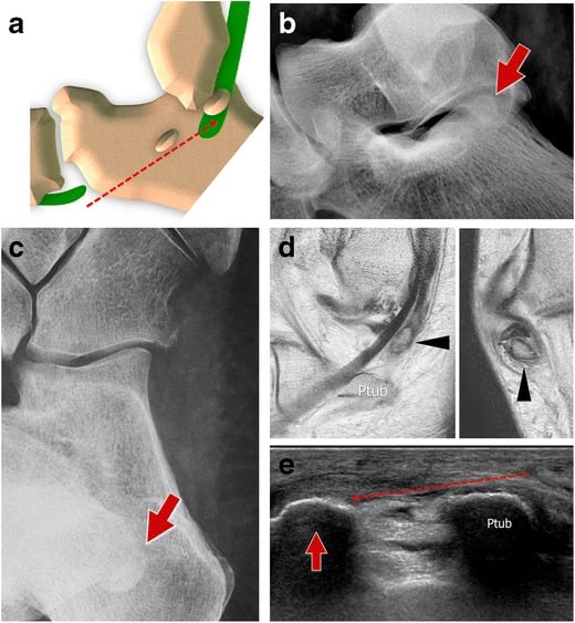
- Peroneal tubercle hypertrophy on the lateral calcaneus - can cause chronic friction and splitting
Classification Systems
Two systems grade different things. Brevis tears are graded by how much of the tendon is involved, which decides between debridement, repair and tenodesis. Acute dislocation is graded by how the retinaculum fails at the fibula.
- Tear Extent
- Less than 50% width
- Tendon Integrity
- Majority intact, functional
- Treatment
- Debridement, synovectomy
- Tear Extent
- 50-100% width, repairable
- Tendon Integrity
- Compromised but reparable
- Treatment
- Tubularisation, side-to-side repair
- Tear Extent
- Over 50% tendon substance loss
- Tendon Integrity
- Severe degeneration, irreparable
- Treatment
- Tenodesis to longus, allograft
The 50% threshold. A tear involving less than 50% of the tendon width can be debrided and retains enough strength for function; over 50%, a tubularisation repair is needed to restore mechanical strength. Grade 3 is defined by over 50% substance loss, tendon tissue actually gone rather than split width, and needs tenodesis because a repair under tension will fail. Grade the tear at operation: MRI often underestimates the extent.
Clinical Assessment
History. The pain is lateral, posterior to the malleolus, and aggravated by activity. An acute SPR tear follows forced dorsiflexion-eversion; a chronic split begins insidiously. The features that point to the tendons:
- A palpable or audible snap with ankle motion (subluxation)
- Difficulty with lateral push-off, cutting and running
- Recurrent lateral ankle pain attributed to ligamentous injury, with failed conservative management and ineffective bracing
Examination. Look for swelling posterior to the lateral malleolus and asymmetry against the other side. Palpate for tenderness along the tendons, over the fibular groove and at the SPR insertion. Record common peroneal nerve function (dorsiflexion, EHL) and the pulses.
The peroneal subluxation test has a specificity of 90%.
- Patient seated, ankle relaxed in neutral
- Examiner passively dorsiflexes and everts the ankle
- Patient actively resists eversion while maintaining dorsiflexion
- Positive: a palpable or visible snap as the tendons subluxate anteriorly over the lateral malleolus
- The tendons reduce with plantarflexion, with an audible or palpable clunk
Circumduction test. Passively circumduct the ankle through its full range. Pain or snapping at a specific arc, usually dorsiflexion-eversion, that reproduces the patient's symptoms is positive.
Resisted eversion. Pain on resisted eversion suggests tendon pathology. Compare strength with the other side: weakness suggests significant pathology (a tear over 50% or chronic degeneration), and pain without weakness a minor tear or tendinopathy.
Chronic lateral ankle pain without an acute injury or obvious instability may be peroneal tendon pathology. Misdiagnosis as a chronic ankle sprain is common and leads to failed conservative treatment. Consider the tendons when:
- The pain lies posterior to the malleolus, not in the anterior ATFL distribution
- Physiotherapy for a presumed sprain has failed for over 3 months
- There is a snapping sensation or audible pop with ankle motion
- Tenderness sits directly over the fibular groove or the peroneal tendons
Image early rather than late, but a normal MRI does not exclude the diagnosis (Investigations). If the clinical picture is convincing, proceed to dynamic ultrasound or tendoscopy rather than reassuring the patient.
- Pain Location / Pattern
- Posterior to lateral malleolus, worse with activity and eversion
- Key Discriminating Feature
- Tenderness along tendon sheath, pain on resisted eversion
- Confirmatory Test
- MRI (split, comma sign) or dynamic ultrasound
- Pain Location / Pattern
- Snapping posterior to malleolus, instability sensation
- Key Discriminating Feature
- Palpable tendon snap on dorsiflexion-eversion against resistance
- Confirmatory Test
- Dynamic ultrasound; positive subluxation test
- Pain Location / Pattern
- Anterolateral, giving way on uneven ground
- Key Discriminating Feature
- Anterior drawer / tilt positive, tenderness anterior to malleolus
- Confirmatory Test
- Stress radiographs, MRI of ATFL/CFL
- Pain Location / Pattern
- Anterolateral, in the sinus tarsi opening
- Key Discriminating Feature
- Point tenderness in sinus tarsi, relief with local anaesthetic injection
- Confirmatory Test
- Diagnostic injection, MRI (oedema/scar in sinus tarsi)
- Pain Location / Pattern
- Deep ankle pain, catching, swelling
- Key Discriminating Feature
- Pain on weight-bearing, effusion, not localised to tendon
- Confirmatory Test
- MRI; CT for bony detail
- Pain Location / Pattern
- Lateral/plantar-lateral pain, may have midfoot component
- Key Discriminating Feature
- Os peroneum tenderness at cuboid, painful resisted 1st ray plantarflexion
- Confirmatory Test
- Radiograph (os peroneum), MRI of peroneus longus
Investigations
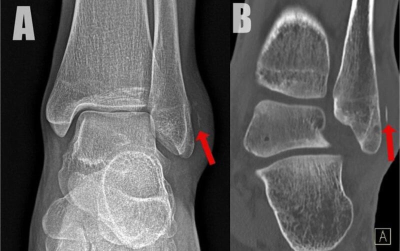
Radiographs. AP, lateral and mortise views of the ankle, with an oblique foot view if the fifth metatarsal base is painful. They have low sensitivity for soft-tissue pathology but are an essential baseline. Look for:
- Fibular groove abnormalities - shallow, convex, spurs
- Os peroneum fragmentation (peroneus longus injury)
- Fifth metatarsal base avulsion (brevis insertion injury)
- Ankle fracture, associated with peroneal injury in 11%
- Lateral calcaneal spurring from chronic friction
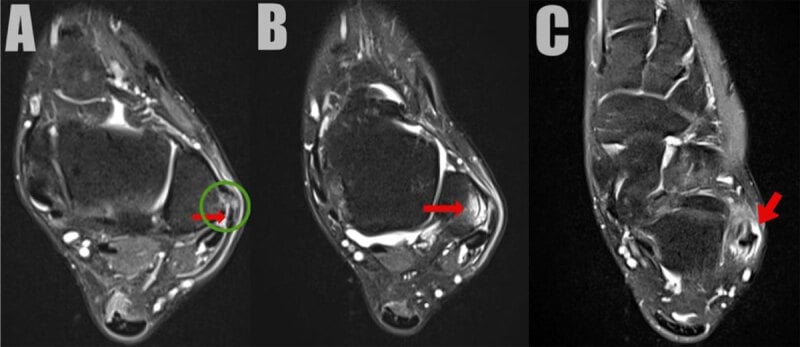
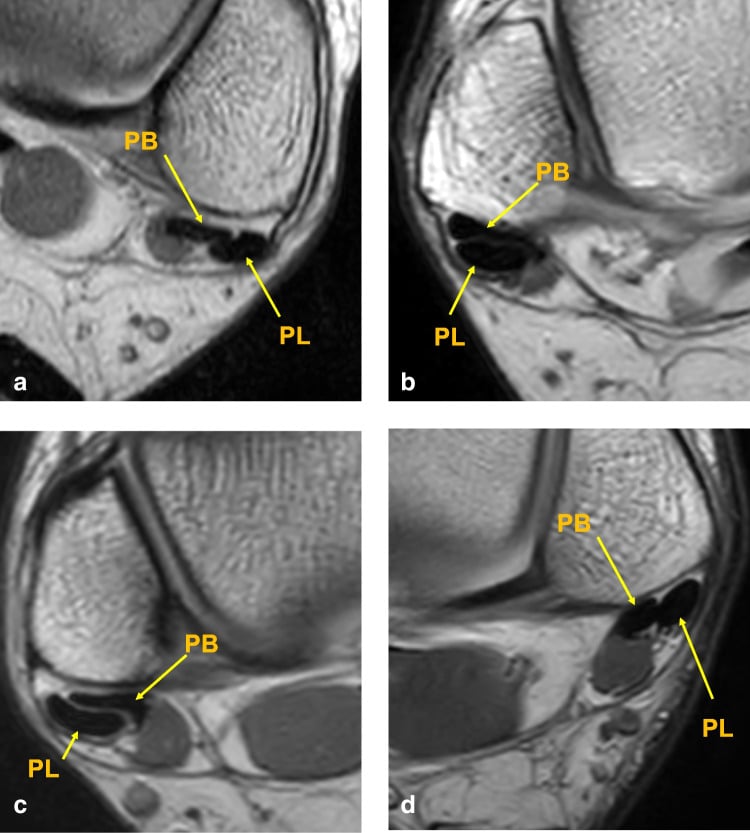
MRI. Axial, sagittal and coronal sequences, T1, T2 and STIR, with fat-suppressed imaging critical. The findings:
- Longitudinal split - linear high signal within the tendon on all sequences
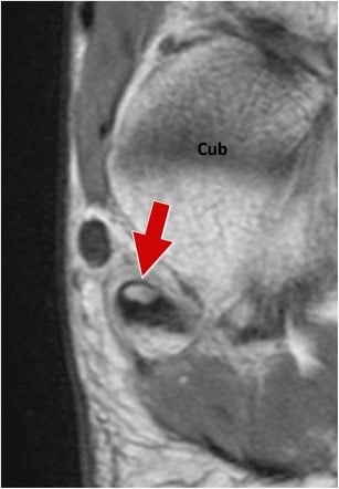
- Comma sign - on axial images the retracted edge of a split brevis makes a C-shaped or comma appearance; pathognomonic, and highly specific for a split tear
- Tendinosis - increased T2 signal, tendon thickening and peritendinous oedema. Diffuse high T2 signal without a discrete linear split suggests tendinopathy, which may respond to conservative treatment
- SPR injury - discontinuity, oedema, avulsion from the fibula. Focal high T2 signal at the fibular insertion with discontinuity indicates an acute injury suitable for primary repair
- Low-lying muscle belly - muscle signal extending into the fibular groove on axial images
- Subluxation - tendons anterior to the lateral malleolus on the axial view
MRI is specific, not sensitive. Pooled sensitivity is 73% for a brevis tear and only 60% for a longus tear, with specificity 88-91% (Ghandour 2025). For the two findings above that concern position rather than substance, routine MRI reporting is close to blind: sensitivity 3% for a low-lying muscle belly and 10% for subluxation (Mirmiran 2015). MRI rules pathology in, not out. Do not expect the report to warn you of a low-lying belly; anticipate it at operation.
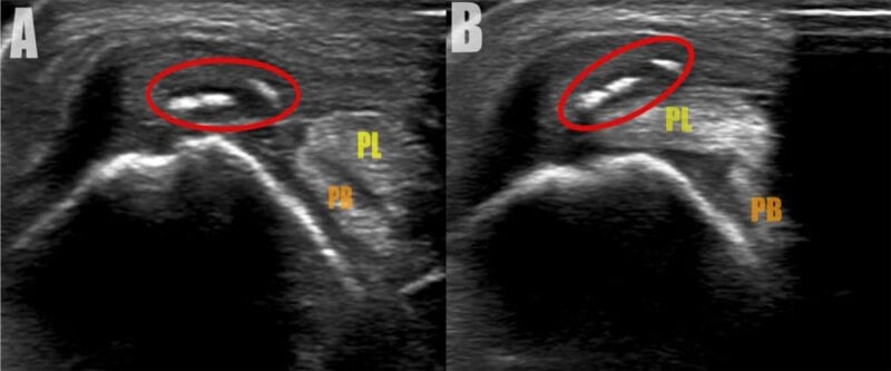
Ultrasound. Real-time dynamic evaluation through ankle motion makes it the only modality that shows a tendon actually subluxing, and the only one that shows intrasheath snapping. It shows a hypoechoic split, tendon thickening, and subluxation on resisted dorsiflexion-eversion. Pooled sensitivity is 93% for the brevis and 94% for the longus, with specificity 85-94%: higher sensitivity than MRI in both tendons (Ghandour 2025).
Its limits and its place. It is operator-dependent, and the pooled estimates rest on only three studies from specialist units, so they do not transfer to an occasional scanner. Use it when the clinical picture is convincing and the MRI is normal, and whenever the question is subluxation or intrasheath instability rather than a substance tear, not only when MRI is unavailable.
CT is rarely needed. It gives precise bony anatomy and a measured groove depth for planning surgery on a shallow or convex groove, but shows nothing of the tendon or the SPR.
Management Algorithm
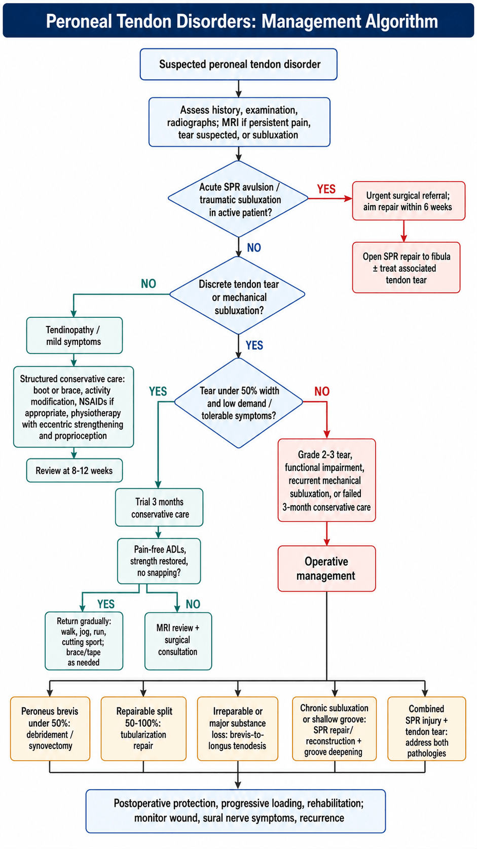
Who is treated without surgery. Conservative care is first-line for every patient without an acute SPR avulsion. It suits:
- Grade 1 tears (under 50% width) in low-demand patients
- Mild subluxation without mechanical symptoms, where the snapping is tolerable
- Tendinopathy without a discrete tear
The 3-month trial runs in phases.
- Weeks 0-2, protect. Reduce inflammation and protect the tendons from further injury: a CAM boot or ankle brace limiting eversion, ice for 15-20 minutes three times a day, a short (2-week) course of NSAIDs if there is no contraindication, and no cutting sports or lateral movements.
- Weeks 2-8, rehabilitate. Restore strength, proprioception and function: eccentric peroneal strengthening and balance training, dorsiflexion mobilisation without eversion stress, single-leg balance, wobble board and sport-specific drills, and athletic taping or bracing for sport if tolerated.
- Weeks 8-12, return to sport. Once daily activities are pain-free, strength equals the other side and there are no subluxation symptoms, progress walk, jog, run, cutting, full sport. External ankle support may be needed long-term. If symptoms persist at 12 weeks, consider MRI and a surgical consultation.
Surgical Technique
Positioning. Supine, with a bump under the ipsilateral hip rotating the leg internally by 10-15 degrees, or lateral decubitus, by surgeon preference. The head rests on a donut with the cervical spine neutral, the torso lies flat on the table, the contralateral leg lies flat and secured with a leg holder or tape, and the operative leg is free-draped from tourniquet to toes.
Tourniquet and preparation. A well-padded high-thigh tourniquet at 250-300mmHg (or limb occlusion pressure plus 100). Elevate the limb for 2 minutes and exsanguinate with an Esmarch bandage from toes to tourniquet. Prepare with Betadine or chlorhexidine from toes to mid-calf and allow it to dry; drape the foot and ankle free, excluding the toes but keeping them visible for positioning checks.
Equipment. Check the kit before starting:
- 2-3 suture anchors (3.0-3.5mm) for the SPR, with extras
- 2-0 FiberWire or Ethibond for tendon repair; 0 or 1 for the SPR
- Small joint set, periosteal elevator and bone rongeur for groove deepening
- Sagittal saw or burr, if groove deepening is planned
- C-arm, not typically needed but available if fracture fixation is required
The incision. An 8-10cm curvilinear incision following the posterior border of the fibula, from 6cm proximal to the tip of the lateral malleolus to 3cm distal, curved slightly posteriorly to avoid the sural nerve anteriorly. Cut sharply through skin and subcutaneous tissue with the knife, preserving the cutaneous nerves.
The sural nerve is the structure most at risk in lateral ankle surgery. It runs with the short saphenous vein in the subcutaneous fat, typically 1-2cm anterior to the posterior fibular border and so anterior to the incision. Place the incision directly over the posterior fibular border or slightly posterior to it, look for the nerve-vessel bundle early in the dissection, and retract it anteriorly with the skin flap, handling it gently and avoiding traction on the flap.
Injury causes lateral foot numbness and a painful neuroma, with an incidence of 5-10% in lateral ankle surgery. If the nerve is injured, consider primary repair if it is a sharp transection, using microsurgical technique (6-0 or 7-0 nylon under loupe magnification), or bury the nerve end in the peroneus brevis muscle to prevent a symptomatic neuroma.
Exposure. The superficial peroneal fascia lies over the tendons just deep to the subcutaneous fat; incise it longitudinally over the palpable tendons, parallel to the skin incision. The SPR appears as a thickened band at the level of the lateral malleolus and is preserved until the pathology is assessed. Then incise it longitudinally directly over the tendons, to be repaired later, and continue proximally and distally to open the sheath fully.
Inspection. A synovectomy clears the view, and both tendons, brevis anterior and longus posterior, are inspected for tears, subluxation and a low-lying muscle belly.
Intraoperative problems and their solutions:
- Cause
- Chronic subluxation, scarring, low-lying muscle belly
- Solution
- Synovectomy, release adhesions, consider groove deepening if shallow
- Cause
- Chronic injury, attenuated SPR
- Solution
- Augment with local fascia or periosteum, consider reconstruction with graft
- Cause
- Over-tensioned repair, insufficient tendon substance
- Solution
- Release tension, consider tenodesis instead of primary repair
- Cause
- Injury during retraction or groove deepening
- Solution
- Identify vessel, bipolar cautery or tie with 3-0 Vicryl, ensure haemostasis before closure
Complications
- Incidence
- 5-10%
- Risk Factors
- Anterior skin incision, rough handling
- Prevention/Management
- Posterior incision, identify and protect nerve; if injured: primary repair or bury in muscle
- Incidence
- 10-15% overall, 40% if groove deepening alone
- Risk Factors
- SPR not repaired, shallow groove not addressed
- Prevention/Management
- Combine SPR repair with groove deepening; revision: SPR reconstruction with graft
- Incidence
- 5-10% after repair
- Risk Factors
- Over-tensioned repair, early mobilisation, low-lying muscle belly variant
- Prevention/Management
- Tension-free repair, protect 4 weeks NWB; revision: tenodesis if re-tear
- Incidence
- 3-5%
- Risk Factors
- Thin subcutaneous tissue, diabetes, smoking
- Prevention/Management
- See below
- Incidence
- 5-10%
- Risk Factors
- Prolonged immobilisation, adhesions
- Prevention/Management
- Early ROM after 4-6 weeks, PT for mobilisation; revision: tenolysis if severe adhesions
- Incidence
- Under 1%
- Risk Factors
- Deep dissection, groove deepening
- Prevention/Management
- Identify and ligate or repair; vascular consult if significant bleeding
Recurrent subluxation is the most common failure mode. Its causes are inadequate SPR repair tension, failure to address a shallow groove, anchor pull-out and chronic tissue attenuation. Prevent it by stress-testing at operation (dorsiflex and evert the ankle and confirm the tendons stay reduced), by combining SPR repair with groove deepening when the groove is shallow, and by using enough anchors (2-3) to distribute the load. Revision is SPR reconstruction with local tissue (a peroneus longus slip) or allograft, with a bone block considered for severe groove deficiency.
Wound problems. Lateral ankle incisions carry a higher wound complication risk because the subcutaneous tissue is thin and the bone superficial; diabetes, smoking, peripheral vascular disease and previous surgery add to it. Prevention is meticulous haemostasis, gentle tissue handling, a tension-free skin closure and postoperative elevation. Recognise dehiscence early (within 2 weeks) and treat it with local wound care, considering VAC therapy if it is deep and antibiotics if infection is suspected; a large defect may require secondary closure or a skin graft.
Postoperative Care and Rehabilitation
The standard protocol follows any tendon or SPR repair.
- Weeks 0-4, protect. A posterior splint in slight plantarflexion-eversion, then at suture removal (2 weeks) a short leg cast or CAM boot: neutral dorsiflexion if the groove was deepened, slight equinus (10-15 degrees of plantarflexion) after tendon repair alone. Non-weight-bearing on crutches, the limb elevated above heart level, DVT prophylaxis with low molecular weight heparin or aspirin per protocol, and a dry dressing with a wound check.
- Weeks 4-8, load. A removable CAM boot from week 4, with progressive weight-bearing: 25% at week 4, 50% at week 5, 75% at week 6 and full by week 8. Gentle plantarflexion-dorsiflexion, avoiding eversion initially, and isometric exercises with no resisted eversion yet. Formal physiotherapy begins at week 6, for range and proprioception.
- Weeks 8-12, strengthen. Full weight-bearing in a supportive shoe, and full range by week 12 (longer if the ankle is stiff). Progressive resistance peroneal exercises with theraband and weights; single-leg balance, wobble board and perturbation training; walking progression, elliptical and swimming, without push-off at first.
- Weeks 12-24, return to sport. Progress once daily activities are pain-free, the range is full and strength is 80% of the other side: jogging at weeks 12-16, cutting drills at 16-20, plyometrics at 20-24, with a lace-up brace or taping for the initial return. The time to competition depends on the sport (Outcomes). A recurrent sensation of subluxation, persistent lateral ankle pain or weakness calls for MRI and specialist review.
Tendon and SPR repairs need 4-6 weeks for initial healing, and weight-bearing before 4 weeks significantly increases the risk of failure: loading puts tensile stress on the repair before it has healed and causes a gap or rupture. Studies show a 20-30% re-tear rate with early weight-bearing against 5-10% with protected weight-bearing. Emphasise compliance with non-weight-bearing to the patient, a short-term inconvenience for long-term success.
The accelerated protocol is only for a Grade 1 tear treated by debridement and synovectomy, with no tendon or SPR repair. The tendon is over 50% intact and needs no prolonged protection; repairs and tenodesis are not suitable and need the standard 4 weeks non-weight-bearing.
- Weeks 0-2 - removable CAM boot, weight-bearing as tolerated with crutches; gentle range of motion avoiding forceful eversion; swelling control with elevation and ice
- Weeks 2-6 - supportive shoe from week 4; progressive resistance peroneal strengthening; full range by week 6; proprioception training throughout
- Weeks 6-12 - sport-specific drills from week 8; return to competition at weeks 10-12 if criteria are met; bracing optional, based on symptoms
Outcomes and Prognosis
- Return to Sport
- 90%
- Patient Satisfaction
- 85-90% satisfied
- Re-operation Rate
- 5-10%
- Key Outcome Factors
- Timing critical - early repair achieves best outcomes
- Return to Sport
- 60-70%
- Patient Satisfaction
- 70-80% satisfied
- Re-operation Rate
- 15-20%
- Key Outcome Factors
- Tissue quality poor, higher failure rate
- Return to Sport
- 80%
- Patient Satisfaction
- 80-85% satisfied
- Re-operation Rate
- 5-10% re-tear
- Key Outcome Factors
- Tension-free repair, early PT important
- Return to Sport
- 60%
- Patient Satisfaction
- 70-75% satisfied
- Re-operation Rate
- Under 5% revision
- Key Outcome Factors
- Accept some eversion weakness, functional limitation
The factors that predict a poor result:
- Delayed surgery, over 6 weeks from an acute SPR injury
- A low-lying muscle belly - recurrent tears are common even after repair
- Severe tendon degeneration - the tissue is poor and a repair under tension fails
- A shallow groove - recurrent subluxation is higher if it is not deepened
- A workers' compensation claim - poorer outcomes regardless of technique
The best case is a young athlete with an acute SPR avulsion repaired within 6 weeks, normal groove anatomy and compliance with the rehabilitation protocol, who can expect over 90% return to their pre-injury level of sport.
Return rates and timelines by sport:
- Cutting sports (soccer, basketball): 80-85% return to the same level, at 5-6 months
- Running sports (track, cross-country): 90%, at 4-5 months
- Contact sports (rugby, AFL): 75-80%, at 6-8 months
- Recreational athletes: 90%, accepting lower intensity if needed
After tenodesis. Isolated peroneus brevis function, eversion through the fifth metatarsal base, is lost, but the longus compensates and preserves overall eversion strength. Patients may notice weakness on single-leg lateral push-off. It is generally well tolerated in non-athletes, and recreational athletes accept the trade-off.
Intrasheath Subluxation
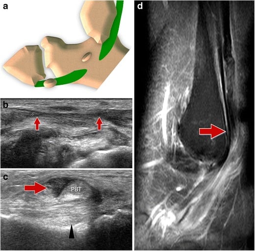
What it is. In classic subluxation the SPR fails and the tendons slip anteriorly over the lateral malleolus. In intrasheath subluxation the SPR is intact and the tendons stay behind the malleolus, but they snap past each other within the sheath during dorsiflexion-eversion: the longus and brevis reverse their relative positions, or a tendon flicks through a split. The snapping is painful, yet there is no visible displacement over the fibula, so the standard subluxation test and static MRI can look normal.
Raikin classification separates the pure swap from the one with a tear:
- Type A - the two tendons simply reverse position within an intact sheath, with no tear
- Type B - a peroneus brevis split, with the longus subluxating into or through it; the tear must be addressed
Catching it. It is a dynamic diagnosis, one of the lesions the tendoscopy review (Tham 2026) lists as missed by imaging. Suspect it when painful snapping is convincing but the frank subluxation test is negative. Dynamic ultrasound or tendoscopy during active eversion shows the intrasheath snap, whereas static MRI often shows only a flattened or split brevis or a convex groove.
Treatment. Groove deepening to give the tendons a deeper channel, repair of any brevis split (Type B), and retinacular tightening, increasingly performed tendoscopically.
Painful Os Peroneum Syndrome and Peroneus Longus Tears
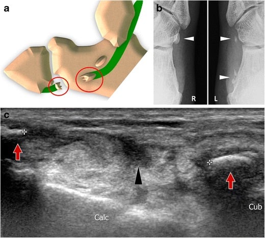
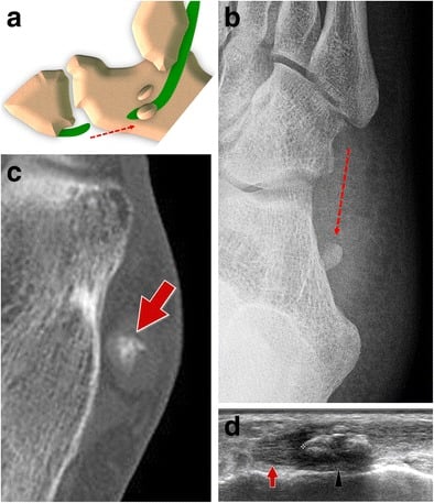
The os peroneum is a sesamoid within the peroneus longus tendon as it turns under the cuboid (the cuboid tunnel) to reach the first ray. It is ossified in about 20% of people and otherwise fibrocartilaginous, and it is a stress riser for the tendon.
Painful os peroneum syndrome (POPS) is a spectrum of peroneus longus pathology centred on the os peroneum. It causes plantar-lateral, not retromalleolar, pain, worse on resisted first-ray plantarflexion. The spectrum:
- An acute os peroneum fracture, or diastasis of a bipartite os
- A chronic fracture with longus tenosynovitis or attrition
- An enlarged or entrapped os catching in the cuboid tunnel
- A frank peroneus longus tear proximal or distal to the ossicle
Diagnosis. Point tenderness over the os peroneum at the lateral cuboid. A radiograph shows the ossicle and any fracture or diastasis; compare sides, because a widely separated bipartite os or a proximally migrated fragment suggests a longus tear. MRI or ultrasound assesses the tendon itself.
Treatment. Non-operative first, with immobilisation and an orthotic to offload the lateral column. For an irreparable longus tear with os peroneum pathology, excise the os peroneum, debride, and tenodese the peroneus longus to the peroneus brevis (Stockton and Brodsky: AOFAS 61 to 92 at about 5 years).
Guidelines, Registries & Global Practice
- Under-recognized: Peroneal pathology is a frequent but missed cause of chronic lateral ankle pain, often labelled "recurrent sprain"
- Acute subluxation/dislocation: Classically in skiers, footballers, basketball and dancers - forced dorsiflexion-eversion
- Coexisting lesions: Low-lying muscle belly (around one third of operated cases) and lateral ligament incompetence are common (Dombek 2003)
- Demographics: Acute subluxation in young athletes (20-40); degenerative splits in older, lower-demand patients
- No dedicated society guideline exists specifically for peroneal tendon disorders; practice is driven by case series and expert review
- Imaging consensus: MRI is specific but can be insensitive (especially for longus tears); dynamic ultrasound is sensitive and shows subluxation in real time (Ghandour 2025)
- Tendoscopy: Increasingly used as a diagnostic-therapeutic adjunct where MRI is equivocal (Tham 2026)
- Emphasis
- Reparable vs irreparable framework
- Position on Imaging
- MRI first-line, US for dynamic subluxation
- Position on Surgery
- Repair single salvageable tendon; tenodese to adjacent tendon if irreparable; reconstruct if both lost
- Emphasis
- Exclude mimics, structured non-operative trial first
- Position on Imaging
- MRI for persistent lateral ankle pain after failed rehab
- Position on Surgery
- Surgery for failed conservative care or mechanical subluxation
- Emphasis
- Recognize subluxation with distal fibula fractures
- Position on Imaging
- CT for groove/bony anatomy, MRI for tendon
- Position on Surgery
- Acute retinacular repair when fracture fixation undertaken
- Emphasis
- Address all components (tendon, retinaculum, groove)
- Position on Imaging
- Ultrasound widely used as first-line in many centres
- Position on Surgery
- Combine groove deepening with retinacular repair for groove deficiency
- No arthroplasty-style registry captures peroneal tendon surgery; the evidence base is Level III-IV case series and a small number of systematic reviews
- Best pooled data: tendoscopy series report AOFAS around 96 and return to sport near 13 weeks with low failure rates (Tham 2026); open athletic series report AOFAS around 97 and return at roughly 3 months (Saxena 2010)
- Practice variation: high-resource centres increasingly favour tendoscopic/minimally invasive techniques and dynamic ultrasound, while diagnosis elsewhere still relies on clinical examination plus MRI where available
- High-resource: MRI plus dynamic ultrasound, tendoscopy, suture-anchor retinacular repair, allograft reconstruction available; structured supervised rehabilitation
- Limited-resource: diagnosis rests on careful examination (subluxation test, focal tenderness) with selective imaging; open repair, groove deepening and local tendon transfer (e.g. tenodesis to peroneus longus) are durable, low-cost options that avoid graft cost and availability problems
- Universal principles: early treatment of acute retinacular injury, tension-free repair, protect the repair before loading, and address coexisting lateral ligament instability
Peroneal tendon pathology is commonly mistaken for a chronic ankle sprain, delaying effective treatment worldwide. Reduce this risk by:
- Documenting findings that distinguish peroneal pathology from ATFL injury (tenderness posterior to the malleolus, positive subluxation test)
- Imaging (MRI or dynamic ultrasound) when symptoms persist beyond 6 weeks despite rehabilitation for a presumed sprain
- Keeping a broad differential for chronic lateral ankle pain (ATFL instability, sinus tarsi syndrome, osteochondral lesion, peroneal pathology)
- Referring to a foot and ankle specialist if structured non-operative care fails at 3 months
Related pages: Peroneal Tendon Tears and Peroneal Tendon Subluxation develop the two main pathologies in full - this page is the umbrella that decides which of them you are looking at; Cavovarus Foot for the hindfoot alignment that loads the peroneals and is the commonest reason a technically sound repair fails - correcting the tendon without correcting the varus invites recurrence; Charcot-Marie-Tooth Disease for the neurological cause of that cavovarus, which must be excluded in bilateral or progressive disease; Lateral Ankle Instability for the ligamentous incompetence that coexisted in a third of Dombek's operative cohort and should be addressed at the same sitting; Chronic Lateral Ankle Pain for the wider differential this is repeatedly mislabelled within; Calcaneal Fractures for the malunited lateral wall that impinges and entraps the tendons; and Tendon Healing for the biology that dictates why a tubularised repair is protected before it is loaded.
Controversies and Areas of Uncertainty
The entire field rests on Level III-IV case series and a handful of systematic reviews - there are no randomised trials and no registry. State this honestly in a viva.
Imaging. The meta-analysis behind the sensitivity figures (Ghandour 2025) qualifies the "gold standard" label for MRI. A normal MRI does not exclude a tear in a convincing clinical picture, and dynamic ultrasound and tendoscopy retain a clear role.
Anatomical variants: cause or coincidence? The low-lying muscle belly is strongly associated with tears in operative series (Mirmiran 2015, Dombek 2003), but MRI studies of asymptomatic ankles fail to confirm several presumed variant-pathology links (Galli 2015). Whether to debulk a low-lying belly routinely, and which groove shape truly predisposes to subluxation, remain debated.
The 6-week window. The soft evidence behind the cut-off and the percentages is set out under Management.
Groove deepening and repair choice. Whether to add groove deepening routinely, when SPR reconstruction beats imbrication, autograft versus allograft, and the role of tendoscopic or minimally invasive stabilisation are all unresolved. Tendoscopy shows excellent pooled scores but lacks a randomised comparison with open surgery (Tham 2026).
MCQ Practice Points
Q: Which peroneal tendon is more commonly torn and why? A: Peroneus brevis is torn in 77 percent of cases. It lies anterior within the fibular groove, directly compressed against the fibula during ankle eversion. This compression generates shear forces on the posterior surface of the tendon, leading to longitudinal splitting. Peroneus longus lies posterior to brevis, protected from direct bony compression, and is less commonly torn (23 percent).
Q: How is acute peroneal tendon dislocation classified? A: By the Eckert-Davis classification (1976), with Oden's Grade IV (1987) addition, graded by how the superior peroneal retinaculum (SPR) fails at the fibula: Grade I SPR + periosteum stripped off the malleolus, tendons between periosteum and bone (commonest, ~50%); Grade II the fibrocartilaginous ridge is elevated with the SPR (~33%); Grade III a cortical bone flake is avulsed with the SPR - the radiographic "fleck sign" (~16%); Grade IV the SPR is torn from its posterior calcaneal attachment. Groove morphology (shallow/convex) and chronicity are separate modifiers, not grades - a shallow groove is the trigger for groove deepening (which must be combined with SPR repair, as deepening alone has a ~40 percent failure rate), and chronic attenuation drives the choice of imbrication vs reconstruction.
Q: What is the comma sign on MRI and what does it indicate? A: The comma sign is a pathognomonic finding for peroneus brevis longitudinal split tear. On axial MRI sequences, the retracted edge of the split tendon creates a C-shaped or comma appearance. This represents the posterior portion of the tendon that has split and retracted laterally within the fibular groove. High specificity for split tear diagnosis - when seen, proceed with surgical planning for debridement or repair.
Q: What is the evidence for timing of acute SPR repair and how does it affect outcomes? A: Acute repair of the superior peroneal retinaculum is favoured over delayed surgery: tissue quality is better and primary reattachment is feasible before retraction and scarring set in. The widely quoted 6-week window is a pragmatic threshold - beyond it, primary repair becomes difficult and reconstruction (groove deepening, retinacular reconstruction, or tendon rerouting) is more often required. Athletic series report high return to sport (AOFAS around 97, return to activity at roughly 3 months) after stabilization of subluxation. Clinical implication: Early diagnosis and referral are essential for optimal outcomes in active patients with acute peroneal subluxation.
Q: What is the threshold for performing tenodesis versus primary repair of a peroneus brevis tear? A: Grade 1 tears (less than 50 percent tendon width) - debridement only, sufficient tendon remains for function. Grade 2 tears (50-100 percent width, repairable) - tubularization repair with side-to-side sutures. Grade 3 tears (over 50 percent tendon substance loss, severe degeneration) - tenodesis to peroneus longus, as primary repair under tension will fail. Intraoperative decision: Assess tissue quality and remaining tendon substance - if repair gaps with ankle ROM or tension excessive, proceed to tenodesis.
Q: What is the most common complication after peroneal tendon surgery and how is it prevented? A: Sural nerve injury occurs in 5-10 percent of lateral ankle surgeries, causing lateral foot numbness or painful neuroma. Prevention: Skin incision along posterior border of fibula (nerve is 1-2cm anterior), identify nerve early in dissection, retract anteriorly with skin flap, avoid rough handling or traction. If injured: Consider primary nerve repair with microsurgical technique if sharp transection, or bury nerve end in peroneus brevis muscle to prevent symptomatic neuroma formation.
Exam Viva Scenarios
Practise clinical reasoning and management decisions out loud
“A 24-year-old basketball player presents with acute onset lateral ankle pain after landing from a jump with forced dorsiflexion and eversion. He reports feeling a pop and now has a painful snapping sensation with ankle motion. On examination, you palpate a subluxating structure posterior to the lateral malleolus during dorsiflexion-eversion. What is your assessment and management?”
“A 35-year-old recreational runner presents with 6 months of lateral ankle pain posterior to the malleolus, worse with activity. Multiple physiotherapy sessions have not helped. MRI demonstrates a longitudinal split tear of the peroneus brevis involving 60 percent of the tendon width. Walk me through your surgical approach and technique for this pathology.”
“A 28-year-old footballer underwent SPR repair 18 months ago but has recurrent painful subluxation of the peroneal tendons. MRI shows attenuated SPR tissue and a shallow fibular groove. He wants to return to competitive sport. How do you manage this complex revision case?”
Key Anatomy
- Peroneus brevis = anterior in groove, inserts 5th metatarsal base, torn 77% of cases
- Peroneus longus = posterior, inserts 1st metatarsal/medial cuneiform, torn 23%
- Superior peroneal retinaculum (SPR) = lateral fibular ridge to calcaneus, prevents subluxation
- Fibular groove = retrofibular, 1cm wide, 2-3mm deep (shallow if less than 2mm)
- Low-lying muscle belly variant = 15-20% prevalence, doubles tear risk, extends into groove
- Sural nerve = 1-2cm anterior to posterior fibular border, at-risk in lateral approach
Classification
- Peroneus brevis tears: Grade 1 (under 50% width) = debridement, Grade 2 (50-100% repairable) = tubularization, Grade 3 (over 50% loss) = tenodesis
- Acute dislocation = Eckert-Davis I-III + Oden IV: I periosteum stripped off fibula (commonest), II fibrocartilaginous ridge elevated, III cortical bone flake avulsed (fleck sign on radiograph), IV SPR torn from posterior calcaneal attachment
- Groove shape (shallow/convex) and chronicity are SEPARATE modifiers, not Eckert-Davis/Oden grades: shallow groove = add groove deepening (with SPR repair); chronic attenuation = imbrication vs reconstruction
- Comma sign on MRI = retracted split tendon edge, pathognomonic for longitudinal tear
Treatment Algorithm
- Acute SPR avulsion (within 6 weeks) = open SPR repair with suture anchors (90% return to sport)
- Delayed SPR injury (after 6 weeks) = SPR reconstruction with autograft or allograft (60% return)
- Grade 1 brevis tear = debridement, synovectomy (90% return, accelerated rehab possible)
- Grade 2 brevis tear = tubularization repair with 2-0 FiberWire (80% good outcomes)
- Grade 3 irreparable tear = tenodesis to longus (60% return, accept eversion weakness)
- Shallow groove = combine groove deepening (2-3mm bone removal) + SPR repair (never alone)
Surgical Pearls
- Lateral approach = posterior fibular border incision, protect sural nerve anteriorly
- Tension-free repair = test with ankle ROM, must not gap or restrict motion
- Groove deepening = remove 2-3mm cortical bone, preserve lateral ridge for SPR
- SPR repair = 2-3 suture anchors to fibular ridge, tie with ankle in neutral-slight plantarflexion
- Postop protocol = 4 weeks NWB in cast (slight plantarflexion-eversion), then progressive WB
- Return to sport = 5-6 months for SPR repair, 4-5 months for tendon repair alone
Complications
- Sural nerve injury = 5-10% (numbness, neuroma), prevent with posterior incision and early identification
- Recurrent subluxation = 10-15% overall, 40% if groove deepening without SPR repair
- Re-tear of brevis = 5-10% after repair (higher if low-lying muscle belly or over-tensioned)
- Wound complications = 3-5% (thin subcutaneous tissue), manage with local care, VAC, antibiotics
- Ankle stiffness = 5-10%, prevent with early ROM after 4-6 weeks, aggressive PT
- Revision surgery needed = 10-15%, lower success rate (70-80% vs 90% primary)
Evidence Base and Key Studies
Peroneal Tendon Tears - Pattern of Pathology and Coexisting Lesions
- Retrospective review of 40 patients undergoing peroneal tendon repair (mean age 42 years)
- Peroneus brevis tears in 88%, combined brevis-and-longus tears in 37%
- Low-lying peroneus muscle belly in 33% and lateral ligament incompetence in 33% coexisted
- Most common cause was ankle sprain or other trauma (58%); peroneal subluxation in 20%
- 98% returned to full activity without pain; clinically significant complications in 10%