Compartments of the Leg
Compartment syndrome is a clinical emergency, but its treatment is anatomical: every one of the four compartments must be opened. Knowing the contents lets you confirm decompression (e.g. you can see tibialis posterior in the deep posterior compartment) and protect the structures at the incisions.
The deep peroneal nerve (anterior) and first web space sensation are early in anterior ACS. At the posteromedial incision protect the saphenous vein and nerve; the deep posterior compartment is the one most often left inadequately released.
Overview
The leg is divided into four osteofascial compartments by the tibia, fibula, the interosseous membrane and the anterior and posterior intermuscular septa. Each is a relatively inelastic box containing a functional muscle group with its motor nerve and (with one exception) a named artery — and it is precisely this inelasticity that makes the leg the classic site of acute compartment syndrome. The exam value of the anatomy is therefore inseparable from the emergency it underlies: you learn the contents so you can recognise which function and sensation fail first, confirm that every box has been opened at fasciotomy, and protect the structures that lie at the decompression incisions. Hold three layers together — the contents of each compartment, the clinical syndrome (pain on passive stretch, the differential-pressure threshold), and the operation (a two-incision four-compartment release). This page is the anatomy and the decision thresholds; the syndrome itself is developed in compartment syndrome of the leg and compartment syndrome, its end-stage in Volkmann's ischaemic contracture, and the exercise-related variant — a different disease with a different threshold — in chronic exertional compartment syndrome. The two nerves that give the compartments their clinical signature have their own pages: the common peroneal nerve and the tibial nerve.
The Four Compartments
Anterior Compartment
- Muscles: tibialis anterior, extensor hallucis longus, extensor digitorum longus, peroneus tertius.
- Nerve: deep peroneal (fibular) nerve.
- Artery: anterior tibial artery.
- Action: ankle dorsiflexion and toe extension.
- Note: the most common compartment to develop acute compartment syndrome; bordered by the tibia, fibula, interosseous membrane and anterior intermuscular septum.
Know which fascial wall separates which compartment — the examiner will make you draw it. The four boxes are bounded not just by the tibia and fibula but by named fascial structures: the deep (crural) fascia forms the outer envelope; the interosseous membrane separates the anterior compartment from the deep posterior; the anterior intermuscular septum (fibula to deep fascia) separates the anterior from the lateral compartment; the posterior intermuscular septum separates the lateral from the superficial posterior compartment; and the transverse intermuscular septum separates the superficial posterior (gastroc-soleus) from the deep posterior compartment. Drawing this correctly also explains the incisions: the anterolateral fasciotomy is placed over the anterior intermuscular septum so one skin incision reaches both the anterior and lateral boxes, and the posteromedial incision lies behind the medial tibia to reach the superficial and (through the transverse septum) the deep posterior boxes. The exam point: each compartment is a fascial box with a specific wall between it and its neighbour — name the septa, not just "intermuscular septa."
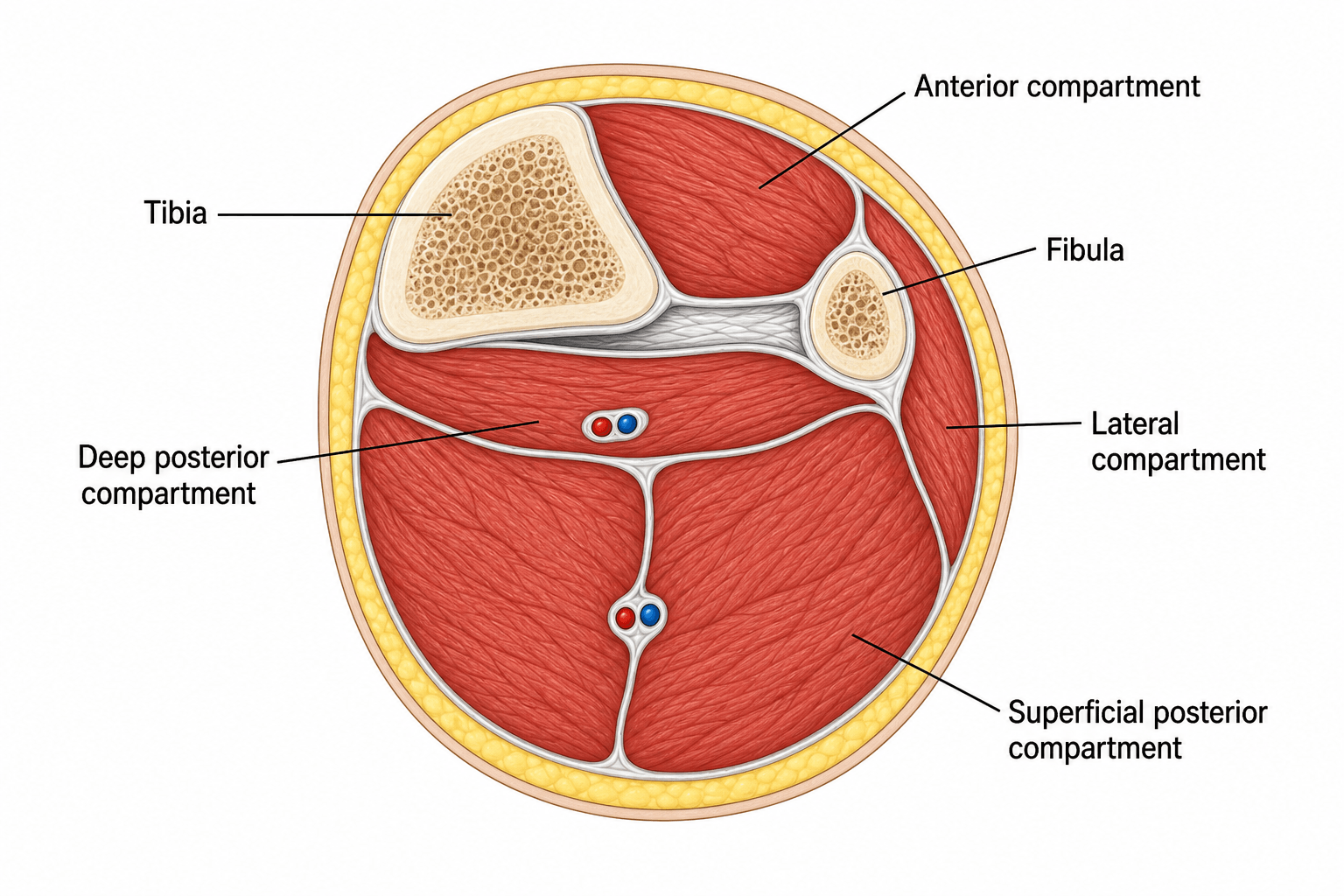
Neurovascular Contents by Compartment
The single highest-yield table in this topic — know the muscles, nerve, artery and action of each compartment cold, because the examiner will ask you to recite them and then link each to a clinical deficit.
- Muscles
- TA, EHL, EDL, peroneus tertius
- Nerve
- Deep peroneal
- Artery
- Anterior tibial
- Action
- Dorsiflexion
- Deficit if affected
- Foot drop; lost first-web-space sensation (earliest in ACS)
- Muscles
- Peroneus longus & brevis
- Nerve
- Superficial peroneal
- Artery
- (perforators)
- Action
- Eversion
- Deficit if affected
- Weak eversion; lost dorsolateral foot sensation
- Muscles
- Gastrocnemius, soleus, plantaris
- Nerve
- Tibial (muscles); sural (cutaneous)
- Artery
- Sural/perforators
- Action
- Plantarflexion
- Deficit if affected
- Weak push-off
- Muscles
- Tibialis posterior, FDL, FHL, popliteus
- Nerve
- Tibial
- Artery
- Posterior tibial + peroneal
- Action
- Inversion, toe flexion
- Deficit if affected
- Clawed toes, lost sole sensation (most often MISSED at fasciotomy)
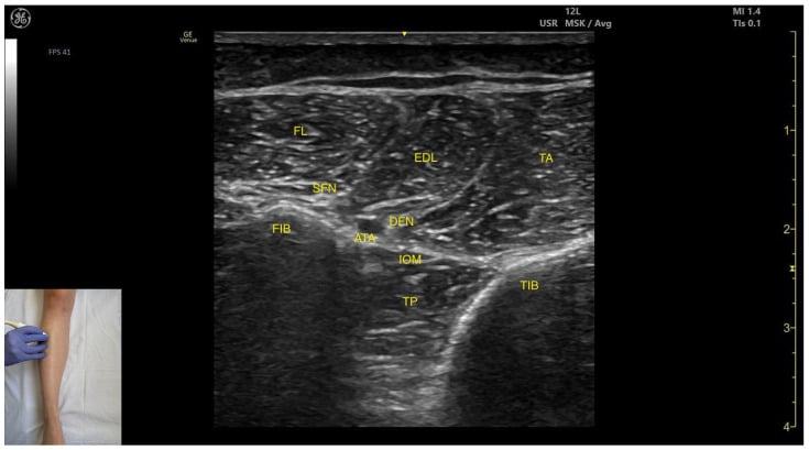
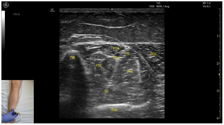
ALSDLeg Compartment Contents
Hook:Four compartments A-L-S-D; the deep peroneal nerve (anterior) and first web space fail first in ACS.
Surgical Relevance: Fasciotomy
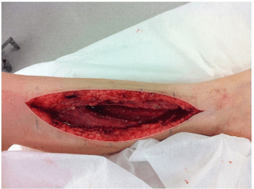
The standard surgical decompression of the leg is the two-incision technique, releasing all four compartments. Failure to release every compartment is the commonest reason a fasciotomy fails.
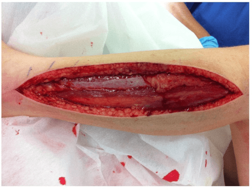
- Anterolateral incision (lateral to the tibial crest, centred over the anterior intermuscular septum): decompresses the anterior and lateral compartments. Identify the septum and the superficial peroneal nerve in the distal lateral compartment.
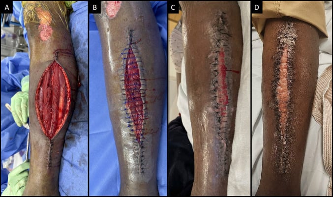
- Posteromedial incision (about 1-2 cm posterior to the medial tibial border): decompresses the superficial and deep posterior compartments - protect the saphenous vein and nerve, and ensure the deep posterior compartment (tibialis posterior) is genuinely released.
- A single-incision perifibular (Matsen) technique can release all four compartments through one long lateral incision over the fibula, but it is more demanding, gives poorer access to the deep posterior compartment, and risks the superficial peroneal nerve — the two-incision technique is preferred for completeness and is the safer answer in a viva.
- Whichever technique, incisions must be generous (typically the length of the leg segment) — short skin incisions cause skin-level constriction and an incomplete release.
- Wounds are left open and managed with delayed closure or skin grafting.
The "fifth compartment" — tibialis posterior in its own fascial sheath. A recurring anatomical reason the deep posterior compartment is the one most often left under-decompressed is that tibialis posterior frequently lies in its own separate fascial subcompartment within the deep posterior space (some describe it as the "fifth compartment of the leg"). Opening the deep posterior fascia and seeing FDL and FHL is therefore not enough — the tibialis posterior sheath must be specifically identified and released along the back of the tibia, or it can remain tight and ischaemic despite an apparently complete fasciotomy. The practical drill at the posteromedial incision: after releasing the superficial posterior (gastroc-soleus) and the main deep posterior fascia, deliberately detach soleus from the posteromedial tibia, follow the neurovascular bundle, and confirm the tibialis posterior fascia itself is opened. The exam point: the deep posterior compartment is functionally subdivided, so "I released the deep posterior compartment" must include the tibialis posterior sheath.
Clinical Correlations: Acute Compartment Syndrome
Acute compartment syndrome (ACS) results from raised pressure within a closed osteofascial compartment that exceeds capillary perfusion pressure, producing ischaemia, muscle and nerve necrosis, and ultimately an unsalvageable limb if not decompressed. It is the clinical reason the compartment anatomy matters.
Causes: most commonly a tibial diaphyseal fracture (the single commonest cause — see tibial shaft fractures), but also crush injury (crush syndrome), reperfusion after vascular injury/ischaemia, a tight cast or dressing, burns with circumferential eschar, high-energy soft-tissue injury, bleeding (anticoagulation/coagulopathy), and extravasation. It can occur with open fractures — an open wound does NOT decompress all compartments and does not exclude ACS.
How often, and in whom
Quoted incidences after a tibial diaphyseal fracture differ several-fold, and the reason is the diagnostic threshold, not the biology — so quote a figure with its denominator and its definition rather than a single number. McQueen & Court-Brown's 1996 prospective series of 116 consecutive fractures with 24-hour anterior-compartment monitoring recorded ACS in 3 patients (2.6%) (PMID 8898137). The same unit's later 13-year cohort of 1,388 tibial diaphyseal fractures, diagnosed on clinical signs, pressure monitoring or both, recorded ACS in 160 patients (11.5%) (PMID 25882967) — a broader ascertainment in a unit with a low threshold to monitor and decompress.
What that larger cohort adds is the risk profile, and it is not the one most candidates offer: youth is the single strongest independent predictor (p < 0.001), with the highest prevalence between 12–19 and 20–29 years. Age, male sex, blue-collar occupation, sporting injury, fracture classification and treatment with an intramedullary nail were all predictive on initial analysis, but after adjusting for age only occupation and implant type remained significant — and implant type lost significance once stratified by Tscherne soft-tissue grade, which argues the nail was a marker of the injury rather than a cause. The translation for the ward round: the young patient with a tibial shaft fracture is the one to monitor, and a benign-looking soft-tissue envelope is not reassurance. The same principle drives the regional variants — see region-specific compartment syndrome, foot and forearm compartment syndrome.
Diagnosis is clinical
The earliest and most reliable findings are pain out of proportion to the injury and pain on passive stretch of the muscles in the affected compartment. A tense, swollen compartment supports the diagnosis. The classic "5 P's" are LATE and unreliable — by the time they appear the limb is often already lost.
- Sign
- Pain out of proportion + pain on passive stretch
- Note
- The two findings to act on; a tense compartment supports it
- Sign
- Paraesthesia (nerve ischaemia)
- Note
- First-web-space numbness in anterior ACS (deep peroneal nerve)
- Sign
- Paralysis, Pallor, Pulselessness
- Note
- Late and unreliable — a palpable pulse does NOT exclude ACS
"Clinical diagnosis" does not mean "one sign is enough" — the signs are weak alone and strong in combination, and this is the number that converts the teaching into a decision. In Ulmer's systematic review of the four eligible prospective studies, the sensitivity of individual clinical findings was only 13–19% and their positive predictive value 11–15%, while specificity and negative predictive value were each 97–98% (PMID 12352566). Two things follow, and they are the whole of the clinical method. First, the probability of ACS rises steeply with the NUMBER of findings present — approximately 25% with one finding, and 93% with three — so the signs are cumulative, and the examination that matters is the serial one that documents a rising count rather than a single snapshot. Second, taken as Ulmer found them, the findings are more useful by their absence in excluding ACS than by their presence in confirming it. Be aware this last point is genuinely contested: Long et al. (PMID 30685220, cited below) conclude that history and examination are unreliable to rule out the diagnosis, and Ulmer himself cautions that with so few eligible studies the predictive value "has yet to be defined." The safe reconciliation — and the one McQueen's group act on — is that a single normal examination in an at-risk limb is not an exclusion, which is precisely why the at-risk patient gets monitored rather than merely re-examined. The exam answer is therefore not "pain on passive stretch means fasciotomy" but "one finding raises suspicion, three findings are near-diagnostic, and a single reassuring examination does not discharge the responsibility."
Intracompartmental pressure measurement
Where the patient is obtunded, anaesthetised, a child, or the picture is equivocal, measure intracompartmental pressure (within about 5 cm of the fracture, all compartments). Act on the differential (delta) pressure rather than the absolute value:
ΔP = diastolic blood pressure − measured compartment pressure. Decompress when the differential pressure drops below 30 mmHg — the original recommendation is a ΔP falling under 30 mmHg, and the widely taught "≤ 30" is the same rule rounded. Using the differential pressure (which accounts for the patient's perfusion pressure) rather than an absolute pressure threshold avoids both missed cases and unnecessary fasciotomies. In McQueen & Court-Brown's prospective series of 116 tibial diaphyseal fractures, a ΔP threshold of 30 mmHg missed no cases, whereas an absolute threshold of 30 mmHg would have sent 50 of the 116 patients (43%) to fasciotomy and an absolute threshold of 40 mmHg still 27 patients (23%) — against a true ACS rate of 2.6%. That is the argument for the differential in one line: the absolute pressure would have operated on roughly one patient in two who did not need it.
Add the duration — a threshold with no time attached is half the rule. The Edinburgh recommendation is decompression on a differential pressure below 30 mmHg sustained for more than 2 hours, read from continuous monitoring (PMID 26814506). A single transient dip below 30 on a one-off needle reading is not the trigger; a sustained fall is. Getting this right cuts both ways — it is what stops the isolated low reading from generating an unnecessary fasciotomy, and, because the criterion is met before signs declare themselves, monitoring on this basis shortens time to definitive treatment compared with waiting for clinical symptoms and signs to develop.
How accurate is the monitor you are being asked to trust? The page above tells you to measure; this is the evidence that the measurement is worth acting on, and it is a fair viva follow-up. In 850 monitored tibial diaphyseal fractures from the Edinburgh unit, continuous intracompartmental pressure monitoring had an estimated sensitivity of 94%, specificity 98%, positive predictive value 93% and negative predictive value 99% (PMID 23595064); 152 patients (17.9%) underwent fasciotomy, of whom 141 had confirmed ACS, 6 did not, and 5 more had ACS at operation despite a normal differential-pressure reading. Set that against the clinical findings' 13–19% sensitivity and the case for monitoring the at-risk limb makes itself. Note the honest limitation in those five false negatives: a normal ΔP does not license you to stop examining the patient — the monitor and the serial examination are complementary, not alternatives.
Timing and complications
Decompression should be as early as possible; irreversible muscle necrosis follows prolonged ischaemia. The familiar "within 6 hours" (and the companion teaching that myonecrosis begins by 6–8 hours) is a conventional benchmark carried through the textbooks rather than a validated threshold derived from a human tibial series — treat it as an urgency instruction, not a deadline you have until. Two things are firmer and more useful in a viva: the clock that matters runs from the onset of the compartment syndrome, not from the injury (so a late-presenting ACS may be hours old on a day-old fracture), and what demonstrably shortens time to decompression is acting on a sustained ΔP below 30 mmHg rather than waiting for the clinical picture to complete itself. Complications of missed or delayed ACS include Volkmann's ischaemic contracture (fibrotic, contracted, functionless muscle), rhabdomyolysis with acute kidney injury and hyperkalaemia, infection, amputation, and death. A very late presentation (established necrosis, typically beyond 36–48 hours) is one situation where fasciotomy may be withheld, because opening dead muscle risks fatal infection — a difficult, senior decision. Missed ACS is one of the commonest sources of successful orthopaedic litigation, so document serial examinations and act decisively.
The P's — but only the first two are earlySigns of acute compartment syndrome
Hook:Act on the first two P's (pain out of proportion + pain on passive stretch). If you wait for pallor, pulselessness and paralysis, the limb is already lost.
Guidelines, Registries & Global Practice
Global Practice Picture
Leg compartment anatomy is universal core knowledge for trauma and the basis of fasciotomy. The internationally consistent teaching: know the four compartments and their contents, diagnose acute compartment syndrome clinically (pain out of proportion, pain on passive stretch) with pressure measurement when equivocal, and decompress all four compartments emergently - most reliably via the two-incision technique. (The full neurovascular contents are tabulated in the Neurovascular Contents by Compartment section above.)
Viva Scenarios
Practise clinical reasoning and management decisions out loud
“Describe the compartments of the leg and how you would perform a fasciotomy for acute compartment syndrome after a tibial fracture.”
“A ventilated, sedated polytrauma patient has a closed tibial shaft fracture. The nursing staff are worried about the swollen calf but the patient cannot report pain. How do you decide whether to perform a fasciotomy?”
Compartments
- Anterior: dorsiflexors | deep peroneal n. | ant. tibial a.
- Lateral: peronei | superficial peroneal n.
- Superficial posterior: gastroc-soleus | sural n.
- Deep posterior: TP/FDL/FHL/popliteus | tibial n. | PT + peroneal a.
Compartment Syndrome
- Commonest cause = tibial diaphyseal fracture; anterior = commonest compartment
- Highest risk = YOUTH (strongest independent predictor, peak 12-29 yrs), blue-collar occupation
- Pain out of proportion + pain on passive stretch (early); 5 P's late/unreliable
- Signs alone insensitive (13-19%); ~25% probability with 1 finding, ~93% with 3 — examine SERIALLY
- Fasciotomy if ΔP (diastolic − compartment pressure) falls under 30 mmHg for over 2 h (McQueen)
- Continuous monitoring: sensitivity 94%, specificity 98%, NPV 99% — but 5 ACS cases had a normal ΔP
- Decompress urgently; the '6 h' is a conventional benchmark, and the clock runs from ACS ONSET
- Missed ACS → Volkmann's contracture, rhabdomyolysis, litigation
Fasciotomy
- Two incisions, all FOUR compartments
- Anterolateral: anterior + lateral
- Posteromedial: superficial + deep posterior
- Protect saphenous v./n.; do not miss deep posterior
Evidence Base
The reviews below frame ACS as a time-critical, medicolegally fraught clinical diagnosis. The pivotal primary paper for the exam is McQueen & Court-Brown's prospective tibial-fracture study, which established the differential-pressure threshold now used worldwide — quote it whenever you are asked how to interpret a compartment pressure, and pair it with McQueen & Duckworth's review for the duration (below 30 mmHg for more than 2 hours) that completes the rule. Read the set as answering four separate questions, because examiners ask them separately: who is at risk (Predictors, 2015 — youth above all), how good are the signs (Ulmer, 2002 — insensitive alone, near-diagnostic in threes), how good is the pressure monitor (2013 — sensitivity 94%, specificity 98%), and what threshold triggers the knife (1996 and 2014). The recurring theme is that a single normal examination cannot reliably exclude ACS, so a low threshold for serial review — and monitoring in the at-risk or obtunded patient — is essential.
Compartment monitoring in tibial fractures. The pressure threshold for decompression
- Prospective continuous anterior-compartment monitoring of 116 tibial diaphyseal fractures
- Acute compartment syndrome occurred in 3 patients (2.6%)
- A differential pressure (diastolic minus compartment pressure) threshold of 30 mmHg missed no cases of ACS
- An absolute threshold of 30 mmHg would have sent 50 of the 116 patients (43 percent) to fasciotomy, and a 40 mmHg threshold 27 patients (23 percent)
The diagnosis of acute compartment syndrome: a review
- Clinical findings have poor sensitivity (13 to 64 percent) compared with intracompartmental pressure monitoring (94 percent)
- Specificity of clinical findings ranges from 63 to 98 percent, against 98 percent for pressure monitoring
- Recommends decompression on a differential pressure of less than 30 mmHg sustained for more than 2 hours
- Deciding on the differential-pressure criterion reduces time to definitive treatment compared with waiting for clinical symptoms and signs to develop