Three Phases | Intrinsic vs Extrinsic | Type III to Type I Transition | Growth Factor Regulation
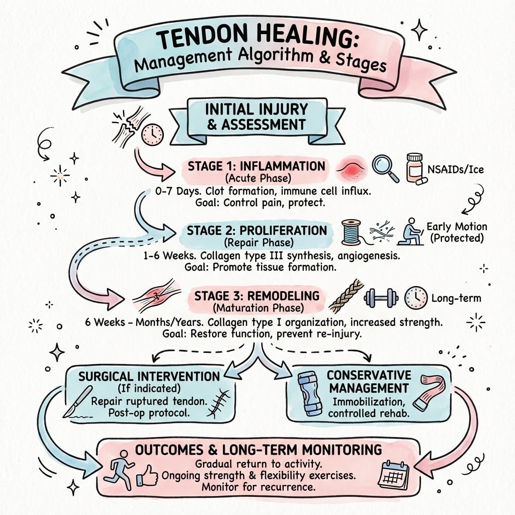
- Three overlapping phases: inflammatory (0-7 days), proliferative (7 days-6 weeks), remodeling (6 weeks-12+ months)
- Intrinsic healing via tenocytes (preferred) vs extrinsic healing via peritendinous fibroblasts (adhesions)
- Collagen type III appears first (disorganized scar), gradually replaced by type I (aligned, strong)
- Growth factors regulate healing: TGF-β (scar formation), VEGF (angiogenesis), IGF-1 and PDGF (proliferation)
- Early controlled motion superior to immobilization - reduces adhesions, promotes intrinsic healing, improves fiber alignment
- “Zone-specific healing in hand flexor tendons: Zone 2 worst prognosis (poor vascularity, synovial sheath)
- “Adhesion formation main complication - balance between healing and motion
- “Gap healing (under 3mm) better outcomes than proliferative healing (larger gaps)
- “Repair quality more important than timing (primary vs delayed) for outcomes
Inflammatory (0-7 days): Hematoma, inflammatory cells, weak fibrin clot. Proliferative (7 days-6 weeks): Tenocyte proliferation, collagen III synthesis, neovascularization. Remodeling (6 weeks-12+ months): Collagen I replaces III, fiber realignment, strength increase.
Intrinsic: Tenocytes and epitenon cells proliferate, synthesize matrix. Results in functional tendon with less adhesions. Extrinsic: Peritendinous fibroblasts invade, form scar tissue. Results in adhesions but provides early mechanical strength.
Type III collagen (thin, disorganized) appears first at 1-2 weeks, peaks at 2-3 weeks. Gradually replaced by Type I collagen (thick, organized) from 6 weeks onward. Type I alignment along stress lines improves tensile strength. Never reaches 100% of normal.
TGF-β: Promotes fibroblast proliferation and collagen synthesis (can cause excessive scarring). VEGF: Angiogenesis (essential early, problematic late). IGF-1 and PDGF: Stimulate tenocyte proliferation. FGF: Matrix synthesis and remodeling.
IPRThree Phases of Tendon Healing
Hook:IPR - I'm Promoting Recovery through three phases!
Overview and Healing Phases
Tendon healing is a complex biological process involving sequential and overlapping phases of inflammation, proliferation, and remodeling. Unlike bone, which heals through regeneration of native tissue, tendon healing occurs through scar tissue formation that never fully restores the original structure and biomechanical properties of healthy tendon.
The healing process involves a delicate balance between intrinsic healing (via tenocytes within the tendon) and extrinsic healing (via peritendinous fibroblasts). Intrinsic healing produces functional tendon with minimal adhesions, while extrinsic healing provides early mechanical strength but at the cost of adhesion formation.
Understanding tendon healing biology explains: why early controlled motion protocols are superior to immobilization; why Zone 2 flexor tendon injuries have poor prognosis; why adhesions form despite modern surgical techniques; why repair strength at 6 weeks allows protected motion but re-rupture risk remains; why biological augmentation strategies (PRP, growth factors) are being investigated.
Concepts and Mechanisms
Fundamental Tendon Healing Biology
Tendon healing represents a fundamental biological process that differs significantly from bone healing. While bone heals through regeneration, restoring native tissue architecture, tendon healing occurs through scar tissue formation that never fully replicates the original structure or mechanical properties.
- Bone regenerates native tissue; tendon forms scar
- Bone healing restores 100% strength; tendon plateaus at 60-80%
- Bone healing reconstitutes normal architecture; tendon remains disorganized
- Bone healing follows predictable timeline; tendon healing highly variable
- Too little inflammation: Delayed healing, poor strength
- Too much inflammation: Excessive scarring, adhesions
- Too little motion: Adhesions, weak disorganized healing
- Too much motion: Gap formation, rupture
Phases of Tendon Healing
Inflammatory Phase (0-7 Days)
Inflammatory Phase Events
Hematoma formation at injury site. Platelets aggregate and release growth factors (PDGF, TGF-β, VEGF) from alpha granules. Fibrin clot provides initial weak scaffold. Vasoactive mediators cause vasodilation and increased vascular permeability.
Neutrophils infiltrate (peak at 24 hours) for phagocytosis of debris and bacteria. Monocytes arrive and differentiate into macrophages (M1 phenotype initially). Inflammatory cells release cytokines (IL-1, IL-6, TNF-α).
Macrophage phenotype shift from M1 (pro-inflammatory) to M2 (pro-healing). M2 macrophages release growth factors promoting angiogenesis and fibroblast recruitment. Neovascularization begins with VEGF stimulation of endothelial cells.
- Platelets: Release growth factors from alpha granules
- Neutrophils: Phagocytosis, debris removal (peak 24h, gone by 3-5 days)
- Macrophages: M1 to M2 transition, orchestrate healing response
- Endothelial cells: Begin angiogenesis in response to VEGF
- Tensile strength: 0-10% of normal
- Fibrin clot provides weak mechanical continuity
- High risk of gapping or re-rupture with loading
- Requires immobilization or protected mobilization
First week after repair: Tendon has minimal strength - relies on suture integrity. Avoid active loading. Early passive motion can begin if repair is strong enough (minimum 4-strand core suture).
Understanding the inflammatory phase explains why NSAIDs should be used cautiously (may impair healing), why diabetes impairs healing (altered macrophage function), and why early infection is devastating (overwhelms nascent healing response).
Intrinsic vs Extrinsic Healing
Two Mechanisms of Tendon Healing
Tendon healing involves contributions from both intrinsic (within-tendon) and extrinsic (peritendinous) mechanisms. The balance between these two determines functional outcome.
- Cell source: Tenocytes within tendon substance and epitenon surface cells
- Mechanism: Tenocytes proliferate and synthesize matrix directly at injury site
- Advantages: Produces organized collagen aligned with tendon axis, minimal adhesions, preserves gliding function
- Disadvantages: Slower initial healing, requires intact vascular supply, vulnerable to complete rupture
- Clinical goal: Promote with early controlled motion and strong surgical repair
- Cell source: Fibroblasts from peritendinous tissue (paratenon, synovial sheath, surrounding fascia)
- Mechanism: Inflammatory response recruits fibroblasts that invade healing site and deposit scar matrix
- Advantages: Rapid mechanical strength, occurs even with complete tendon disruption
- Disadvantages: Disorganized collagen, adhesions bind tendon to surrounding structures, loss of gliding function
- Clinical problem: Adhesions are the main complication of flexor tendon repair
- Intrinsic-Dominant Healing
- Minimal - smooth gliding maintained
- Extrinsic-Dominant Healing
- Severe - tendon bound to sheath or surrounding tissue
- Intrinsic-Dominant Healing
- Aligned along tendon axis
- Extrinsic-Dominant Healing
- Disorganized, multidirectional
- Intrinsic-Dominant Healing
- Higher (70-80% of normal)
- Extrinsic-Dominant Healing
- Lower (50-60% of normal)
- Intrinsic-Dominant Healing
- Excellent - near normal gliding
- Extrinsic-Dominant Healing
- Poor - restricted by adhesions
- Intrinsic-Dominant Healing
- Early controlled motion promotes intrinsic
- Extrinsic-Dominant Healing
- Immobilization promotes extrinsic (avoid)
Zone 2 (no man's land) has worst prognosis because: (1) Poor intrinsic healing potential (avascular), (2) Enclosed synovial sheath promotes extrinsic healing and adhesions, (3) Long finger flexion excursion (8-9cm) requires extensive gliding, (4) Critical balance between early motion (prevent adhesions) and protection (prevent rupture). Requires 4-strand core suture minimum for early motion protocols.
Management Algorithm
Clinical Relevance
Clinical Applications and Implications
Understanding tendon healing biology directly informs clinical decision-making and patient management across multiple scenarios.
- Zone 2 requires 4-strand core suture minimum for early motion
- Duran, Kleinert, Indiana, Mayo protocols all based on healing biology
- Early motion (weeks 0-6) prevents adhesions during proliferative phase
- Protected loading until 6 weeks when remodeling begins
- Immobilization in equinus for inflammatory phase (0-2 weeks)
- Controlled dorsiflexion starting at 2 weeks (proliferative phase)
- Progressive loading from 6 weeks (remodeling phase)
- Full weight-bearing by 8-12 weeks
- Return to sport at 6-9 months (60-80% strength restoration)
- Strong initial fixation allows early passive motion
- Avoid active loading during inflammatory phase (0-6 weeks)
- Progressive strengthening during remodeling (6+ weeks)
- Biological augmentation (PRP, patches) targets proliferative phase
- Re-tear risk highest in first 3 months (weak collagen III phase)
- Healing never restores 100% of normal strength
- Re-rupture risk persists lifelong (60-80% final strength)
- Smoking cessation essential (impairs all phases)
- Diabetes control critical (affects macrophages, collagen synthesis)
- Early compliance with rehabilitation determines final outcome
This topic covers healing of an acute tendon injury, but the examinable counterpart is chronic overuse tendinopathy, which is best understood as a failed/incomplete healing response rather than an inflammatory "tendinitis." Histology shows a degenerative picture - collagen disarray and fibre disorganisation, increased ground substance (proteoglycan/water), neovascularisation with accompanying nerve ingrowth, and rounded tenocytes - with few or no inflammatory cells ("angiofibroblastic/mucoid degeneration"). Hence the modern term tendinopathy (or tendinosis) over tendinitis.
The Cook and Purdam continuum describes three (potentially reversible early, with a degenerative endpoint) stages:
- Reactive tendinopathy - a non-inflammatory proliferative cell/matrix response to acute overload; the tendon thickens to reduce stress; reversible if load is reduced.
- Tendon dysrepair - failed healing with greater matrix breakdown, more cells and early neovascularity; some reversibility remains.
- Degenerative tendinopathy - areas of cell death and disorganised matrix with neovessels; largely irreversible and the substrate for spontaneous rupture.
Treatment implications (why this matters): because the problem is degeneration/failed healing, not inflammation, the first-line is load management and progressive mechanotherapy (eccentric or heavy-slow-resistance loading) to stimulate tenocyte matrix remodelling - not anti-inflammatories or repeated corticosteroid injection (which give short-term analgesia but worsen tendon structure and increase rupture risk).
Exam point: tendinopathy = failed healing / degeneration (tendinosis), not tendinitis; recognise the Cook-Purdam reactive → dysrepair → degenerative continuum, and treat with graded loading, reserving corticosteroid for short-term symptom control with caution.
Molecular Biology and Growth Factors
Growth Factors Regulating Tendon Healing
Growth factors released from platelets, inflammatory cells, and healing tissue cells orchestrate the healing process. Understanding their roles explains therapeutic targets and potential augmentation strategies.
- Most abundant growth factor in tendon healing
- Three isoforms: TGF-β1 (pro-fibrotic), TGF-β2 (intermediate), TGF-β3 (anti-scarring)
- Stimulates fibroblast proliferation and collagen synthesis
- High levels associated with adhesion formation
- Potential target: reduce TGF-β1 or supplement TGF-β3 to minimize scarring
- Master regulator of angiogenesis
- Peaks at 1-2 weeks, essential for delivering nutrients and cells
- Biphasic role: beneficial early (healing), detrimental late (chronic tendinopathy)
- Anti-VEGF therapies investigated for chronic tendinopathy
- Promotes tenocyte proliferation and Type I collagen synthesis
- Enhances matrix protein production
- Anti-apoptotic (supports cell survival)
- Component of growth hormone-stimulated healing
- Released from platelet alpha granules
- Chemotactic for fibroblasts and inflammatory cells
- Stimulates cell proliferation
- Component of PRP preparations
- Stimulates fibroblast proliferation and angiogenesis
- Enhances collagen synthesis
- Improves tensile strength in animal models
- Investigated as therapeutic agent
TV PIG-FGrowth Factors in Tendon Healing
Hook:Watch TV with a PIG for healing - growth factors drive tendon repair!
Factors Affecting Healing
Clinical and Biological Factors Influencing Outcome
- Better blood supply correlates with better healing
- Zone 2 flexor tendons (avascular) heal poorly
- Achilles watershed zone (2-6cm proximal to insertion) ruptures frequently
- Compromised vascularity slows all phases of healing
- Zone 1: Good vascularity, favorable outcomes
- Zone 2: Poor vascularity, enclosed sheath, worst outcomes
- Zone 3-5: Excellent vascularity, favorable outcomes
- Rehabilitation protocols must be zone-specific
- Early controlled motion: Promotes intrinsic healing, aligns fibers, reduces adhesions, improves final strength
- Immobilization: Promotes extrinsic healing, adhesions, disorganized collagen, weaker outcome
- Optimal window: Enough motion to stimulate healing, not so much to cause gapping
- Gap healing (less than 3mm) produces organized tendon with minimal adhesions
- Proliferative healing (greater than 3mm gap) requires extensive granulation tissue and adhesions
- Strong repair (4-strand core suture, 40-60N) allows early motion
- Weak repair (2-strand, 20-30N) requires longer immobilization
- Children and adolescents: Faster healing, better intrinsic response, lower adhesion rates
- Older adults: Slower healing, reduced cellularity, higher adhesion rates
- Healing time increases approximately 10% per decade after age 30
- Impaired macrophage function (delayed M1 to M2 transition)
- Reduced tenocyte proliferation and collagen synthesis
- Impaired cross-linking (advanced glycation end products)
- Higher re-rupture rates (2-3x for Achilles)
- Vasoconstriction reduces blood flow
- Tissue hypoxia from carbon monoxide
- Reduced fibroblast proliferation and collagen synthesis
- Cessation 4+ weeks pre-op reduces complications
- NSAIDs: May impair early inflammatory phase (evidence mixed)
- Corticosteroids: Inhibit collagen synthesis, delay healing
- Quinolone antibiotics: Associated with tendon ruptures
VZMQBLFactors Affecting Tendon Healing
Hook:VZMQBL - Very Zealous Motion Quite Beneficial for Living tendons!
Healing Across Tendon Types (Comparison)
Why the Same Biology Behaves Differently by Site
The three-phase healing cascade is universal, but local anatomy, vascularity and mechanical demand mean outcomes and rehabilitation differ markedly between tendons. The flexor tendon heals as tendon-to-tendon within a synovial environment; the rotator cuff and Achilles enthesis must regenerate a graded tendon-to-bone interface that is rarely fully restored.
- Zone 2 Flexor
- Tendon-to-tendon in synovial sheath
- Achilles (mid-substance)
- Tendon-to-tendon, paratenon-covered
- Rotator Cuff (enthesis)
- Tendon-to-bone (fibrocartilage enthesis)
- Zone 2 Flexor
- Avascular zone, diffusion-dependent
- Achilles (mid-substance)
- Watershed 2-6 cm above insertion
- Rotator Cuff (enthesis)
- Hypovascular footprint, often degenerate
- Zone 2 Flexor
- Adhesions then rupture
- Achilles (mid-substance)
- Re-rupture, elongation (heel-rise weakness)
- Rotator Cuff (enthesis)
- Re-tear at footprint (up to ~50% in large tears)
- Zone 2 Flexor
- Early motion to prevent adhesions
- Achilles (mid-substance)
- Functional loading, avoid elongation
- Rotator Cuff (enthesis)
- Protect repair, delayed loading to allow enthesis healing
- Zone 2 Flexor
- Good if intrinsic promoted
- Achilles (mid-substance)
- Good for mid-substance, scar fills gap
- Rotator Cuff (enthesis)
- Poor - fibrovascular scar, not true enthesis
A stiff or weak finger/limb after tendon repair is not always failed healing. Distinguish: (1) adhesions (passive ROM greater than active ROM, gradual onset, no sudden pop); (2) re-rupture (sudden loss of active motion, often a palpable gap or audible pop); (3) suture failure or gapping (early loss of correction without true rupture); (4) joint contracture (limited passive and active ROM equally); (5) infection (pain, erythema, systemic signs out of proportion). The management differs completely - therapy and possible tenolysis for adhesions, revision for rupture, and aggressive treatment for infection.
The table contrasts tendon-to-tendon and tendon-to-bone healing, but the examinable basic-science detail is the structure of the normal enthesis and why surgery cannot rebuild it. A direct (fibrocartilaginous) enthesis has four graded zones:
- Tendon proper (dense type I collagen, tenocytes),
- Uncalcified fibrocartilage (type II collagen, aggrecan; begins to dissipate stress),
- The tidemark (the abrupt basophilic line separating uncalcified from calcified fibrocartilage),
- Calcified fibrocartilage, blending into bone.
This graded soft-to-hard transition mineralises stress over a short distance and minimises stress concentration at the interface. After repair (e.g. rotator cuff to the footprint), the body does not recreate these zones - it heals by disorganised fibrovascular scar with Sharpey-like fibres anchoring tendon to bone, which is mechanically inferior. This is the biological reason tendon-to-bone repairs (cuff, especially large/massive tears in older patients) re-tear so commonly and why biological augmentation and graded-interface tissue engineering are active research areas.
Exam point: the normal enthesis is a four-zone (tendon → uncalcified fibrocartilage → tidemark → calcified fibrocartilage → bone) graded transition; healing replaces it with fibrovascular scar / Sharpey fibres, not regenerated fibrocartilage, which underlies the high re-tear rate of tendon-to-bone repairs.
Guidelines, Registries & Global Practice
Global Epidemiology
- Tendon and ligament injuries account for a substantial share of musculoskeletal presentations; overuse tendinopathy is estimated to underlie roughly 30% of running-related injuries, and lateral elbow tendinopathy affects up to 40% of tennis players (Sharma & Maffulli).
- Acute Achilles rupture incidence has risen in many high-income settings to the order of 20-30 per 100,000 person-years, concentrated in active middle-aged adults ("weekend warriors").
- Flexor tendon lacerations are predominantly young working-age males with sharp hand trauma; outcome is heavily zone- and rehabilitation-dependent worldwide.
Side-by-Side Guidance (Principles, Not Country Frames)
- Emphasis
- Acute Achilles rupture
- Practical Recommendation
- Either operative or non-operative acceptable; functional early rehabilitation reduces re-rupture across both
- Emphasis
- Open fractures and soft-tissue trauma
- Practical Recommendation
- Early senior decision-making, meticulous soft-tissue handling to protect the healing environment
- Emphasis
- Repair mechanics
- Practical Recommendation
- Stable repair construct that permits early protected motion - mechanical stability enables biological healing
- Emphasis
- Tendinopathy and biologics
- Practical Recommendation
- Loading-based rehabilitation first-line; biologics (PRP) not routinely endorsed given inconsistent evidence
Registry and High-Level Evidence Notes
- There is no dedicated international tendon registry equivalent to arthroplasty registries (NJR, AJRR, AOANJRR, SHAR). Best evidence comes from condition-specific RCTs (e.g. PATH-2 for PRP) and large trauma databases.
- Achilles and rotator cuff cohorts within national audit and shoulder/elbow datasets consistently report re-rupture/re-tear as the dominant healing-failure endpoint, reinforcing that biology, not technique alone, limits outcomes.
High- vs Limited-Resource Practice Variation
- High-resource settings: ready access to hand therapy, ultrasound/MRI for integrity assessment, and structured early-motion protocols - the main determinants of good flexor and cuff outcomes.
- Limited-resource settings: where supervised therapy is scarce, simpler, more protective immobilization protocols may be chosen to reduce rupture risk, accepting a higher adhesion burden. Strong, reproducible repair constructs and clear patient self-rehabilitation instructions become even more important.
- Biologics (PRP, growth factors) are not a priority where evidence is weak and cost is high; investment in rehabilitation infrastructure yields greater returns on healing outcomes.
Controversies and Areas of Uncertainty
Where Evidence Is Evolving or Conflicting
NSAIDs after tendon repair - Experimental work consistently shows COX inhibitors impair early proliferative healing and reduce tensile strength, yet they remain widely used for analgesia and ectopic-ossification prophylaxis. The pragmatic position is short-course, minimal-dose NSAIDs and avoidance during the first 1-2 weeks after a tendon repair, accepting that high-level human tendon-outcome data are lacking.
Biological augmentation (PRP, growth factors, stem cells) - Despite a strong biological rationale, the best controlled data (PATH-2 for acute Achilles rupture) show no benefit of PRP. Heterogeneous preparations (leukocyte-rich versus leukocyte-poor, activated versus not, variable platelet concentration) make pooled interpretation difficult. There is no consensus to use these routinely.
Inflammation - friend or foe? - The older view that inflammation is purely harmful has shifted. Macrophage M1-to-M2 transition is now seen as essential for orderly healing, and excessive suppression of early inflammation may impair repair. The target is modulation, not abolition.
Operative versus non-operative Achilles management - Modern functional rehabilitation with early weight-bearing has narrowed the re-rupture gap between surgical and conservative care, shifting many units toward non-operative management for selected patients - a direct clinical application of loading biology.
Type III collagen - obstacle or scaffold? - Persistent Type III collagen is associated with weaker tissue, yet it is also the necessary early scaffold. Strategies aimed simply at suppressing Type III risk destabilising early repair; the goal is timely Type I conversion and alignment, not Type III elimination.
Key Exam Points and MCQ Practice
Q: What are the three phases of tendon healing and their approximate durations? A: Inflammatory (0-7 days), Proliferative (7 days to 6 weeks), Remodeling (6 weeks to 12+ months). Phases overlap and exact timing varies by tendon type and injury severity.
Q: Which collagen type appears first during tendon healing and what is the final Type I:III ratio? A: Type III collagen appears first (1-2 weeks), forming disorganized scar. Gradually replaced by Type I over months. Normal ratio is 19:1 but healed tendon achieves only 2:1 to 3:1 at 12+ months.
Q: What is the difference between intrinsic and extrinsic tendon healing? A: Intrinsic: Tenocytes proliferate and synthesize organized matrix - minimal adhesions, better function. Extrinsic: Peritendinous fibroblasts invade and deposit scar - adhesions form, poorer function. Early motion promotes intrinsic over extrinsic.
Q: Which growth factor is most abundant in tendon healing and what is its dual role? A: TGF-β (particularly β1 isoform) is most abundant. Dual role: essential for collagen synthesis and healing, but excess causes scarring and adhesions. TGF-β3 has anti-scarring properties.
Q: What percentage of normal tendon strength is achieved by healed tendon at 12 months? A: 60-80% of normal tensile strength. Never reaches 100% due to persistent Type III collagen, suboptimal fiber alignment, and altered matrix composition. Explains ongoing re-rupture risk.
Q: Why do Zone 2 flexor tendon injuries have the poorest prognosis? A: Four reasons: (1) Avascular - poor intrinsic healing, (2) Enclosed flexor sheath - promotes extrinsic healing and adhesions, (3) High excursion (7-8cm gliding) - adhesions severely limit function, (4) Critical balance - need aggressive early motion (prevent adhesions) but higher re-rupture risk.


Exam Viva Scenarios
Practise clinical reasoning and management decisions out loud
“Examiner asks: Describe the biology of flexor tendon healing after surgical repair. Why do Zone 2 injuries have poor outcomes?”
“A 35-year-old laborer sustains a complete Zone 2 FDP laceration. After 4-strand core suture repair, should you immobilize or use early motion protocol? Justify your answer with the healing biology.”
“The patient asks about platelet-rich plasma injection to speed healing of his Achilles tendon repair. Discuss the growth factors involved in tendon healing and the evidence for PRP use.”
Three Phases
- Inflammatory (0-7 days): Hematoma, neutrophils then macrophages (M1 to M2), growth factors released, minimal strength (0-10%)
- Proliferative (7d-6wk): Tenocyte proliferation, Type III collagen synthesis, neovascularization peaks (3-4wk), strength to 30%
- Remodeling (6wk-12+mo): Type I replaces Type III, fiber alignment, MMP remodeling, strength to 60-80%
Intrinsic vs Extrinsic
- Intrinsic: Tenocytes synthesize organized collagen, minimal adhesions, better function (promote with early motion)
- Extrinsic: Peritendinous fibroblasts deposit scar, adhesions, poorer function (minimize by early motion)
- Early controlled motion promotes intrinsic over extrinsic healing
Collagen Transition
- Type III (thin, weak) appears first at 1-2 weeks, peaks at 2-3 weeks
- Type I (thick, strong) replaces Type III from 6 weeks onward
- Type I:III ratio: Normal 19:1, Healed 2:1 to 3:1 (never returns to normal)
- Tensile strength correlates with Type I content and fiber alignment
Key Growth Factors
- PDGF: Chemotactic, recruits cells (early phase)
- TGF-β: Collagen synthesis, cell proliferation (but excess causes scarring)
- VEGF: Angiogenesis (peaks 1-2 weeks, needed early, problematic if persistent)
- IGF-1: Tenocyte proliferation, Type I collagen synthesis
- bFGF: Cell proliferation, matrix synthesis, synergy with VEGF
Factors Affecting Healing
- Vascularity: Better blood supply equals better healing (Zone 1 better than Zone 2)
- Zone: Zone 2 flexor tendons worst (avascular, synovial sheath, high excursion)
- Motion: Early controlled motion better than immobilization (intrinsic healing, fiber alignment, prevent adhesions)
- Repair quality: Strong repair (4-strand, 40-60N) allows early motion without gapping
- Biologics: Age, diabetes, smoking impair healing; NSAIDs may impair (controversial)
Clinical Pearls
- Gap healing (under 3mm) better than proliferative healing (over 3mm, adhesions)
- 4-strand minimum for early active motion (2-strand passive only)
- Zone 2 requires aggressive early motion protocols (Kleinert, Duran, Indiana, Mayo)
- Final strength 60-80% explains ongoing re-rupture risk
- PRP evidence mixed, not routine, may consider in high-risk patients
Evidence Base
Tendon Injury and Tendinopathy: Healing and Repair (landmark review)
- Healing is a continuous process described in three overlapping phases: inflammatory, proliferative (collagen-producing), and remodeling (consolidation then maturation)
- Healing occurs via both intrinsic (epitenon and endotenon tenocyte proliferation) and extrinsic (sheath and synovial cell invasion) mechanisms
- Type III collagen predominates early and is progressively replaced by aligned Type I collagen during remodeling
- Biochemical and mechanical properties of healed tendon never fully match those of intact tendon
Early Controlled Passive Mobilization Superior to Immobilization (canine landmark)
- Canine flexor tendon repair model comparing immediate motion, delayed motion and immobilization over 12 weeks
- Immediately mobilized tendons at 3 weeks had roughly twice the ultimate load and nearly three times the linear slope (stiffness) of immobilized repairs
- At 12 weeks distal interphalangeal angular rotation was 95% of intact controls with immediate motion versus only 19% with immobilization
- Early protected passive motion augments the physiologic processes governing strength and excursion (intrinsic healing, fewer adhesions)
Neutralizing Antibody to TGF-β1 Increases Postoperative Range of Motion
- Rabbit zone II flexor tendon transection-repair model with intraoperative anti-TGF-β1 neutralizing antibody
- Anti-TGF-β1 increased combined IP joint flexion from 15 plus or minus 6 degrees (control) to 32 plus or minus 9 degrees (p = 0.002)
- Adding anti-TGF-β2 nullified the benefit (18 plus or minus 4 degrees), implying isoform-specific roles
- TGF-β1 is implicated in pathological scar formation, so its modulation reduces adhesions
PATH-2: Platelet-Rich Plasma Confers No Benefit in Acute Achilles Rupture
- Multicentre, placebo (dry needle) controlled, double-blinded superiority RCT across 19 UK units; 230 adults with acute Achilles rupture managed non-operatively
- Primary outcome (limb symmetry index for heel-rise work at 24 weeks) showed no difference: adjusted mean difference -3.9% (95% CI -10.5% to 2.7%)
- No difference in patient-reported function (Achilles tendon rupture score), pain, quality of life or adverse events
- Injected PRP was confirmed good quality with expected growth factor content, so failure was not due to a poor preparation
NSAIDs (Parecoxib, Indomethacin) Impair Early Tendon Healing
- 60 rats with a 3 mm Achilles defect randomised to parecoxib, indomethacin or saline for 7 days, tested at 14 days
- Both the COX-2-selective (parecoxib) and non-selective (indomethacin) NSAID significantly reduced tensile strength versus control
- Parecoxib also significantly reduced stiffness; both drugs reduced the cross-sectional diameter of the healing callus
- The negative effect was most pronounced with parecoxib, implicating COX-2-mediated prostaglandins in early proliferative healing
Tendon-to-Bone Healing Fails Often in Large/Massive Cuff Tears - Age and Tear Size Predict Failure
- 27 shoulders with large or massive cuff tears repaired (mini-open) and assessed by ultrasound at 3-5 years
- Recurrent defects in 14 of 27 (51.9%) despite repair, illustrating biologically poor tendon-to-bone healing
- Patients with an intact repair were on average 15 years younger than those who re-tore (49.9 versus 64.1 years, p less than 0.005)
- Larger preoperative tear size was negatively associated with healing, and large re-tears correlated with worse function