Comprehensive overview of tendon ultrastructure, molecular composition, hierarchical organisation, and biomechanical properties essential for understanding tendon pathology and healing.
- Type I collagen comprises 95% of total collagen with type III increasing in healing and tendinopathy
- Hierarchical organisation spans tropocollagen (300 nm) to tendon unit with 67 nm D-period fibrils
- Crimp pattern (50-100 μm) enables toe region (0-2% strain) elastic deformation
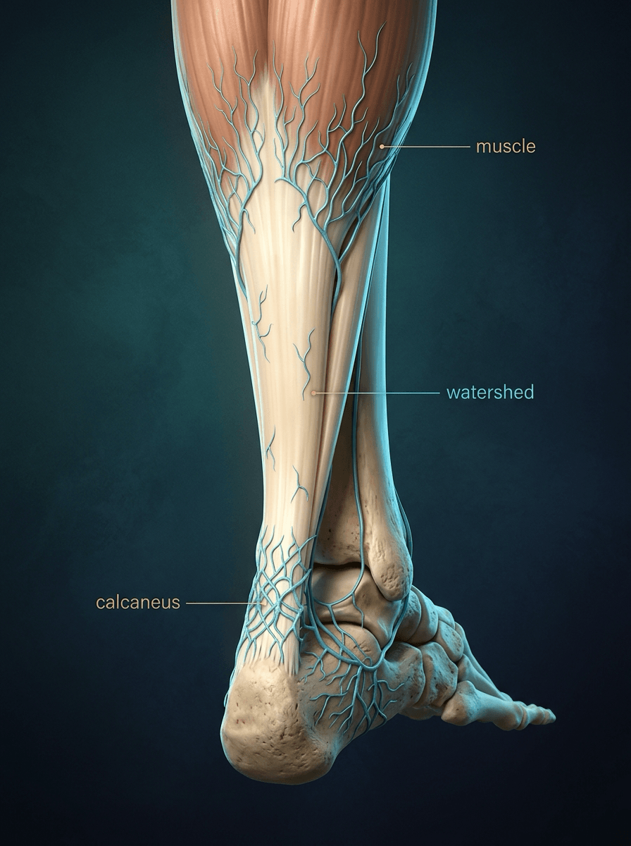
- Vascular watershed zones in mid-substance (Achilles 2-6 cm proximal) predispose to pathology
- Enthesis transitions through four zones, two of them fibrocartilage, over ~1 mm (200 MPa to 20 GPa)
- “Stress-strain curve: toe (0-2%), linear (2-4%, 1-2 GPa), yield (~4%), failure (8-15%, 50-100 MPa)
- “Healed tendon achieves only 50-80% of original strength due to persistent type III collagen and disorganised matrix
- “Decorin regulates fibril diameter; decreased in tendinopathy leads to irregular enlarged fibrils
- “Tenocytes form mechanosensitive networks via gap junctions and cytoplasmic processes
High-yield for basic science vivas. Examiners commonly ask about hierarchical organisation, collagen composition and biomechanical properties. Know the stress-strain curve and its clinical correlations with tendon healing and pathology.
Hierarchical Organisation
Tendon is built as a hierarchy spanning seven orders of magnitude, from nanometres to centimetres, and each level adds a further degree of organisation. The arrangement transmits force efficiently while allowing some internal compliance to accommodate variations in loading pattern and direction, and it is the key to both normal function and disease.
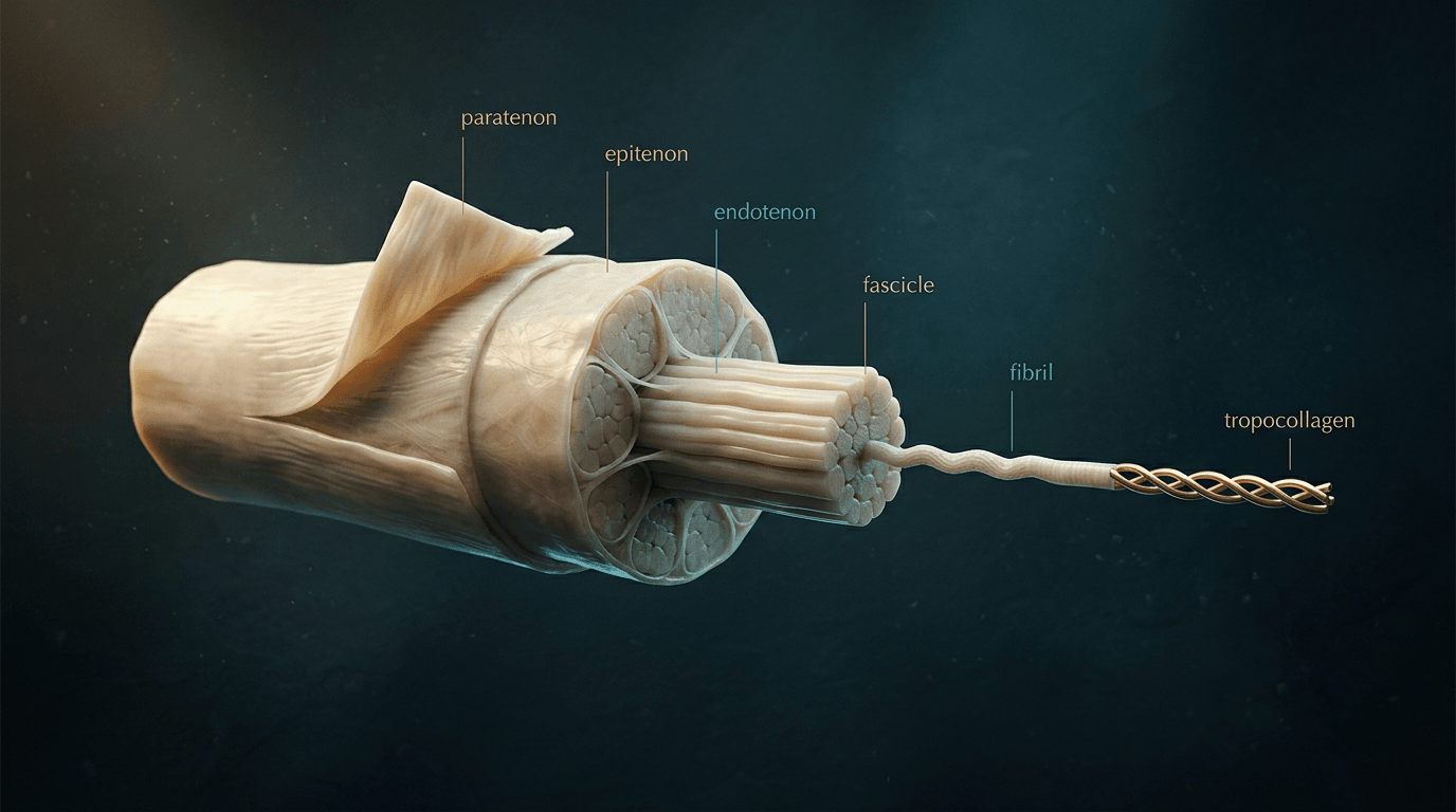
The molecule. The building block is tropocollagen, a right-handed triple helix about 300 nm long and 1.5 nm across, made of two alpha-1 chains and one alpha-2 chain. Its sequence repeats Gly-X-Y: glycine at every third position allows tight helical packing, and proline and hydroxyproline commonly occupy X and Y and stabilise the helix. Post-translational hydroxylation of proline and lysine needs vitamin C as a cofactor, which explains the tendon pathology of scurvy.
The fibril. Tropocollagen molecules assemble into fibrils 20-400 nm in diameter, each molecule offset 67 nm from its neighbour in a quarter-stagger array. The stagger produces the characteristic 67 nm D-period banding; fibrils are visible on electron microscopy. Lysyl oxidase cross-links the molecules by oxidative deamination of lysine and hydroxylysine residues; these covalent cross-links give the fibril its tensile strength and stability, and their density and maturity increase with age and loading history.
The fibre. Fibrils aggregate into fibres 1-20 μm across, the smallest unit visible on light microscopy. The interfibrillar matrix holds the small leucine-rich proteoglycans, decorin and biglycan in particular, which bind specific sites on the fibrils, control lateral fusion and so set fibril diameter and spacing. The crimp is seen at this scale, and the fibre is the unit that straightens through the toe region of the stress-strain curve.
The fascicle. Fibres bundle into fascicles 50-300 μm across, each wrapped in endotenon, a fine connective tissue sheath carrying blood vessels, lymphatics and nerves. The endotenon lets fascicles slide on one another, reducing internal friction as the tendon moves.
The tendon. Fascicles combine into the whole tendon, covered by the epitenon, a thin connective tissue layer on the tendon surface, and outside that by the paratenon, loose areolar tissue that lets the tendon glide against its neighbours. Together they form the peritenon of a non-synovial tendon. Where a tendon runs through high angular deviation, as the hand flexors do, a synovial sheath replaces the paratenon: visceral and parietal layers separated by synovial fluid, which reduce friction.
Why the hierarchy matters for healing. Collagen synthesis restores the molecule quickly, but re-establishing fibril diameter distribution, crimp and fascicular alignment is slow. Incomplete remodelling at any level leaves biomechanically inferior repair tissue, which is why tendon healing takes months rather than weeks and rarely achieves pre-injury mechanical properties.
Crimp. In a relaxed tendon the collagen fibres lie in a wave-like planar pattern, seen under polarised light microscopy, with a wavelength of 50-100 μm and an amplitude that varies with location and loading history. Crimp is the structural basis of the toe region of the stress-strain curve: it straightens under initial load with minimal stress, and it lets stress be distributed evenly across all fibres despite minor variations in fibre length. Loss or disorganisation of crimp is a feature of tendinopathy and ageing and may contribute to altered mechanics and injury susceptibility.
Collagen Types and Distribution
Type I carries the load. It makes up about 95% of the collagen in mature healthy tendon and provides the tensile strength and stiffness needed to transmit force from muscle to bone. Its two alpha-1(I) chains and one alpha-2(I) chain are encoded by COL1A1 and COL1A2; mutations in these genes cause osteogenesis imperfecta, which frequently presents with tendon and ligament laxity as well as bone fragility. Its half-life in healthy adults is estimated at 50-100 years or longer.
Type III is the repair collagen. Normally under 5% of tendon collagen, it increases dramatically in healing and in chronic tendinopathy. It forms thinner fibrils than type I, with reduced mechanical strength, and turns over more rapidly. It appears early in the proliferative phase of healing as a rapid but mechanically inferior repair; over months to years the ratio of type III to type I falls as remodelling proceeds, but healing tendon rarely regains the normal 95:5 ratio, which explains the persistent mechanical deficit after repair.
In tendinopathy type III stays persistently elevated, sometimes approaching 50% or more, which reduces tensile strength and increases the risk of rupture. A proportion greater than 5% is elevated and pathological, and a high type III to type I ratio marks inferior tissue quality.
- Structure
- Heterotrimer (2 α1, 1 α2 chains)
- Share of collagen
- 95%
- Fibrils
- Large, 50-400 nm; heterotypic fibrils with type V
- Role and location
- Primary tensile strength; throughout tendon substance
- Clinical correlation
- Decreased in tendinopathy; OI mutations cause laxity
- Structure
- Homotrimer (3 α1 chains)
- Share of collagen
- Less than 5% (normal)
- Fibrils
- Thin, less than 50 nm
- Role and location
- Early healing response; increased in healing and pathology
- Clinical correlation
- Elevated in chronic tendinopathy and repair tissue
- Structure
- Heterotypic with type I
- Share of collagen
- 2-3%
- Fibrils
- Forms the fibril core at nucleation sites
- Role and location
- Regulates fibril diameter
- Clinical correlation
- Appears critical for initiating fibrillogenesis in development and healing
- Structure
- Beaded microfibrils
- Share of collagen
- Less than 1%
- Fibrils
- Not applicable (microfibrillar)
- Role and location
- Interfibrillar matrix; likely contributes to interfibrillar cohesion and mechanical integration
- Clinical correlation
- May mediate cell-matrix communication
- Structure
- -
- Share of collagen
- -
- Fibrils
- -
- Role and location
- Specifically at the fibrocartilaginous enthesis, contributing to the transitional zone
- Clinical correlation
- -
- Structure
- FACIT collagens
- Share of collagen
- -
- Fibrils
- Associated with the fibril surface
- Role and location
- -
- Clinical correlation
- -
Non-Collagenous Matrix
Proteoglycans. A core protein carrying covalently attached glycosaminoglycan (GAG) chains. Proteoglycans make up about 1-5% of tendon dry weight but have effects out of proportion to their mass, and they fall into two classes.
Decorin. The small leucine-rich proteoglycans (SLRPs) are the dominant class in tendon, and decorin is the most abundant. It binds type I fibrils at sites aligned with the D-period. It limits lateral fusion, which sets fibril diameter and keeps interfibrillar spacing optimal. Decorin knockout mice develop irregular, enlarged fibrils with reduced mechanical properties: decorin is a brake on fusion, so its loss produces fibrils that are too large and irregular rather than too few. In human tendinopathy decorin is often decreased, which may contribute to abnormal fibril geometry.
Biglycan, fibromodulin and lumican. Biglycan is structurally homologous with decorin but binds different sites on collagen and may serve distinct regulatory functions. It is prominent early in development and at the enthesis, and increases during healing and in response to mechanical loading. Fibromodulin binds collagen and regulates fibrillogenesis, with effects overlapping decorin, and lumican also controls fibril organisation, particularly fibril number in early assembly.
Large aggregating proteoglycans. Aggrecan is present in compressed regions of tendon, particularly at entheses and at sites of angular deviation where tendons wrap around bony prominences. Its large GAG chains (chondroitin sulphate and keratan sulphate) give compressive resilience through hydration and electrostatic repulsion, and its upregulation at entheses and around pulleys explains fibrocartilaginous metaplasia under compression. Versican is present in lower concentrations and may have a role in cell adhesion and tissue hydration.
Glycoproteins.
- Tenascin-C is expressed in development, in healing and in response to mechanical loading; it modulates cell adhesion and migration and may regulate fibrillogenesis.
- Thrombospondins take part in cell-matrix interactions and have been implicated in mechanotransduction.
- Cartilage oligomeric matrix protein (COMP) is found in compressed regions and during healing, may contribute to fibril assembly and stabilisation, and has been investigated as a serum biomarker of tendon pathology and healing.
Together these components regulate fibrillogenesis, mechanical properties, hydration and cell behaviour, and their dysregulation contributes to tendinopathy and impaired healing.
Elastin: the figure to quote. Elastin is conventionally quoted at 1-2% of tendon dry weight, and that is the figure to give. The range reported across tendons and ligaments is wider, from 0.25% to 10%, because content tracks function: energy-storing tendons carry more than positional ones, and the equine superficial digital flexor tendon, which stores and returns energy through the gait cycle, has about 75% more elastin than the common digital extensor from the same animal. The elastin-rich spinal ligaments (ligamentum nuchae, ligamentum flavum) are excluded from tendon figures because their elastin content is in another class altogether. The nuchal ligament is a spinal ligament and must not be cited as an "elastic tendon".
Where elastin sits and what it does. Elastin provides elastic recoil and contributes to toe-region behaviour. Elastic fibres form two structurally distinct populations, and the distinction matters more than the percentage:
- Sparse fibres running along the long axis within fascicles, largely following the collagen crimp and lying alongside rows of tenocytes
- A denser, mesh-like network in the interfascicular matrix that appears to tether adjacent fascicles together
Given how little elastin there is, it is unlikely to bear load directly. The current view is that it governs how collagen uncrimps and reorganises under low strain and resists interfascicular sliding, which is why elastin loss shows most clearly in shear and transverse loading rather than in straight tension.
Why elastin loss is permanent. Elastin is essentially not renewed after development: its mean carbon residence time in human lung is around 74 years, and degraded fibres are not replaced. Functional elastic fibre content therefore falls with age and is reduced in chronic tendinopathy, and in severely degenerate tendon it may be absent altogether. The loss accumulates irreversibly across a lifetime and is one plausible contributor to age-related tendon degeneration.
Tenocytes and Tenoblasts
Tenocytes. Specialised fibroblasts, about 90-95% of the cells in mature tendon. They lie in longitudinal rows parallel to the collagen fibres, seen on histology as elongated nuclei aligned with the long axis. Cellularity varies with site and age, from about 15% in highly loaded tendons to 25% in less mechanically active regions.
A connected network. Each tenocyte extends long cytoplasmic processes to its neighbours, and gap junctions between adjacent cells allow intercellular communication. The cells form a mechanosensitive network spanning the tissue, able to respond to mechanical stimuli in a coordinated way.
Synthesis. Tenocytes make all the major matrix components, including type I collagen, proteoglycans and glycoproteins. Matrix turnover in healthy adult tendon is slow, but tenocytes keep the capacity to upregulate synthesis after injury or altered loading.
Mechanotransduction. Tenocytes sense load through integrin-mediated cell-matrix adhesions, stretch-activated ion channels and primary cilia. Mechanical stimuli activate intracellular signalling cascades, including FAK, ERK and Rho/ROCK, that regulate gene expression and matrix synthesis.
Degradation. Tenocytes produce matrix metalloproteinases, particularly MMP-1 (collagenase-1), MMP-3 (stromelysin-1) and MMP-13 (collagenase-3), balanced by tissue inhibitors of metalloproteinases (TIMPs). Dysregulation of the MMP/TIMP balance is implicated in tendinopathy.
Tenoblasts. The immature, proliferative precursors of tenocytes, prominent in development, growth and healing, with higher metabolic activity, synthetic capacity and proliferation rates than mature tenocytes. In healing, resident tenocytes de-differentiate towards a tenoblast-like phenotype, proliferate and lay down abundant matrix. Re-differentiation to mature tenocytes may be incomplete, which contributes to the inferior mechanical properties of healed tendon.
Other cells.
- Tendon stem/progenitor cells occupy specialised niches, particularly in the peritenon and at vascular sites. They are multipotent and can become tenocytes, adipocytes, chondrocytes or osteocytes under appropriate stimuli; their role in homeostasis and healing is an active area of research.
- Synovial cells line the inner surface of the synovial sheaths of tendons with high angular deviation and produce synovial fluid components, including hyaluronic acid and lubricin, that reduce friction.
- Vascular and neural cells are sparse in the tendon substance and more abundant in the surrounding tissues. Endothelial cells line the limited vascular network, and nerve fibres, primarily sensory and sympathetic, provide nociceptive and proprioceptive innervation.
Blood Supply, Nutrition and Innervation
How little blood there is. Vessels occupy only 1-2% of a tendon's cross-sectional area, generally poor compared with other musculoskeletal tissues. The sparse supply reduces metabolic demand during repetitive loading, but it slows healing, leaves tendon susceptible to ischaemic injury and constrains healing potential after injury.
Three sources. The dominant one varies with anatomical site.
- Musculotendinous junction: vessels from the muscle belly extend into the proximal tendon for a variable distance
- Osseous insertion: periosteal and bone vessels at the enthesis, a modest contribution in most tendons
- Peritendinous tissues: the paratenon or synovial sheath, the dominant supply in most tendons and the most important for mid-substance nutrition. Vessels run longitudinally in the epitenon and send perpendicular branches into the substance through the endotenon.
Perfusion. Tendons are nourished by a dual system, and perfusion is only half of it. In non-sheathed tendons such as the Achilles and patellar tendon, the paratenon carries longitudinal vessels that send transverse branches through the epitenon and endotenon. In sheathed flexor tendons the supply is segmental, entering through the vincula, condensations of mesotenon (a vinculum longum and a vinculum breve to each of FDS and FDP), rather than running the whole length.
Diffusion. Between the vincula, large avascular segments of a sheathed flexor tendon are nourished by diffusion of nutrients from the synovial fluid of the sheath, known as imbibition, rather than by blood vessels. The cyclical pressure changes of tendon excursion act as a pump that drives it, which is why a flexor tendon can survive and heal across a relatively avascular zone.
Why zone II is precarious. Flexor zone II depends heavily on diffusion and on the vincula, so sheath or vincular injury, prolonged immobilisation (no pumping) and an over-tight repair all compromise nutrition and promote adhesion. That is the rationale for preserving the sheath and vincula at repair and for using early controlled motion to drive synovial diffusion. Flexor-tendon repair technique itself is covered in the dedicated flexor-tendon topics.
Vascular perfusion (paratenon vessels in non-sheathed tendons, segmental vincular vessels in sheathed ones) and synovial diffusion, pumped by tendon excursion. Avascular segments of sheathed flexor tendons survive chiefly by diffusion.
Watershed zones. Where supply from different sources is marginal, some tendons have poorly vascularised watershed zones:
- Achilles: 2-6 cm proximal to the calcaneal insertion, the most common site of rupture in adults
- Supraspinatus: the critical zone near the insertion, whose relative hypovascularity may contribute to the high prevalence of rotator cuff pathology and tears there
- Flexor pollicis longus: avascular segments that may predispose to rupture or delayed healing
These patterns bear on healing potential and on the surgical choice between repair and reconstruction.
Innervation. Tendon innervation is more extensive than previously recognised and serves sensory and proprioceptive functions. Three classes of fibre are present:
- Type I and II sensory fibres (myelinated) form mechanoreceptors, the Golgi tendon organs, Ruffini corpuscles and Pacinian corpuscles, which provide proprioceptive feedback on tension and position
- Type III and IV sensory fibres (thinly myelinated or unmyelinated) mediate nociception; substance P and calcitonin gene-related peptide (CGRP) are neurotransmitters associated with these pain pathways
- Sympathetic fibres may regulate vascular tone and have been implicated in some chronic pain syndromes
Neural density is highest in the peritenon and enthesis and sparser in the mid-substance. Painful tendinopathy often correlates with neovascularisation accompanied by neoinnervation, suggesting that neurovascular ingrowth is a substrate for pain.
The Enthesis: Tendon-Bone Interface
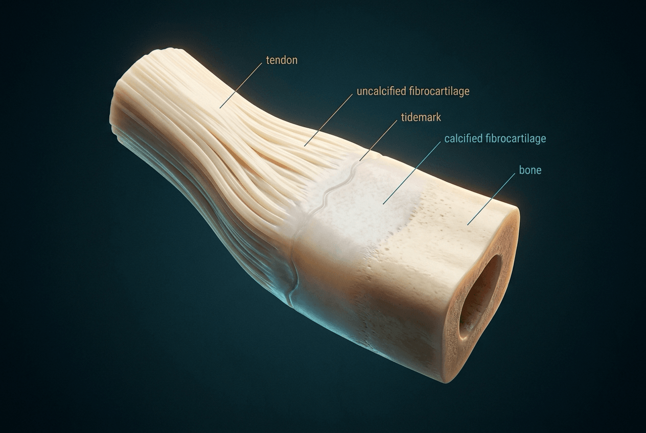
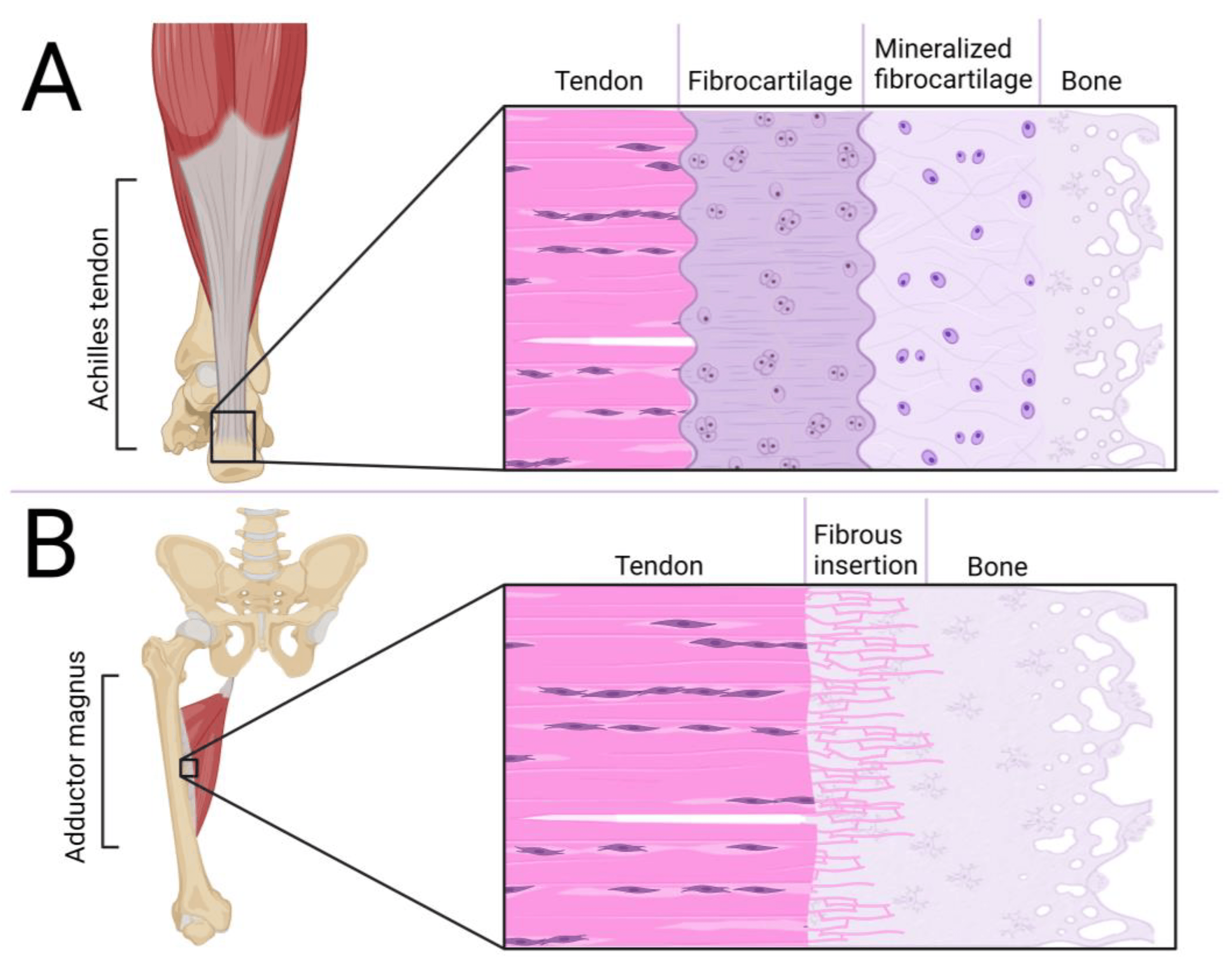
Fibrocartilaginous enthesis. Most major load-bearing tendons, including the Achilles and the rotator cuff footprint, insert through a fibrocartilaginous enthesis. It is a transitional zone about 1 mm long in which material properties change gradually from compliant tendon to rigid bone, the elastic modulus rising from about 200 MPa in tendon to 20 GPa in cortical bone. The gradual transition minimises the stress concentration an abrupt interface would create. Histology shows four zones:
- Tendon: aligned type I collagen fibres with elongated tenocytes in longitudinal rows
- Uncalcified fibrocartilage: rounded, chondrocyte-like cells in a matrix of type II collagen, aggrecan and other cartilage-associated molecules, providing compressive resilience
- Calcified fibrocartilage: similar cells and matrix with hydroxyapatite mineral deposition; the tidemark, a basophilic line on histology, marks the boundary between zones 2 and 3
- Bone: mineralised bone with osteocytes in lacunae, into which tendon collagen fibres continue as Sharpey fibres and anchor the tendon
Fibrous (periosteal) enthesis. Some tendons attach directly to periosteum with no intervening fibrocartilage. These attachments are more common where there is little compressive loading and at low-load sites, include portions of the rotator cuff insertion on the greater tuberosity, and are generally weaker and more susceptible to avulsion.
Enthesis disease.
- Insertional tendinopathy: degeneration of the enthesis fibrocartilage, often with calcification, bone marrow oedema and sometimes an enthesophyte; common at the Achilles insertion and the common extensor origin at the lateral epicondyle
- Enthesitis: inflammation at the enthesis, characteristic of the seronegative spondyloarthropathies (ankylosing spondylitis, psoriatic arthritis, reactive arthritis); several sites may be affected at once
- Avulsion: acute failure through bone (avulsion fracture, most common in children, whose bone is weaker), through fibrocartilage (common in adults) or through tendon substance (rare, and a sign of severe degeneration)
Why enthesis repair disappoints. The zonal architecture cannot be recreated. A repaired enthesis heals with fibrovascular scar in which zones 2 and 3 are absent, the gradient in mechanical properties is not restored and stress concentrates at the repair site. This explains the persistent mechanical deficits and high re-tear rates after rotator cuff repair.
Biomechanics
How it is measured. Standard testing grips the tendon ends and applies a uniaxial tensile load while recording force and displacement. The resulting stress-strain curve defines the material properties:
- Elastic (Young's) modulus: the slope of the linear region, typically 1-2 GPa
- Ultimate tensile strength: the maximum stress sustained, typically 50-100 MPa in healthy young adults
- Toe-region modulus: the initial low stiffness, which is highly variable
- Energy absorption: the area under the curve to failure
Toe region (0-2% strain). The crimp straightens and the tendon elongates easily with little stress, in a nonlinear, compliant way. This allows initial joint motion without high muscle forces, and the crimp buffers the fibrils against high stress as loading begins.
Linear region (2-4%). With the crimp gone, the fibres bear load directly and stress rises approximately linearly; the slope is the modulus. Deformation here is recoverable, and most physiological loading occurs in this elastic range.
Yield (4-8%). Beyond about 4% microscopic damage accumulates: fibrils and fascicles slide on one another, some cross-links rupture and permanent deformation develops. This is subclinical injury that may heal with rest or, with continued loading, progress to clinical tendinopathy or rupture.
Failure (over 8%). Fibres rupture progressively and macroscopic tearing develops. Complete rupture typically occurs between 8% and 15% strain, with considerable variation by age, conditioning and prior pathology.
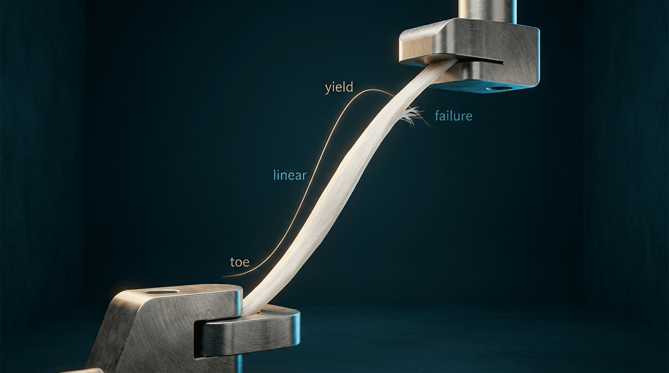
Strength and the point of failure. Tensile strength varies considerably with anatomical site, age and loading history, and the Achilles, which carries up to 3 kN during running, is particularly strong. Failure does not happen at the crimp-to-linear transition, which at about 2% strain lies well inside the elastic range; rupture comes beyond the yield point, once microscopic damage has accumulated.
Most ruptures are not this curve. Spontaneous rupture is rarely a healthy tendon meeting an excessive load. In the largest histopathological series of its kind, 891 patients with spontaneously ruptured tendons against 445 age- and sex-matched cadaveric controls, not one ruptured tendon showed a healthy structure, and 97% of the changes were degenerative: hypoxic degenerative tendinopathy, mucoid degeneration, tendolipomatosis and calcifying tendinopathy, alone or combined. The same changes appeared in 34% of control tendons, but significantly less often.
So the healthy curve is the wrong model for most clinical ruptures. In degenerate tissue the material properties are already compromised by more type III collagen, matrix disorganisation and loss of cross-links, so rupture occurs at lower absolute loads and often at lower strain. In the young, with genuine sudden overload, the tear is usually mid-substance; with chronic degeneration it tends to occur in the degenerate zone or near the insertion.
Creep. Tendon is viscoelastic: its response depends on time and on loading rate. Under constant load the tendon continues to elongate slowly, at a rate that depends on load magnitude and tissue composition. Bracing, splinting and serial casting use sustained gentle tension to achieve gradual lengthening and correct contracture.
Stress relaxation. Held at a fixed length, the stress needed to maintain that length falls as the tissue relaxes. At surgical repair, initial suture tension decreases over minutes to hours, which may lead to gap formation if inadequate tension was applied.
Hysteresis and rate dependence. The loading and unloading curves do not coincide, and about 10-15% of the energy of each cycle is lost as heat through internal friction. Tendon is also stiffer and stronger when loaded rapidly, which is why explosive movements generate higher forces and greater injury risk than slow controlled ones.
What changes the properties.
- Age: strength peaks in the third to fourth decade and then declines; stiffness may increase despite the loss of strength
- Loading history: training increases cross-sectional area, stiffness and strength, and immobilisation causes rapid deterioration
- Hydration: water content affects viscoelastic properties, and dehydrated tendon is stiffer and more brittle
- Temperature: warm-up increases compliance and may reduce injury risk; cold tendon is stiffer
- Anatomical site: high-load tendons such as the Achilles and patellar tendon are stronger than low-load tendons
These principles guide rehabilitation, surgical technique and counselling about return to activity.
Positional and energy-storing tendons. This functional division predicts which tendons are injury-prone. Positional tendons, such as the digital extensors and many forearm tendons, chiefly transmit muscle force to position a joint precisely; they operate at relatively low strains, are comparatively stiff for their size and rarely rupture spontaneously. Energy-storing tendons, the Achilles in humans and the equine superficial digital flexor tendon (SDFT), act as biological springs: they store elastic strain energy in the stretch phase of gait and return it at push-off, markedly improving the economy of locomotion.
Why energy-storing tendons fail. They are loaded to high strains close to their functional limit, show greater hysteresis, and depend on an efficient, compliant interfascicular matrix that lets fascicles slide and recoil after each cycle. With little safety margin they accumulate fatigue microdamage and are disproportionately prone to tendinopathy and rupture; the equine SDFT is the classic experimental model. Ageing and degeneration stiffen the interfascicular matrix and reduce its sliding and recoil, eroding the margin further.
Positional tendons transmit force at low strain and rarely rupture. Energy-storing tendons (Achilles, equine SDFT) are springs working at high strain with little safety margin, relying on a compliant, fatigue-resistant interfascicular matrix for recoil, and they are the tendons that most commonly become tendinopathic and rupture.
Maturation, Ageing and Degeneration
Development. In fetal life and early childhood tendons are highly cellular, with abundant tenoblasts actively synthesising matrix; fibril diameter is small and relatively uniform, and the ratio of type III to type I collagen is higher than in adults. With skeletal maturation tenocyte density falls, cells become more elongated and synthetic activity declines, while fibril diameter increases and becomes more heterogeneous and cross-links become denser and more mature, enhancing tensile strength. By late adolescence composition and mechanical properties are those of the adult.
Adult homeostasis. In healthy young adults, approximately 20 to 40 years old, matrix turnover is slow, with synthesis and degradation in balance. Strength, stiffness and fatigue resistance are optimal, and physiological loading maintains tenocyte homeostasis and matrix integrity.
Ageing. From approximately the fourth decade, progressive degenerative change sets in:
- Cells: tenocyte density falls by 30-50%, and the remaining cells become more rounded, less metabolically active and show senescent phenotypes, with decreased responsiveness to mechanical stimuli
- Organisation: total collagen may decrease slightly, but more importantly interfibrillar and interfascicular disarray increases and fibril diameter becomes more heterogeneous. Some cross-links degrade while others form aberrantly, leaving areas of both increased stiffness and weakness.
- Composition: type III collagen increases in some regions, particularly around microdamage; decorin falls and large aggregating proteoglycans rise, altering hydration and mechanical properties
- Vessels: vascularity decreases further in already hypovascular regions, which may impair healing, although vascular density may paradoxically increase in pathological regions (neovascularisation)
- Mechanics: the elastic modulus may increase while ultimate tensile strength falls, so the tendon is both less compliant and more fragile, and declining fatigue resistance increases susceptibility to cumulative microdamage
The rupture epidemiology follows. Tendon rupture is bimodal. Young athletes suffer acute traumatic ruptures of normal tendons under extreme loads; middle-aged and older people rupture degenerate tendons under moderate loads, sometimes spontaneously during routine activity. Degeneration is not uniform: the Achilles, rotator cuff and patellar tendon are particularly susceptible, whereas most flexor tendons rarely rupture even in the elderly, reflecting differences in loading, vascular supply and possibly genetic factors.
Mechanobiology
Tendon adapts to load. Tendons are exquisitely responsive to mechanical stimuli, and their loading pattern profoundly shapes structure, composition and mechanical properties, so that architecture is optimised for the demands placed on it. Optimal loading, of appropriate magnitude, frequency and duration, maintains homeostasis and promotes adaptation:
- Tenocytes upregulate type I collagen synthesis
- Fibril diameter increases and becomes more uniform
- Cross-link density and maturity improve
- Proteoglycan expression is optimised for current loading
- Ultimate tensile strength, elastic modulus and fatigue resistance increase
Athletes who train consistently have tendons with 10-30% greater cross-sectional area, higher stiffness and superior mechanical properties compared with sedentary controls. The adaptation is tendon-specific: only loaded tendons show it.
Underloading. Without mechanical stimulus, tendon degrades rapidly:
- Matrix synthesis falls while degradation continues, with net collagen loss and a smaller cross-sectional area
- Fibril diameter becomes smaller and more uniform
- Cross-links degrade, reducing tensile strength
- Glycosaminoglycan content increases, altering viscoelastic properties
- Mechanical properties deteriorate within weeks
In animal studies, 8-12 weeks of immobilisation reduced ultimate tensile strength by 30-50%. Recovery takes months of rehabilitation and some deficits may be permanent. This explains tendon complications after prolonged casting or bedrest, and it is why early controlled mobilisation after tendon injury and repair is emphasised when feasible.
Overloading. Acute overload beyond yield strain causes microscopic damage, including fibril rupture, interfibrillar separation and matrix disruption, and if severe progresses to macroscopic tearing or complete rupture. Chronic repetitive loading without adequate recovery can produce cumulative microdamage that exceeds repair capacity, and the result is tendinopathy. The transition from physiological adaptation to pathological degeneration remains incompletely understood, but likely involves exceeding cellular repair capacity, ischaemia-reperfusion injury, inflammatory mediator release and genetic susceptibility.
Tendinopathy and Healing
Tendinopathy is a failed healing response to cumulative microdamage. Its features:
- Matrix: collagen disorganisation with loss of parallel fibre alignment; increased type III collagen; an altered proteoglycan profile with more aggrecan and other large aggregating proteoglycans; mucoid or hyaline degeneration in chronic cases and fatty infiltration in severe ones; regional matrix necrosis creating focal defects
- Cells: hypercellularity in early (reactive) tendinopathy and hypocellularity in chronic degeneration; an altered tenocyte phenotype with increased MMP production; chondrocyte-like cells indicating metaplasia; increased apoptosis creating hypocellular areas
- Vessels and nerves: neovascularisation, particularly at the margins of pathological tissue, with neoinnervation as the pain substrate; the increase in vascularity is paradoxical given ischaemic theories of its aetiology
- Mechanics: ultimate tensile strength reduced by 30-50%, stiffness increased in some cases despite the loss of strength, impaired fatigue resistance and increased rupture risk
Healing passes through overlapping phases:
- Inflammatory (0-7 days): haematoma with platelet activation; neutrophils and then macrophages infiltrate; cytokines and growth factors (TGF-β, PDGF, VEGF) are released; tenocytes activate and begin to proliferate
- Proliferative (7 days to 6 weeks): abundant type III collagen, rapid but weak; high cellularity with tenoblast-like cells; neovascularisation; disorganised matrix
- Remodelling (6 weeks to 12 months or more): type III collagen gradually and incompletely replaced by type I; cellularity falls towards normal; fibres align along lines of stress; cross-links mature; strength improves but rarely reaches normal
The ceiling on repair. Healed tendon achieves only 50-80% of normal strength. The collagen ratio rarely returns to normal, cross-link patterns and fibril diameter distribution stay altered, crimp is disrupted or absent, and the scar lacks the normal hierarchical organisation. The reasons are vascular (vessels only 1-2% of the cross-section, watershed zones in critical areas, limited delivery of nutrients and oxygen) and cellular (low tenocyte density that falls further with age, and a limited stem cell population).
Classification
Tendons can be classified by anatomical, functional and structural characteristics. The functional division into positional and energy-storing tendons is set out under Biomechanics, because it predicts which tendons rupture.
- Categories
- Synovial vs non-synovial
- Examples
- Synovial: flexor tendons (hand), peroneal tendons. Non-synovial: Achilles, patellar, quadriceps tendons
- Clinical Relevance
- Determines blood supply pattern and tenosynovitis risk
- Categories
- Flexor vs extensor vs stabiliser
- Examples
- Flexors: FDP, FDS, biceps, Achilles. Extensors: EDC, quadriceps, triceps
- Clinical Relevance
- Flexors generally thicker with higher tensile loads
- Sheath
- Synovial
- Loading
- High tension + friction
- Blood Supply
- Vincula (limited)
- Healing Challenge
- Adhesion formation in sheath
- Sheath
- Paratenon only
- Loading
- Highest tensile loads (up to 12 x body weight)
- Blood Supply
- Watershed zone 2-6 cm from insertion
- Healing Challenge
- Mid-substance hypovascularity
- Sheath
- Bursal surface
- Loading
- Tension + compression (acromion)
- Blood Supply
- Critical zone near insertion
- Healing Challenge
- Enthesis cannot regenerate zonal structure
- Sheath
- Paratenon (anterior knee fat pad)
- Loading
- High eccentric loads
- Blood Supply
- Fat pad contribution
- Healing Challenge
- Jumper's knee at inferior pole
Clinical Assessment
History. Each symptom maps onto a structural change:
- Activity-related pain suggests loading beyond the yield point
- Morning stiffness reflects altered proteoglycan content affecting tissue hydration
- Weakness reflects partial disruption or inhibition
- Swelling reflects the inflammatory phase or neovascularisation
- A sudden pop means macroscopic fibre rupture
Risk factors are age (reduced cellularity and matrix quality), previous tendon pathology (pre-existing type III collagen), fluoroquinolone use (inhibited collagen synthesis) and systemic disease such as diabetes, rheumatoid arthritis and renal disease.
Inspection. Swelling may be localised (sheath) or diffuse (paratenon). Look for deformity from tendon discontinuity or retraction, muscle wasting from chronic denervation or disuse, and skin changes such as steroid injection sites or surgical scars.
Palpation.
- Tenderness localises the pathology to a segment; the hypovascular Achilles watershed, 2-6 cm from the insertion, is commonly tender in tendinopathy
- A palpable gap means rupture
- Thickening reflects chronic tendinopathy with increased type III collagen
- Crepitus reflects tenosynovitis, inflammation of the synovial sheath
- Technique
- Squeeze calf, observe plantar flexion
- Positive Finding
- No plantar flexion = rupture
- Structural Basis
- Tendon discontinuity disrupts force transmission
- Technique
- Resist abduction in 90° forward flexion, internal rotation
- Positive Finding
- Weakness or pain
- Structural Basis
- Enthesis pathology at critical zone
- Technique
- Block adjacent fingers, test DIP/PIP flexion
- Positive Finding
- Loss of active flexion at specific joint
- Structural Basis
- Individual tendon integrity assessment
Pain and the stress-strain curve. When in the loading cycle the pain arises is a guide to what is damaged:
- Pain on initial movement (toe region) suggests crimp pattern disruption
- Pain under sustained load (linear region): a collagen fibre loading abnormality
- Symptoms at high load (yield region): the microscopic damage threshold exceeded
Dynamic tests. Pain with eccentric load suggests the matrix cannot handle normal deformation; the tests are heel drops for Achilles tendinopathy, eccentric wrist extension for lateral epicondylitis and decline squats for patellar tendinopathy. Proprioception depends on the tendon mechanoreceptors, and impaired proprioception may indicate neural involvement; balance testing assesses it in lower limb tendon disorders.
Differential diagnosis. A painful tendon region has several mimics. Distinguishing degenerative tendinopathy from inflammatory enthesitis, complete rupture and referred pain is essential because management differs fundamentally.
- Key Feature
- Load-related pain, focal thickening, intact function
- Structural Basis
- Increased type III collagen, matrix disorganisation, neovascularisation
- Discriminator
- Hypoechoic/neovascular on ultrasound; no gap; pain with eccentric load
- Key Feature
- Sudden pop, weakness, palpable gap
- Structural Basis
- Macroscopic fibre discontinuity (usually on degenerate tendon)
- Discriminator
- Positive Thompson/Simmonds test; gap on ultrasound or MRI
- Key Feature
- Multiple sites, inflammatory pattern, morning stiffness
- Structural Basis
- Inflammation at the fibrocartilaginous enthesis
- Discriminator
- HLA-B27, systemic features, bone marrow oedema at enthesis on MRI
- Key Feature
- Acute severe pain, often shoulder
- Structural Basis
- Hydroxyapatite deposition within tendon substance
- Discriminator
- Calcific deposit on radiograph; hyperechoic focus with shadowing
- Key Feature
- Pain with crepitus along sheathed tendon
- Structural Basis
- Inflammation of synovial sheath, not tendon substance
- Discriminator
- Sheath fluid on ultrasound; positive Finkelstein (de Quervain)
- Key Feature
- Pain not reproduced by direct tendon loading
- Structural Basis
- Nerve root or proximal pathology, tendon structurally normal
- Discriminator
- Normal tendon imaging; neural tension signs; dermatomal pattern
Investigations
Imaging findings correlate directly with the structural changes in tendon composition.
- Principle
- Echogenicity reflects collagen organisation
- Normal Finding
- Hyperechoic, fibrillar pattern
- Pathological Finding
- Hypoechoic areas (matrix disruption), neovascularisation (Doppler)
- Advantages
- Dynamic assessment, low cost, no radiation
- Principle
- Water content and collagen orientation
- Normal Finding
- Low signal on all sequences (organised collagen)
- Pathological Finding
- Increased T2 signal (oedema, mucoid change), discontinuity (rupture)
- Advantages
- Excellent soft tissue detail, multiplanar imaging
- Principle
- Only detects calcification/ossification
- Normal Finding
- Soft tissue shadow only
- Pathological Finding
- Calcific tendinopathy, enthesophytes, avulsion fragments
- Advantages
- Readily available, low cost, bone assessment
Ultrasound. Hypoechoic areas represent collagen disorganisation, increased type III collagen with thinner fibrils, mucoid degeneration and loss of parallel fibre alignment. Hyperechoic foci are calcific deposits or chronic degenerative change.
Power Doppler. Normal tendon is avascular on Doppler. In tendinopathy the Doppler signal shows neovascularisation, which accompanies the neoinnervation that is the pain source and correlates with symptom severity. The signal is a target for sclerosing injections and may guide eccentric loading protocols.
- Normal Tendon
- Low signal (dark)
- Tendinopathy
- Intermediate signal (mucoid change)
- Rupture
- Gap with retraction, haemorrhage (variable signal)
- Normal Tendon
- Low signal (dark)
- Tendinopathy
- Increased signal (oedema, proteoglycan change)
- Rupture
- Fluid signal in gap, surrounding oedema
- Normal Tendon
- Low signal
- Tendinopathy
- Subtle signal changes, good for morphology
- Rupture
- Excellent for partial thickness tears
Magic angle artefact. At 55° to the main magnetic field, normal tendon shows false-positive signal. It matters at entheses and in curved tendon regions, and is avoided by imaging at 0° or with appropriate sequences.
Research techniques.
- Ultrasound tissue characterisation (UTC) quantifies the distribution of echo types; types I-IV correlate with collagen integrity, allowing objective monitoring of the healing response
- Shear wave elastography measures tissue stiffness (elastic modulus); a stiffer tendon may indicate chronic change, and it can detect subclinical pathology
Laboratory tests. Serum biomarkers remain research tools, not yet validated for clinical use: COMP (elevated in tendinopathy), PICP and PINP (collagen synthesis) and MMP-3 (matrix degradation). Histopathology of surgical specimens shows the tendinopathy changes described above: an elevated type III to type I ratio, altered decorin and aggrecan, cellularity that is high early and low late, neovascularisation and mucoid degeneration.
Management
Load management. Management follows the healing phase and the stress-strain curve. Keep loads within the toe and linear regions, under 4% strain, avoid yield-zone loading during active pathology, and progress load gradually to stimulate matrix synthesis.
Eccentric loading. It stimulates tenocyte collagen production, promotes type I over type III synthesis and improves fibril alignment and cross-linking, and the evidence is strongest for Achilles and patellar tendinopathy. Conservative management still fails to restore normal structure, which is why recurrence is common and why athletes may not return to pre-injury performance.
Drugs.
- NSAIDs may impair collagen synthesis in early healing. Use them short term for pain and avoid them during the proliferative phase if possible.
- Corticosteroid injection gives short-term pain relief but directly inhibits tenocyte collagen synthesis, reduces proteoglycan production and shifts the MMP/TIMP balance towards degradation. It is associated with tendon weakening and rupture, the risk is highest with repeated injections, and repeated injections should be avoided.
- Goal
- Control inflammation, protect repair
- Management
- Relative rest, ice, compression, elevation
- Biological Basis
- Allow cytokine signalling and cell recruitment
- Goal
- Stimulate matrix synthesis
- Management
- Protected early motion, gentle loading
- Biological Basis
- Mechanical stimulation promotes type I collagen
- Goal
- Improve matrix organisation, restore strength
- Management
- Progressive loading, eccentric exercise, sport-specific training
- Biological Basis
- Stress along fibre axis promotes alignment and cross-linking
Platelet-rich plasma. Biological adjuvants such as PRP and stem cells have theoretical benefits but limited clinical evidence. PRP concentrates growth factors (TGF-β, PDGF, VEGF), stimulates tenocyte proliferation and matrix synthesis, and may promote angiogenesis. Trial results conflict; it may benefit chronic tendinopathy, the optimal preparation is unclear, and it is not standard of care.
Stem cells. The sources are bone marrow aspirate concentrate (BMAC), adipose tissue and tendon-derived progenitor cells. The multipotent cells may differentiate into tenocytes, and they act through paracrine effects on resident cells and modulation of the inflammatory response. The data are primarily preclinical and early clinical, and regulatory considerations limit widespread use.
Physical modalities.
- Extracorporeal shockwave therapy (ESWT) mechanically stimulates tissue and may promote neovascularisation and growth factor release. The evidence supports it in calcific tendinopathy, with mixed results in non-calcific tendinopathy.
- Low-level laser therapy may modulate cellular activity; high-quality evidence is limited, and it is generally considered safe.
Indications for surgery.
- Failed conservative management, typically after 6 months or more
- Complete tendon rupture
- Significant structural pathology, such as large partial tears
- Mechanical symptoms requiring debridement
- Insertional pathology with bony impingement
Surgical Technique
Strength starts with the suture. Initial repair strength depends entirely on the suture-tendon interface, which is the weakest link at first, and biological healing adds strength incrementally over months. Early mobilisation protocols balance the stimulus to healing against the risk of gap formation.
Core suture.
- More strands give more strength (2-strand vs 4-strand vs 6-strand)
- Grasping sutures are stronger than locking sutures
- A purchase of 2-3 mm from the cut end is optimal
Epitendinous suture. A running circumferential suture that prevents bunching and gap formation, adds 10-50% to repair strength and improves the gliding surface.
Handling. Avoid crushing the tendon with forceps, which damages tenocytes. Preserve the paratenon or sheath for blood supply and, in flexor tendon repair, the vincula, and limit dissection to maintain the vascular pedicles.
Gaps. A gap of more than 2-3 mm is associated with adhesion and weakness, so suture tension at repair has to be adequate.
- Excessive suture tension causes tissue necrosis and failure
- Inadequate suture purchases lead to pullout
- Sheath damage promotes adhesion formation
- Gap formation greater than 3 mm correlates with poor outcomes
- Ignoring stress relaxation leads to late gap formation
- Strength Source
- Suture only
- Biological Contribution
- Minimal (inflammatory phase)
- Clinical Implication
- Protected motion, avoid resistance
- Strength Source
- Suture + early callus
- Biological Contribution
- Type III collagen synthesis (weak)
- Clinical Implication
- Gradual active motion, still protected
- Strength Source
- Biological healing dominant
- Biological Contribution
- Type I collagen increasing, cross-linking
- Clinical Implication
- Progressive resistance, light activity
- Strength Source
- Remodelled matrix
- Biological Contribution
- Improved organisation, mature cross-links
- Clinical Implication
- Return to sport, max 50-80% original strength
- Strength
- High tensile strength
- Handling
- Good knot security
- Disadvantage
- May cause tissue reaction
- Strength
- Very high tensile strength
- Handling
- Excellent knot security
- Disadvantage
- Stiff, potential abrasion
- Strength
- Moderate
- Handling
- Smooth, less tissue reaction
- Disadvantage
- Knot may loosen
Enthesis repair. The technical approaches are:
- Double-row repair increases footprint coverage
- Suture-bridge techniques compress tendon to bone
- Transosseous-equivalent configurations
- Biological augmentation (PRP, patches) is under investigation
Mechanical augmentation with suture tape or mesh can reduce the risk of failure but may stress-shield the healing tissue.
Grafts.
- Autograft: palmaris longus (if present) and plantaris for hand reconstruction, hamstrings for ACL reconstruction, and an Achilles turndown for Achilles reconstruction. Initial strength varies by donor site, and patellar tendon is stronger than hamstring.
- Allograft requires incorporation and remodelling, with a temporary loss of strength during revascularisation and slower healing than autograft, but no donor site morbidity.
- Synthetic grafts lack the capacity for biological integration.
Complications
Repair failure. Sutures pull out through degenerative tissue, stress relaxation opens a gap, biological healing is inadequate or early loading is excessive. Type III-rich tissue in chronic tendinopathy cannot hold sutures, and repair strength is suture-dependent for the first 6 weeks.
Adhesions. Scar bridges form between the tendon and the surrounding tissues when gliding surfaces are lost through sheath disruption or immobilisation is excessive. Tendon healing produces fibrovascular scar, a disrupted synovial sheath loses its lubricating function, and motion is required to prevent adhesions maturing.
- Incidence
- 5-25% (varies by location)
- Structural Cause
- Incomplete matrix remodelling, persistent type III collagen
- Prevention
- Protected loading, adequate rehabilitation time
- Incidence
- 10-30% (higher in sheathed tendons)
- Structural Cause
- Adhesion between tendon and sheath/surrounding tissues
- Prevention
- Early controlled motion protocols
- Incidence
- Common (most repairs)
- Structural Cause
- Healed tendon 50-80% original strength, altered collagen ratio
- Prevention
- Progressive loading to optimise remodelling
Fluoroquinolone-associated tendinopathy. Fluoroquinolones inhibit tenocyte metabolic activity, reduce collagen and proteoglycan synthesis and increase MMP production. The risk factors are age, steroid use and renal impairment.
Postoperative Care
Postoperative protocols are built on the phases of tendon healing and the stress-strain curve. The timing balances the risk of adhesion from too little motion against the risk of repair failure from too much load.
- Goals
- Protect repair, prevent adhesions
- Activities
- Immobilisation or controlled passive motion
- Biological Rationale
- Suture-only strength; early motion prevents adhesion
- Goals
- Stimulate healing, prevent stiffness
- Activities
- Active-assisted motion, gentle active motion
- Biological Rationale
- Mechanical loading promotes type I collagen synthesis
- Goals
- Progressive loading, restore function
- Activities
- Progressive resistance, light activity
- Biological Rationale
- Stress along fibre axis promotes alignment and cross-linking
- Goals
- Sport-specific training, full function
- Activities
- Gradual return to sport, plyometrics
- Biological Rationale
- Continued remodelling, max 50-80% original strength achieved
Early controlled motion. It prevents adhesions from forming and maturing, stimulates tenocyte collagen synthesis, promotes aligned collagen deposition and maintains the gliding surface. In practice this means Kleinert or modified Duran protocols for flexor tendons, a controlled ankle motion (CAM) boot for the Achilles and passive motion protocols for the rotator cuff.
Progressive loading. Loading increases collagen cross-linking, promotes type I over type III collagen, improves fibril alignment and increases ultimate tensile strength, and eccentric loading may be particularly beneficial. Toe-region loading is safe and stimulates without damaging, so start there; progress to the linear region once biological healing is established; and avoid the yield zone until late remodelling, at 12+ weeks.
The loading window.
- Too little loading: matrix degradation and adhesion formation
- Optimal loading: type I collagen synthesis and organised matrix
- Excessive loading: yield-zone damage, gap formation and failure
By tendon.
- Flexor tendons (zone II): early active motion is superior to immobilisation, with fewer adhesions; true active motion protocols such as Manchester show improved outcomes; place-and-hold techniques minimise gap formation; and 6-strand repairs allow earlier active motion
- Achilles: functional rehabilitation with early weight-bearing gives re-rupture rates similar to cast immobilisation with a faster return to activity; progressive eccentric loading (the Alfredson protocol) is used for tendinopathy
- Rotator cuff: early passive motion to prevent stiffness, active motion delayed to 6 weeks to protect the repair, and larger tears may require longer protection
Outcomes
- Return to Activity
- 6-12 months
- Re-rupture Rate
- 5-10% (surgical)
- Functional Outcome
- Good; most return to sport
- Limiting Factor
- Watershed zone vascularity
- Return to Activity
- 6-12 months
- Re-rupture Rate
- 40-60% (imaging)
- Functional Outcome
- Variable; pain relief often achieved despite re-tear
- Limiting Factor
- Enthesis healing, fatty infiltration
- Return to Activity
- 3-6 months
- Re-rupture Rate
- 5-10%
- Functional Outcome
- Good with modern protocols; adhesions common
- Limiting Factor
- Adhesion formation in sheath
Counselling. Expectations must be realistic because the biology sets a ceiling. Healing is prolonged, with remodelling continuing for 12 months or more, and full recovery of pre-injury strength is unlikely because collagen composition and organisation stay altered. Discuss activity modification if needed, warn that high-level athletes may not return to their previous level, and acknowledge that the risk of re-injury remains elevated compared with uninjured tissue.
What predicts the result. Outcome is better with young age (higher cellularity, better vascularity), an acute injury with less pre-existing degeneration, good tissue quality at repair, no smoking, compliance with rehabilitation and no systemic disease. It is worse with older age (decreased tenocyte function), chronic tendinopathy (elevated type III collagen), poor tissue quality from degeneration, smoking (which impairs collagen synthesis), diabetes (impaired healing), a large tear, and muscle atrophy or fatty infiltration.
Outcome measures.
- Clinical scores: VISA-A (Achilles tendinopathy), DASH (upper limb function), Constant score (shoulder), Buck-Gramcko score (flexor tendon)
- Objective measures: range of motion (adhesion assessment), strength testing (dynamometry), hop testing (lower limb function), ultrasound tissue characterisation (research)
Guidelines, Registries & Global Practice
Global Epidemiology of Tendon Disorders
Tendon disease is one of the commonest reasons for musculoskeletal consultation worldwide. The clinically dominant condition affecting tendon structure is the Achilles tendon, where nationwide registry data show a clear rise in rupture incidence over recent decades, concentrated in an ageing population - a direct epidemiological consequence of the age-related degenerative changes described above.
- Period
- 1997 to 2019
- Incidence
- Rose from 17.3 to 32.3 per 100,000 person-years
- Trend
- Rise greatest in the elderly; operative rate fell
- Period
- 2010 to 2017
- Incidence
- Stable at 12.8 to 13.9 per 100,000 (men higher than women)
- Trend
- Incidence flat; surgical proportion rose to 72%
Practice variation: Achilles rupture incidence and the operative-versus-nonoperative balance differ markedly by region. Scandinavian and Finnish registries show rising incidence with falling surgery rates, whereas Japanese national data show stable incidence but rising surgery rates - emphasising that treatment culture, not just biology, drives practice.
Guideline Positions on Tendinopathy and Tendon Injury (Side by Side)
Tendon structure and composition is a basic-science topic, so few bodies issue disease-specific guidelines, but several professional organisations publish positions relevant to tendon-loading pathology and rupture.
- Scope
- Acute Achilles tendon rupture management standards
- Position
- Functional rehabilitation; shared decision-making on operative vs non-operative
- Evidence level
- Consensus standard informed by RCT evidence
- Scope
- Clinical practice guidance on Achilles rupture / rotator cuff
- Position
- Both operative and non-operative acceptable; early motion encouraged
- Evidence level
- Mixed (limited to moderate) recommendations
- Scope
- Tendinopathy and soft-tissue injury pathways
- Position
- Exercise-based loading first-line; restrict corticosteroid injection
- Evidence level
- Guideline / pathway level
- Scope
- Load-induced tendinopathy
- Position
- Progressive loading (eccentric/isometric) first-line; injections adjunctive
- Evidence level
- Level I-II for eccentric loading in Achilles/patellar
- Progressive mechanical loading is first-line for tendinopathy
- Corticosteroid injection discouraged for long-term tendon management
- Functional rehabilitation increasingly preferred over rigid immobilisation
- Shared decision-making for Achilles rupture (operative vs non-operative)
- Operative rate for Achilles rupture varies widely by country
- Role of biologics (PRP, BMAC) unresolved and regionally variable
- Imaging access (early MRI/ultrasound) differs by health system
- Rehabilitation protocol intensity and supervision differ internationally
MCQ Practice Points
Q: What is the hierarchical structure of tendon and what is the predominant collagen type?
A: Tendon has hierarchical structure: Collagen molecule (tropocollagen) assembles into microfibrils, which form subfibrils, then fibrils (visible on EM), then fibers (visible on light microscopy), then fascicles (surrounded by endotenon), then the whole tendon (surrounded by epitenon and paratenon). The predominant collagen is Type I (95%) with small amounts of Type III (increases in healing/degeneration), Type V (regulates fibril diameter), and Type XII. Collagen fibrils have a characteristic crimp pattern (wave-like) that allows initial elongation without structural damage.
Q: What is the role of the endotenon, epitenon, and paratenon in tendon structure?
A: Endotenon: Loose connective tissue surrounding individual fascicles; carries blood vessels, lymphatics, and nerves; allows fascicles to glide against each other. Epitenon: Dense connective tissue surrounding the entire tendon; continuous with endotenon; contains blood vessels. Paratenon: Loose areolar tissue external to epitenon; present in tendons without synovial sheath (Achilles, patellar); allows gliding against surrounding tissues. Tendons with synovial sheath (flexor tendons in hand) have visceral and parietal layers instead of paratenon. The mesotenon (vinculum) carries blood supply.
Q: What is the crimp pattern in tendons and what is its functional significance?
A: The crimp pattern is the characteristic wavy or sinusoidal arrangement of collagen fibrils in tendon. It represents a "toe region" in the stress-strain curve - when tendon is loaded, the crimp straightens first (low stiffness), then the straightened fibers resist load (linear region, high stiffness). Functional significance: 1) Acts as shock absorber allowing initial elongation (1-2% strain) without structural damage; 2) Provides energy storage for explosive movements; 3) Loss of crimp (during chronic loading) leads to tendinosis with decreased shock absorption. Crimp is re-established during healing but may be disorganized.
Q: What is the blood supply to tendons and how does it vary along the tendon length?
A: Blood supply comes from: 1) Musculotendinous junction - muscle vessels extend into tendon; 2) Bone-tendon junction (enthesis); 3) Along the tendon - via paratenon (unsheathed tendons) or vincula/mesotenon (sheathed tendons). Watershed zones (hypovascular areas) occur where supplies meet, making these areas prone to degeneration: Achilles tendon 2-6cm proximal to insertion, supraspinatus near insertion ("critical zone"), FPL at level of sesamoids. Tendon nutrition also occurs by diffusion from synovial fluid (especially in sheathed tendons).
Q: What are the cellular components of tendon and their functions?
A: Tenocytes (90-95% of cells): Specialized fibroblasts arranged in longitudinal rows between collagen fibers; synthesize collagen and extracellular matrix; connected by gap junctions for mechanotransduction; elongated with wing-like cytoplasmic extensions. Tendon stem/progenitor cells: Small population that can differentiate into tenocytes; important for healing and regeneration. Synovial cells: Line tendon sheath, produce synovial fluid. Vascular cells: Endothelial cells, pericytes. In tendinopathy, there is increased cellularity, neovascularization, and changes from Type I to Type III collagen with disorganized matrix.
Summary
Tendons are sophisticated composite materials with hierarchical organization spanning from molecular to tissue levels. Type I collagen provides the primary structural framework, regulated by proteoglycans that control fibril assembly and mechanical properties. Tenocytes maintain matrix homeostasis and respond to mechanical loading through complex mechanotransduction pathways.
The characteristic stress-strain behavior reflects underlying structural features, with the toe region corresponding to crimp straightening and the linear region representing elastic collagen loading. Viscoelastic properties including creep, stress relaxation, and rate dependence have important clinical implications for rehabilitation and surgical decision-making.
Age-related degeneration, pathological loading, and inadequate healing capacity contribute to tendinopathy and rupture. Understanding tendon structure and composition at multiple scales is essential for comprehending disease mechanisms, interpreting imaging findings, optimizing surgical techniques, and developing effective rehabilitation protocols.
For examination purposes, focus on hierarchical organization, collagen composition (especially type I versus type III), the stress-strain curve with specific strain values, enthesis zonal anatomy, and the structural basis of tendinopathy. These topics are consistently emphasized in basic science vivas and MCQs.
Clinical Decision Scenarios
Practise clinical reasoning and management decisions out loud
“An examiner presents an electron micrograph showing striated collagen fibrils and asks you to explain the molecular basis of the banding pattern, then proceeds to question you about the hierarchical organization of tendon structure from molecular to tissue level.”
Hierarchical Organization (Smallest → Largest)
- Tropocollagen: 300 nm × 1.5 nm, triple helix, Gly-X-Y pattern
- Fibril: 20-400 nm diameter, 67 nm D-period from quarter-stagger
- Fiber: 1-20 μm diameter, crimp pattern visible (50-100 μm wavelength)
- Fascicle: 50-300 μm diameter, surrounded by endotenon
- Tendon: Multiple fascicles, epitenon + paratenon layers
Collagen Types - Know the Percentages
- Type I: 95% of total, heterotrimeric (2α1 + 1α2), primary tensile strength
- Type III: Less than 5% normally, increases in healing/tendinopathy (can reach 50%)
- Type V: 2-3%, regulates fibril diameter, in fibril core
- Type VI: Less than 1%, interfibrillar cohesion, beaded microfibrils
- Type X: At enthesis, fibrocartilaginous insertion zone
Key Proteoglycans - Regulators of Structure
- Decorin: Most abundant SLRP, regulates fibril diameter by binding at D-period sites
- Biglycan: Similar to decorin, distinct regulatory functions
- Aggrecan: In compressed regions (enthesis, wraparound zones), compressive resistance
- COMP: Compressed regions, potential healing biomarker
- All SLRPs decreased in tendinopathy → abnormal fibril geometry
Stress-Strain Curve - Must Know for Viva
- Toe region (0-2% strain): Crimp straightening, nonlinear, compliant
- Linear region (2-4% strain): Elastic collagen loading, modulus 1-2 GPa
- Yield point (~4% strain): Microscopic damage begins, permanent deformation
- Failure (8-15% strain): Macroscopic rupture, UTS 50-100 MPa
- Viscoelastic: Creep, stress relaxation, hysteresis (10-15% energy loss), rate-dependent
Enthesis - Four Zones of Insertion
- Zone 1: Tendon proper - type I collagen, longitudinal tenocytes
- Zone 2: Uncalcified fibrocartilage - type II collagen, aggrecan, chondrocytes
- Zone 3: Calcified fibrocartilage - same as zone 2 but mineralized, tidemark boundary
- Zone 4: Bone - osteocytes, Sharpey fibers anchor tendon to bone
- Gradient: Modulus 200 MPa (tendon) → 20 GPa (bone) over ~1 mm
Vascular Supply - Watershed Zones
- Generally poor vascularity (1-2% of cross-sectional area)
- Three sources: Musculotendinous junction, osseous insertion, peritendinous tissues
- Achilles watershed: 2-6 cm proximal to insertion (common rupture site)
- Supraspinatus critical zone: Near insertion, hypovascular (common tear location)
- Poor vascularity → slow healing, ischemic injury susceptibility
Age-Related Changes - Degeneration Pattern
- Decreased tenocyte density (30-50% reduction), cells become rounded
- Increased collagen disorganization, heterogeneous fibril diameter
- Increased type III collagen in damaged areas
- Decreased decorin, increased aggrecan (altered proteoglycan profile)
- Mechanical: Increased stiffness BUT decreased strength (stiff + fragile = injury-prone)
Tendinopathy Pathology - Structural Changes
- Type III collagen up to 50% (normally less than 5%)
- Loss of parallel fiber alignment, matrix disorganization
- Altered proteoglycans (decreased decorin, increased aggrecan)
- Neovascularization + neoinnervation (pain substrate)
- Mechanical deficits: 30-50% reduced UTS, impaired fatigue resistance
High-Yield Numbers for MCQs
- Tropocollagen: 300 nm long, 1.5 nm diameter
- D-period: 67 nm (quarter of 300 nm = 75 nm, but actual is 67 nm due to molecular overlap)
- Type I collagen: 95% of total collagen
- Elastic modulus: 1-2 GPa (linear region)
- UTS: 50-100 MPa, Failure strain: 8-15%
- Crimp wavelength: 50-100 μm, Toe region: 0-2% strain
Evidence Base
Key Evidence on Tendon Structure and Healing
The understanding of tendon biology is built on foundational studies in biomechanics, biochemistry, and clinical research.
Structure of the tendon connective tissue
- Tropocollagen molecules (300 nm) assemble in quarter-stagger array creating 67 nm D-period
- Type I collagen comprises 95% of normal tendon collagen content
- Decorin regulates collagen fibril diameter by binding at D-period sites
- Crimp pattern (50-100 μm wavelength) creates mechanical buffer in toe region
Biomechanics of ligaments and tendons
- Toe region (0-2% strain) represents crimp straightening
- Linear region (2-4% strain) reflects elastic collagen fiber loading with modulus 1-2 GPa
- Yield region begins around 4% strain with microscopic damage accumulation
- Failure occurs at 8-15% strain with ultimate tensile strength 50-100 MPa
Targeted Disruption of Decorin Leads to Abnormal Collagen Fibril Morphology and Skin Fragility
- Decorin-null mice are viable but have skin of markedly reduced tensile strength
- Tendon and skin collagen fibrils show coarse, irregular outlines on electron microscopy
- Quantitative scanning transmission electron microscopy showed UNCONTROLLED LATERAL FUSION of fibrils - decorin acts as a brake on fibril diameter rather than a builder of it
- Establishes a direct genotype-phenotype link between a small leucine-rich proteoglycan and matrix mechanics
Histopathological Changes Preceding Spontaneous Rupture of a Tendon - a Controlled Study of 891 Patients
- 891 patients treated between 1968 and 1989: 397 Achilles, 302 biceps brachii, 82 quadriceps and patellar, 40 extensor pollicis longus and 70 other tendons
- A HEALTHY STRUCTURE WAS NOT SEEN IN ANY spontaneously ruptured tendon, while two-thirds of the 445 controls were structurally healthy (p less than 0.001)
- 97 per cent of the pathological changes were DEGENERATIVE - hypoxic degenerative tendinopathy, mucoid degeneration, tendolipomatosis and calcifying tendinopathy, alone or combined
- The remaining 3 per cent (26 tendons) is the differential worth knowing: intratendinous foreign body, rheumatoid tendinitis, xanthoma, tumour, or a tumour-like lesion such as an intratendinous ganglion
- The same degenerative changes were found in 34 per cent of CONTROL tendons, significantly less often but far from rare - degeneration is common and silent
- The authors conclude that degenerative change is common in the tendons of people over THIRTY-FIVE, at least in an urban population