Wear Debris → Macrophage Activation → Osteolysis | HXLPE Reduces Wear 95% | Early Detection Critical
- Polyethylene wear was the primary cause of late THA failure and aseptic loosening before HXLPE shifted the failure mode to instability and infection
- Particle size 0.1-1μm is most biologically active - triggers macrophage response
- Highly cross-linked PE (HXLPE) reduces wear by over 90% compared to conventional
- Osteolysis is silent - often massive bone loss before symptoms develop
- Annual surveillance radiographs are mandatory for early detection
- “Osteolysis = biological response to wear particles, not mechanical loosening
- “Effective joint space (EJS) concept - particles access bone via capsular defects
- “HXLPE trade-off: reduced wear but lower fracture toughness and oxidation
- “Isolated liner exchange only if the shell is well fixed and the osteolysis is focal and accessible for curettage
Overview and Epidemiology
Polyethylene wear and the osteolysis it provokes were the primary mode of late failure in total hip arthroplasty. Understanding the mechanisms, prevention and management of particle disease remains fundamental to arthroplasty practice.
The conventional era. First-generation THA (Charnley) used conventional ultra-high molecular weight polyethylene (UHMWPE), and linear wear rates of 0.1-0.2 mm per year were common. By 10-15 years significant osteolysis had developed in 10-30% of cases. In the 1990s-2000s revision for osteolysis became the leading indication in many series, accounting for up to 30% of revision procedures.
Cross-linking. Highly cross-linked polyethylene (HXLPE) was introduced in the late 1990s and is now the standard bearing surface for primary THA. Osteolysis rates have fallen dramatically, to under 5% at 10-15 years, and wear-related failure is now uncommon in modern primary THA. HXLPE has shifted the failure mode from wear to instability and infection, and in the AOANJRR, which tracks bearing performance, revision for wear and osteolysis has declined significantly since HXLPE adoption.
Why it still matters. Conventional polyethylene THAs implanted from the 1980s to the 2000s are still in situ and continue to present with osteolysis, and their likelihood of revision increases with time. Many of these patients are asymptomatic until catastrophic failure, so surveillance is mandatory.
Accelerated wear. Wear is faster with any of the following:
- Young, active patients with a high activity level
- Larger femoral heads, which travel a greater linear distance
- Thin polyethylene liners, under 6 mm minimum thickness
- Malpositioned components causing impingement or edge loading
- Third-body debris: PMMA, metal, bone cement fragments
Anatomy and Pathophysiology of Particle Disease
Osteolysis is the biological response to wear particles, not mechanical loosening, which is why it is called particle disease. Sub-micron polyethylene (or metal) wear particles generated at the bearing surface are phagocytosed by macrophages, and the cascade below follows.
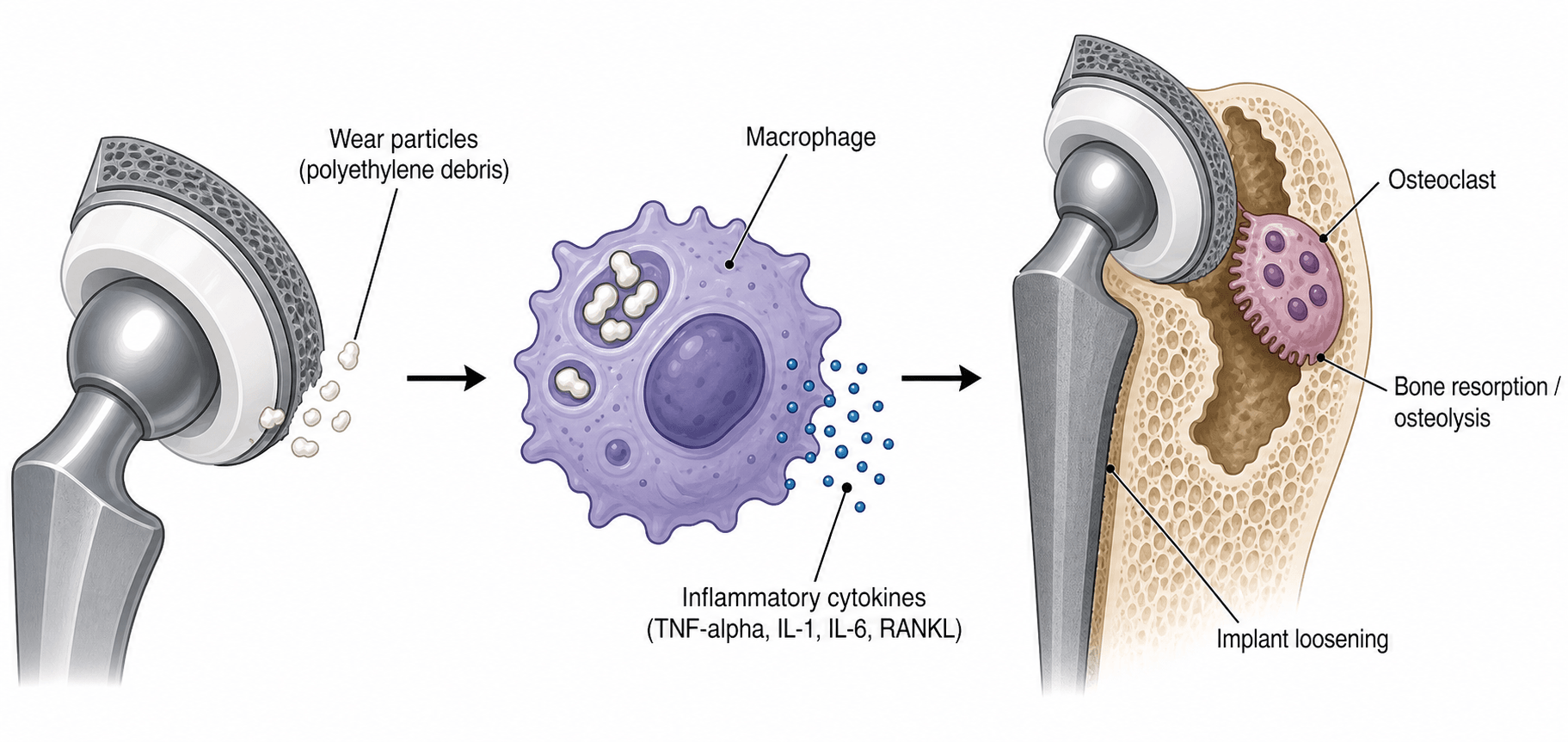
Three mechanisms. Polyethylene particles are generated at the articular surface in three ways, which contribute different shares of total wear:
- Adhesive wear, 30-40%. Molecular bonding between the bearing surfaces during contact transfers material from the polyethylene to the metal or ceramic head, and continuous loading and unloading releases particles.
- Abrasive wear, 40-50%. The hard femoral head ploughs through the soft liner, creating scratches, grooves and deformation. A roughened femoral head accelerates it.
- Third-body wear, 10-30% but highly variable. Interposed particles (PMMA cement, metal debris, bone) act as abrasives and can increase wear rates 10-100 fold. Common sources are cement from acetabular preparation and metal from impingement.
Particle size. Particles in the 0.1-1 micrometre range are the most biologically active: this size optimally activates macrophages. Smaller particles (under 0.1 μm) are phagocytosed less, and larger particles (over 1 μm) trigger less inflammatory response. Conventional polyethylene wear generates billions of particles in this critical range, and particle number, size and total volume together drive the response.
Cross-linking reduces the volume of polyethylene lost, but it also shifts the debris smaller, further into the 0.1-1 micrometre band that macrophages respond to most strongly. Per unit volume, HXLPE debris is therefore at least as biologically potent as conventional debris, and the review cited on this page warns explicitly that reducing particle size may increase biological activity. HXLPE works despite this: the fall in total volume, roughly five-fold in the 20-year randomised data, is large enough to outweigh any gain in potency per particle, so the net biological burden drops.
Two things follow. A bearing is judged on total biological burden, not linear wear rate alone, which is why volumetric wear rather than head penetration is the quantity that matters. And a low-wearing bearing still wears: patients with HXLPE still require surveillance, because osteolysis has been described with cross-linked liners where wear was elevated by malposition, impingement or third-body damage.
Backside Wear: The Other Particle Source
The three classic wear mechanisms describe the articular bearing surface. A modular acetabular liner also has a non-articular backside that abuts the metal shell, and micromotion between liner and shell generates particles from this surface: backside wear.
It is easily forgotten yet clinically important, because these particles are delivered straight to bone through the acetabular screw holes, the very conduits of the effective joint space. That makes backside wear a major and often-underappreciated driver of acetabular osteolysis. Monoblock or cemented-liner constructs remove the micromotion interface altogether.
- Articular (bearing) wear
- Head-on-liner articulation
- Backside wear
- Non-articular liner-shell interface
- Articular (bearing) wear
- Adhesive, abrasive, third-body
- Backside wear
- Micromotion of the liner against the metal shell
- Articular (bearing) wear
- Via the effective joint space generally
- Backside wear
- Delivered directly to bone through acetabular screw holes
- Articular (bearing) wear
- Bearing couple, head size, component position
- Backside wear
- Locking-mechanism quality, screw-hole number, liner-shell conformity
- Articular (bearing) wear
- HXLPE, hard bearings, optimal positioning
- Backside wear
- Robust locking mechanism, fewer or plugged screw holes, polished congruent shell, monoblock or cemented liner
Classification Systems
Polyethylene wear is not formally classified. It is measured and characterised.
Linear wear is the total head penetration into the liner, measured in millimetres on radiographs. Dividing total penetration by years in situ gives an annualised rate, but that over-estimates the rate early on; take it from the steady-state slope instead (see Interpreting Wear below). HXLPE is expected to wear under 0.02 mm/year.
Volumetric wear is calculated from linear wear with geometric formulas and is typically reported in mm³/year. It is a more accurate representation of total material loss, and it accounts for head size, since larger heads travel more distance.
Wear pattern. Three patterns are described:
- Concentric: uniform wear around the entire liner, the normal pattern
- Eccentric: focal wear in one area, suggesting malposition or impingement
- Accelerated: a wear rate increasing over time, suggesting third-body or component issues
The Paprosky classification describes the pattern of bone loss and guides reconstruction when revising for osteolysis.
- Defect
- Minimal: intact rim and columns
- Reconstruction
- Standard cementless cup, morselised graft
- Defect
- Superior migration: superior and medial wall defect
- Reconstruction
- High hip centre cup, or standard position with grafting
- Defect
- Ischial osteolysis: ischium and inferior wall loss
- Reconstruction
- Standard cup with medial augment
- Defect
- Teardrop osteolysis: medial wall defect
- Reconstruction
- Mesh or augment for medial support
- Defect
- Superior and medial migration: less than 50% host bone contact
- Reconstruction
- Structural graft, augments, jumbo cup
- Defect
- Superior and medial migration: less than 50% host bone contact, ischial lysis
- Reconstruction
- Custom triflange, cage + liner
Cavitary type II loss can be managed with a cementless cup and impaction grafting, lateral wall loss with augments, a jumbo cup or structural graft, and structural allograft is an option across type III.
- Metaphyseal bone
- Minimal loss, intact
- Canal
- Normal or minimally expanded
- Reconstruction
- Standard-length cementless stem
- Metaphyseal bone
- Metaphyseal damage, compromised
- Canal
- Expanded but intact diaphysis
- Reconstruction
- Extensively coated or modular stem
- Metaphyseal bone
- Severe loss, severely compromised
- Canal
- More than 4 cm intact diaphyseal tube
- Reconstruction
- Modular stem, allograft-prosthetic composite
- Metaphyseal bone
- Severe loss, severely compromised
- Canal
- Less than 4 cm intact diaphyseal tube
- Reconstruction
- Modular stem, allograft-prosthetic composite
- Metaphyseal bone
- Extensive loss, absent
- Canal
- Isthmus gone, canal widened extensively
- Reconstruction
- Allograft-prosthetic composite, tumour prosthesis
Highly Cross-Linked Polyethylene (HXLPE)
Manufacture. HXLPE is made by gamma or electron beam irradiation of conventional UHMWPE at 50-100 kGy, against under 25 kGy for sterilisation. The radiation creates free radicals that form covalent bonds between polymer chains, and the increased cross-link density restricts chain mobility, dramatically increasing wear resistance.
Dealing with free radicals. Residual free radicals cause oxidative degradation, and each generation has eliminated them differently:
- First generation (1990s to early 2000s). Remelting above 150°C eliminated the free radicals but reduced crystallinity, lowering fracture toughness and mechanical properties, and some oxidation occurred in vivo.
- Second generation (mid 2000s to present). Annealing below the melting temperature (130-150°C) preserves more crystallinity, with better mechanical properties. Vitamin E doping adds antioxidant stabilisation, and oxidation resistance is superior.
- Third generation (2010s to present). Sequential irradiation and annealing, with vitamin E blended in before cross-linking, gives mechanical properties closer to conventional PE and excellent oxidation resistance.
- Conventional UHMWPE
- 0.1-0.2 mm/year
- HXLPE (100 kGy)
- 0.01-0.02 mm/year
- Clinical Implication
- 80-90% wear reduction
- Conventional UHMWPE
- 10-30%
- HXLPE (100 kGy)
- Under 5%
- Clinical Implication
- Dramatic reduction in particle disease
- Conventional UHMWPE
- Higher (more resistant)
- HXLPE (100 kGy)
- Lower (more brittle)
- Clinical Implication
- Rim fractures reported with large heads, thin liners
- Conventional UHMWPE
- Good (if shelf-aged properly)
- HXLPE (100 kGy)
- Variable (first-gen poor, newer excellent)
- Clinical Implication
- Second/third-gen HXLPE superior
- Conventional UHMWPE
- 6mm
- HXLPE (100 kGy)
- 8-10mm
- Clinical Implication
- Need thicker liner for same mechanical strength
Multiple studies now have 15-20 year follow-up on HXLPE, and they consistently show wear rates 90-95% lower than conventional PE, with no increase in revisions for fracture or other complications. HXLPE is now the standard of care for primary THA bearing surfaces.
Where caution is warranted. The trade-offs are reduced fracture toughness and potential oxidation, and HXLPE is not recommended for young, high-demand patients with large heads. These are the scenarios to think about:
- Concern
- Thin liner, rim fracture risk
- Recommendation
- Use 36mm or smaller, ensure adequate liner thickness
- Concern
- Theoretical oxidation over 40-50 years
- Recommendation
- Consider ceramic-on-HXLPE or ceramic-on-ceramic
- Concern
- Mechanical stress on brittle material
- Recommendation
- Some surgeons prefer ceramic bearings
- Concern
- Cannot achieve adequate liner thickness
- Recommendation
- May need conventional PE or ceramic bearings
Clinical Presentation and Assessment
Osteolysis typically presents in one of four ways:
- Asymptomatic (70-80%, the most common). Discovered on routine surveillance radiographs in a patient with no complaints whose THA is functioning well clinically. Detecting it at this stage, before progression, is critical.
- Pain (15-20%). Gradual-onset groin or thigh pain, worse with activity, sometimes with mechanical symptoms (clunking, instability). It often indicates implant loosening or impending failure, and unexplained pain warrants work-up rather than being put down to arthritis.
- Instability (5-10%). Recurrent dislocation, associated with liner wear (which can reduce the effective head size), abductor damage from acetabular osteolysis, or malposition from component migration.
- Catastrophic failure (under 5%). A periprosthetic fracture through osteolytic bone, with acute severe pain and inability to weight-bear. This is the worst case, often with massive bone loss.
Bone resorption is painless until mechanical failure occurs, and early osteolysis is almost always asymptomatic. Patients remain well while massive osteolysis develops, and by the time pain appears there may be catastrophic bone loss or an impending fracture needing complex reconstruction. Annual radiographs for life are mandatory for all THA patients: detecting osteolysis early allows intervention before catastrophic failure.
Investigations and Imaging
Plain radiographs are the mandatory baseline and the annual surveillance study. Three views are taken:
- AP pelvis: bilateral comparison, cup position, acetabular osteolysis
- AP hip: a close-up of the affected hip
- Lateral hip: frog-leg or cross-table lateral, for femoral osteolysis
What to look for. The signs of osteolysis and loosening are:
- Radiolucent lines at the bone-implant interface, progressive and wider than 2 mm
- Expanding lesions: scalloped, geographic bone loss
- Implant migration compared with baseline, with cup inclination and version measured
- Component loosening: a radiolucent line over 2 mm or migration over 2 mm
- Thin or absent polyethylene, shown by an eccentric head position
Always compare to baseline immediate postoperative radiographs. Osteolysis is a progressive process - documenting progression over time is more important than a single time point. Measure lesion size, document zones involved, assess component position changes.
CT is the gold standard for quantifying the extent of osteolysis. 3D reconstructions show the true volume of bone loss and guide surgical planning (bone graft needs, structural support), and modern protocols reduce metal artefact. It is recommended for large lesions, surgical planning and unclear plain-film findings.
MRI with metal artefact reduction sequences (the MARS protocol) is excellent for soft tissue and can detect early osteolysis not visible on plain films. It is not routine but is valuable in selected cases: abductor tears, fluid collections and soft-tissue masses.
Nuclear medicine is mainly used when infection is suspected. A Tc-99m MDP bone scan is non-specific, showing increased uptake, whereas an In-111 white cell scan differentiates infection from aseptic loosening.
Measuring wear. Three radiographic methods are in use.
- Principle
- Computer-assisted measurement of head penetration
- Advantages
- Accurate, reproducible
- Limitations
- Requires specialised software
- Principle
- Measurement from centre of head to reference points
- Advantages
- Simple, widely available
- Limitations
- Less accurate for small amounts
- Principle
- Automated edge detection software
- Advantages
- Very accurate, minimal observer error
- Limitations
- Requires high-quality images
Interpreting Wear: Bedding-in, Creep, and the Osteolysis Threshold
Measured head penetration on serial radiographs is not all true wear. It is biphasic and has two components, and misreading it leads to over-calling wear in the first year or two.
Two phases. An early bedding-in (running-in) phase, roughly the first year, shows higher apparent penetration, and a lower, linear steady-state phase follows. The wear rate should be taken from the steady-state slope, not from total penetration divided by years, which over-estimates the rate early on.
- Creep (cold flow)
- Plastic/viscoelastic deformation under load (head beds in without material loss)
- True wear
- Removal of material from the surface
- Creep (cold flow)
- None
- True wear
- Billions of submicron particles
- Creep (cold flow)
- Dominates the early bedding-in phase (first ~1 year)
- True wear
- Continues at a steady linear rate thereafter
- Creep (cold flow)
- No
- True wear
- Yes - the driver of particle disease
- Creep (cold flow)
- Inflates apparent early penetration
- True wear
- Measure from the steady-state slope, not total penetration over years
The osteolysis threshold. Osteolysis is uncommon when the steady-state linear wear rate is below approximately 0.05 mm/year, the commonly cited osteolysis threshold. Conventional polyethylene exceeds it, whereas HXLPE, at approximately 0.01-0.03 mm/year, sits well below it: the mechanistic reason cross-linking nearly abolishes osteolysis.
Management Algorithm
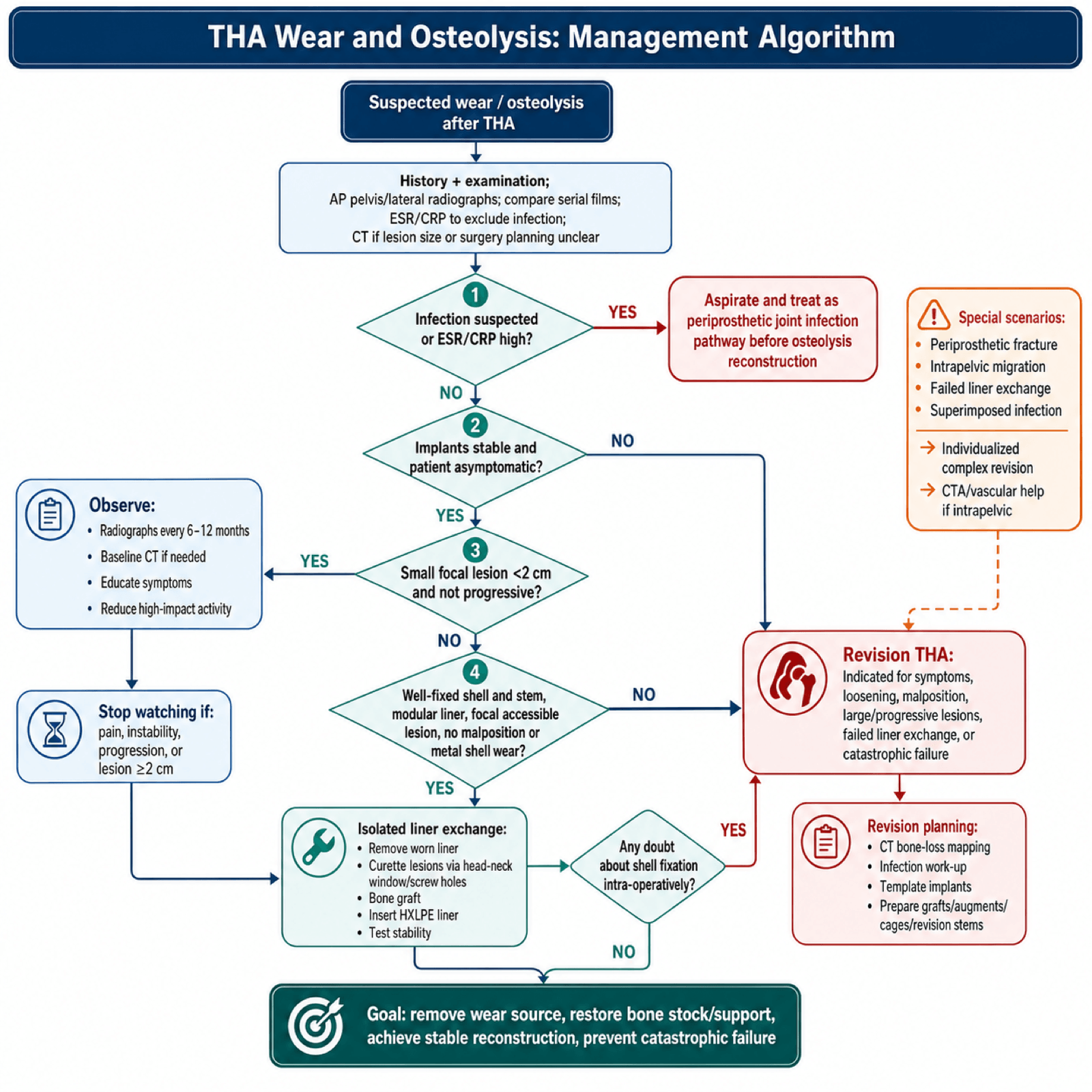
Who to observe. Observation is indicated for:
- An asymptomatic patient
- Focal lesions under 2 cm in diameter
- Stable implants, well fixed with no migration
- A low activity level or an elderly patient
- Significant medical comorbidities
How to observe. Obtain radiographs every 6-12 months, more often if the lesion is progressing, and a baseline CT to quantify its extent, repeated if progression is suspected. Teach the patient which symptoms to watch for (pain, instability), reduce high-impact activity if possible, and set the threshold for early intervention at documented progression.
Observation is not indefinite. If lesions are progressively enlarging, or cross the 2cm threshold, or if any symptoms develop, surgical intervention should be strongly considered. Don't wait for catastrophic failure.
Surgical Technique for Revision with Osteolysis
Posterior approach. The most common for revision: familiar anatomy, extensile proximally and distally, and excellent visualisation of the acetabulum and femoral canal. Its dislocation risk is higher, so the repair must be meticulous.
Anterolateral approach. Less dislocation risk, but it can compromise the abductors, which are important to preserve, and its distal extension is limited.
Extended trochanteric osteotomy (ETO). Used for difficult stem extraction or severe femoral osteolysis. It preserves the abductors, allows cement removal and canal preparation, and is fixed with cables or wires at closure.
The choice depends on the prior approach and the extent of reconstruction needed. Whatever the approach, four principles apply:
- Protect the neurovascular structures: the sciatic nerve posteriorly, the femoral vessels anteriorly
- Preserve bone stock by removing implants without additional bone loss
- Expose the osteolytic lesions, windowing if needed to reach the posterior column or calcar
- Mark component positions to help with templating and final positioning
Complications
- Incidence
- 5-10% after isolated liner exchange
- Prevention/Management
- Complete revision if shell loose, ensure adequate fixation
- Incidence
- 10-20% (higher than primary)
- Prevention/Management
- Large heads, dual mobility, repair soft tissues, correct malposition
- Incidence
- 3-5% (higher than primary)
- Prevention/Management
- Prolonged antibiotics, meticulous technique, rule out infection preop
- Incidence
- 5-10% intraop, 2-5% postop
- Prevention/Management
- Gentle technique, protect osteoporotic bone, bypass weak areas
- Incidence
- 1-3% after revision
- Prevention/Management
- Know anatomy, protect during retraction, avoid excess traction
- Incidence
- Under 1% but catastrophic
- Prevention/Management
- Identify vessels on CT, careful medial wall work, available vascular backup
- Incidence
- 10-20% at 10 years
- Prevention/Management
- Meticulous technique, adequate bone grafting, optimize patient factors
- Incidence
- 10-15%
- Prevention/Management
- Set realistic expectations, rule out infection, PT and pain management
Catastrophic osteolysis. Massive bone loss before detection may require complex reconstruction with allografts and cages, potentially over multiple operations, and function may never return to baseline. Prevention, through annual surveillance radiographs, is the key.
HXLPE liner fracture. Rare, but reported with large heads and thin liners, typically as rim fractures at impingement sites. It causes pain, instability and metallosis from shell-head contact, and is prevented by adequate liner thickness and by avoiding large heads with small cups.
Failed bone grafting. Graft resorption or non-incorporation leads to recurrent bone loss or component loosening. It is more common with structural than with morselised allograft and may require re-revision with an alternative fixation strategy.
Leg length discrepancy. Common after revision because of bone loss or component positioning. Patients often tolerate some lengthening better than shortening, a shoe lift may be needed, and an excessive discrepancy (over 2 cm) can cause back pain and gait abnormality.
Postoperative Care and Rehabilitation
Hip abduction pillow or brace, DVT prophylaxis (chemoprophylaxis plus mechanical), multimodal analgesia and early mobilisation, sitting to a chair and standing. Check drains and neurovascular status.
Physiotherapy progresses weight-bearing per surgeon protocol, within the restrictions set out below, which depend on the bone grafting and fixation. Gait training with a walker or crutches, observing hip precautions after a posterior approach.
Wound check at 2 weeks, removing sutures if non-absorbable. Weight-bearing restrictions continue, with outpatient physiotherapy 2-3 times per week, hip abductor strengthening, and monitoring for infection and dislocation.
Radiographs at 6 weeks assess component position and rule out early loosening. Progress to full weight-bearing if healing is appropriate, wean assistive devices and increase strengthening; return to driving at 6-8 weeks (if right hip and adequate control).
Radiographs at 3 months. Most patients are off all assistive devices and return to low-impact activities, continuing the strengthening programme while any persistent pain or functional limitation is addressed.
Radiographs at 1 year, then annually for life, to detect early failure. Full activity as tolerated within the high-impact restriction below, maintaining musculoskeletal fitness and a healthy weight, with patient education on the symptoms that require earlier follow-up.
Weight-bearing depends on the reconstruction:
- Standard revision without major graft: weight-bearing as tolerated from day 1
- Morselised allograft for cavitary defects: weight-bearing as tolerated, because the graft is impacted
- Structural allograft for segmental defects: toe-touch for 6 weeks, progressing to full by 12 weeks
- Pelvic discontinuity repair: toe-touch for 8-12 weeks minimum
- Periprosthetic fracture fixation: depends on construct stability, often 6-12 weeks protected
Activity. Hip precautions after a posterior approach (no flexion over 90°, no adduction, no internal rotation) last 6-12 weeks. High-impact activities (running, jumping) are avoided indefinitely, and low-impact activities (swimming, cycling, golf) are encouraged. Return to work is at 4-6 weeks for a desk job and 3-6 months for manual labour.
Outcomes and Prognosis
- 10-Year Survival
- 80-90% stable
- Main Failure Mode
- Progression requiring surgery
- Notes
- Success depends on patient compliance with surveillance
- 10-Year Survival
- 70-80%
- Main Failure Mode
- Unrecognised shell loosening
- Notes
- Best outcomes with strict selection criteria
- 10-Year Survival
- 80-90%
- Main Failure Mode
- Aseptic loosening, infection
- Notes
- Superior to isolated liner exchange in most cases
- 10-Year Survival
- 75-85%
- Main Failure Mode
- Aseptic loosening, dislocation
- Notes
- Depends on bone loss severity and fixation achieved
Better prognosis goes with early detection (small lesions, stable components), adequate bone stock or successful grafting, well-fixed revision components, young age (better healing, and a longer life expectancy in which to benefit), absence of comorbidities and compliance with restrictions.
Worse prognosis goes with delayed detection (massive bone loss), pelvic discontinuity or severe defects, prior failed revisions, infection, medical comorbidities (diabetes, smoking, osteoporosis) and obesity.
The best outcomes are in preventing osteolysis from occurring in the first place. This is achieved through:
- HXLPE for all primary THAs (reduces wear 90%)
- Optimal component positioning (avoid impingement, edge loading)
- Annual surveillance radiographs (detect early, intervene before catastrophic failure)
- Patient education on lifelong follow-up importance
Guidelines, Registries & Global Practice
Global epidemiology and burden:
Total hip arthroplasty is one of the most commonly performed and successful elective operations worldwide, and primary volumes are rising steeply. In the United States, primary THA demand was projected to grow by 174% to roughly 572,000 procedures per year by 2030, with hip revision demand projected to double by 2026 (Kurtz et al., J Bone Joint Surg Am 2007). As the implanted population grows and ages in situ, the cumulative number of patients at risk of late polyethylene wear and osteolysis increases correspondingly, even though the per-implant risk has fallen sharply with cross-linked bearings. Osteoarthritis is the dominant indication globally (the large majority of primaries), with post-traumatic arthritis, inflammatory arthritis, osteonecrosis and dysplasia accounting for the remainder.
Registry evidence on bearing surfaces (the global picture):
National joint registries — including the AOANJRR (Australia), the NJR (England, Wales, Northern Ireland and the Isle of Man), the AJRR (USA), the Swedish (SHAR), Norwegian and NZJR registries — have been decisive in bearing-surface governance. Two registry-driven lessons dominate:
- Registry signal
- Now the dominant primary bearing; markedly reduced revision for wear/osteolysis since widespread adoption
- Practical consequence
- Reference bearing for most primary THA
- Registry signal
- Historically higher late revision for wear/osteolysis (3x revision vs XLPE in the 20-year RCT, Devane et al. 2025)
- Practical consequence
- Largely superseded for primary THA
- Registry signal
- Registries identified high early revision for adverse reaction to metal debris (pseudotumour, ALTR)
- Practical consequence
- Largely withdrawn from routine practice
- Registry signal
- Very low wear; small risks of squeaking and ceramic fracture
- Practical consequence
- Selective use, often in younger patients
Because cross-linked PE has nearly removed wear as a dominant failure mode, registries now report infection and instability/dislocation as the leading causes of revision in contemporary THA.
Guidelines and consensus, side by side:
There is broad international agreement and few hard inter-guideline conflicts on this topic; the practical recommendations align across regions:
- Position relevant to wear/osteolysis
- Cross-linked PE preferred over conventional PE to reduce wear and osteolysis
- Evidence basis
- Supported by RCT/registry data
- Position relevant to wear/osteolysis
- Use bearings/implants with ≥10-year revision benchmarks (ODEP ratings); routine implant choice favours well-evidenced low-wear bearings
- Evidence basis
- Benchmark- and registry-driven
- Position relevant to wear/osteolysis
- Cross-linked (incl. antioxidant-stabilised) PE endorsed as standard for most primaries; bearing individualised for young/high-demand patients
- Evidence basis
- RCT and registry evidence
- Position relevant to wear/osteolysis
- Define cross-linking, sterilisation and oxidation-resistance testing for UHMWPE bearings
- Evidence basis
- Materials/laboratory standards
Practice variation:
- Bearing choice in young, high-demand patients is the main area of genuine debate: ceramic-on-XLPE and ceramic-on-ceramic are used variably by region and surgeon preference, weighing wear reduction against squeaking/fracture risk and cost.
- Resource setting: in limited-resource settings, conventional UHMWPE and metal-on-PE remain in wider use for cost reasons, so wear-related osteolysis remains a more prominent late problem than in high-income systems that have transitioned almost entirely to cross-linked bearings.
- Pharmacological prevention: bisphosphonates (e.g. alendronate) reduce early periprosthetic bone loss after cementless THA but have not been shown to durably prevent osteolysis, so they are not recommended as a substitute for low-wear bearings (Zeng et al., Orthopedics 2011, systematic review).
Frame this topic globally: cross-linked polyethylene is the international standard bearing, validated by both long-term RCTs (Devane et al. 20-year RCT) and multiple national registries (AOANJRR, NJR and others). The same registries withdrew metal-on-metal after detecting high revision for adverse reaction to metal debris. The remaining live controversy is bearing choice in the young, high-demand patient (ceramic-on-XLPE vs ceramic-on-ceramic).
MCQ Practice Points
Q: What size polyethylene wear particles are most biologically active in causing osteolysis? A: 0.1-1 micrometer. This size range optimally activates macrophages and triggers the inflammatory cascade. Smaller particles (under 0.1μm) are less readily phagocytosed. Larger particles (over 1μm) elicit less inflammatory response.
Q: By approximately what percentage does highly cross-linked polyethylene (HXLPE) reduce wear compared to conventional polyethylene? A: Over 90% (typically 90-95%). HXLPE wear rates are approximately 0.01-0.02mm per year compared to 0.1-0.2mm per year for conventional PE.
Q: What key cytokines are released by macrophages in response to polyethylene wear particles that drive osteolysis? A: TNF-α (tumor necrosis factor alpha), IL-1β (interleukin-1 beta), IL-6, and PGE2. These pro-inflammatory cytokines upregulate RANKL expression, which drives osteoclast differentiation and activation, leading to bone resorption.
Q: What does the term 'effective joint space' refer to in the context of THA osteolysis? A: The pathways by which wear particles access the bone-implant interface. Common pathways include screw holes in acetabular shells, gaps in press-fit fixation, osteotomies, and capsular defects. Particles migrate through these pathways to reach bone and trigger osteolysis.
Q: What is the main trade-off of highly cross-linked polyethylene compared to conventional polyethylene? A: Lower fracture toughness (more brittle). The cross-linking process that increases wear resistance also reduces the material's resistance to crack propagation. However, clinical studies show no increase in mechanical failures when HXLPE is used with appropriate liner thickness and head size.
Q: What is the recommended radiographic surveillance interval for patients with total hip arthroplasty? A: Annual radiographs for life. Osteolysis is typically asymptomatic until late-stage failure. Annual AP and lateral pelvis radiographs allow early detection of osteolysis, implant loosening, or other complications before catastrophic failure occurs.
Q: What is the most important prerequisite for isolated liner exchange in a patient with osteolysis? A: Well-fixed acetabular shell with absolutely no loosening. The shell must be completely stable with no radiolucent lines and no migration. Intraoperative stress testing should confirm stability. If there is any doubt, complete acetabular revision is safer and more durable.
Clinical Decision Scenarios
Practise clinical reasoning and management decisions out loud
“A 68-year-old woman presents for routine annual follow-up of her right THA performed 12 years ago with conventional polyethylene. She is completely asymptomatic and very active. AP pelvis radiograph shows a 2.5cm radiolucent lesion in DeLee-Charnley zone I (superior acetabulum). There are no radiolucent lines around the cup or stem. How do you manage this?”
“A 72-year-old man with a 15-year-old right THA (conventional PE) presents to the emergency department with sudden onset severe right hip pain and inability to weight-bear after a minor fall at home. Radiograph shows a Vancouver B3 periprosthetic femoral fracture with massive osteolysis around the femoral stem and acetabular component. The stem is clearly loose. How do you manage this complex problem?”
“You are planning a primary THA in a 38-year-old male manual laborer with post-traumatic osteoarthritis. He is very active, plays recreational sports, and has 40-50 years of expected implant service life. He asks why you're recommending HXLPE when he's heard it's 'more brittle' and 'might break.' Discuss your bearing surface decision and address his concerns.”
WEAR MECHANISMS
- Adhesive wear: surface-to-surface molecular bonding and transfer
- Abrasive wear: hard surface plowing through soft PE (scratching)
- Third-body wear: interposed particles (PMMA, metal) acting as abrasives
- Critical particle size: 0.1-1μm (most biologically active)
PARTICLE DISEASE CASCADE
- 1. PE particles generated at bearing surface
- 2. Particles access bone via effective joint space (screw holes, gaps)
- 3. Macrophages recognize and phagocytose particles
- 4. Cytokine release: TNF-α, IL-1β, IL-6, PGE2
- 5. RANKL upregulation drives osteoclast differentiation
- 6. Osteoclast-mediated bone resorption (osteolysis)
HXLPE PROPERTIES
- Manufacturing: 50-100 kGy radiation creates cross-links
- Wear reduction: over 90% vs conventional PE
- Wear rate: 0.01-0.02mm/year (vs 0.1-0.2mm/year conventional)
- Trade-off: lower fracture toughness (more brittle)
- Prevention: adequate thickness (8-10mm), appropriate head size (32-36mm)
- Long-term data: 15-20 year follow-up confirms durability, under 5% osteolysis
SURVEILLANCE PROTOCOL
- Annual AP and lateral pelvis radiographs for life (mandatory)
- Compare to baseline postoperative films
- Look for: radiolucent lines, expanding lesions, component migration
- CT scan to quantify osteolysis if suspected on plain films
- Osteolysis is typically asymptomatic - imaging essential for detection
MANAGEMENT ALGORITHM
- Small lesions (under 2cm), stable components: observe with close surveillance
- Large lesions (over 2cm), stable components: isolated liner exchange vs revision
- Isolated liner exchange: strict criteria (well-fixed shell, accessible lesions)
- Complete revision: symptomatic, loose components, large/progressive osteolysis
- Curettage and bone graft all osteolytic lesions
- Use HXLPE liner to prevent recurrent wear
SURGICAL PRINCIPLES
- Acetabular: assess shell stability (stress test intraop), revise if any doubt
- Femoral: bypass osteolytic areas with long stem (4-6cm contact in good bone)
- Bone grafting: morselized for cavitary, structural for segmental defects
- Impaction grafting technique for contained defects
- HXLPE liner essential to prevent recurrence
- Large head (32-36mm) to minimize dislocation risk
KEY EXAM PEARLS
- Osteolysis is biological (particle disease), not purely mechanical
- Prevention is superior to treatment: HXLPE for all primary THAs
- Annual surveillance radiographs are non-negotiable for life
- Asymptomatic osteolysis is the ideal time to intervene (before disaster)
- AOANJRR data: wear revisions declined dramatically with HXLPE adoption
- Metal-on-metal abandoned due to adverse reactions and pseudotumor
Evidence Base
HXLPE vs Conventional PE - 20-Year Double-Blind RCT (Landmark)
- Longest-term RCT to date: 122 patients randomised to cross-linked (Marathon) versus conventional (Enduron) liners with otherwise identical components, minimum 20-year follow-up.
- After bedding-in, steady-state wear was 0.034 mm/year for XLPE versus 0.181 mm/year for conventional PE.
- Conventional-PE liners had a revision rate roughly three times higher than XLPE (28 versus 9 of the 37 revisions), and of the 43 patients alive and unrevised at 20 years, 28 were in the XLPE arm against 15 in the conventional arm.
- Wear-rate confidence intervals are tight and do not overlap: 0.181 mm/year (95% CI 0.175-0.188) conventional against 0.034 (0.028-0.039) XLPE.
Multicentre Wear of Remelted HXLPE in THA (2012 John Charnley Award)
- 768 primary THAs (head sizes 26-36 mm) across eight centres followed to 7-13 years.
- Serial radiographs showed no periprosthetic osteolysis in any of the three studies.
- Mean femoral head penetration did not correlate with time in vivo for standard head sizes; 36 mm heads trended higher but stayed below the osteolysis threshold.
Cellular and Molecular Biology of Periprosthetic Osteolysis
- Wear debris primarily targets macrophages and osteoclast-precursor cells, with osteoblasts, fibroblasts and lymphocytes also involved.
- Particles activate MAP-kinase pathways and NF-κB, up-regulating pro-inflammatory signalling and RANKL while suppressing protective cytokines.
- Reducing particle size to lower wear may paradoxically increase biological activity; no approved pharmacological treatment for osteolysis exists.
Vitamin E-Blended HXLPE Cup - 6-Year RCT
- 199 patients randomised to a vitamin E-blended HXLPE cup versus a conventional UHMWPE monoblock cup.
- Femoral head penetration was lower with vitamin E HXLPE (0.028 mm/year) than UHMWPE (0.035 mm/year).
- Six-year survival to revision was 98% for both cups with no aseptic loosening and no adverse reactions attributable to vitamin E.