Isolated shear fractures of the humeral trochlea
AO/OTA Classification (13-B3)
Critical Must-Knows
- Rare Entity: Isolated trochlea fractures are rare. Usually part of a transcondylar or capitellum fracture.
- Shear Injury: Like the capitellum, these are coronal shear fractures with no soft tissue attachments (free floating).
- Double Arc Sign: On lateral X-ray, seen as a second arc behind the capitellum (often missed).
- Surgical Approach: Requires a medial approach (Over-the-top or Osteotomy) for visualization.
Clinical Pearls
- "Missed Diagnosis: Often misdiagnosed as a medial epicondyle fracture or 'sprain'.
- "Ulnar Nerve: High risk of injury due to proximity.
- "Instability: Loss of the trochlea causes ulno-humeral instability.
Clinical Imaging
Imaging Gallery


Critical Diagnostics
The Hidden Fracture
Isolated trochlea fractures are invisible on AP X-ray (overlapped by ulna). The lateral view shows the "Double Arc" sign but is subtle.
The Free Fragment
The fragment has NO soft tissue attachments. It is purely articular. Blood supply is entirely retroactive from subchondral bone (which is fractured). High AVN risk.
Associated Injuries
Look for associated Elbow Dislocation or Capitellum fracture (creates a Type IV capitellum fracture).
CT is Mandatory
You cannot plan this surgery without a CT scan. It defines Articular comminution.
Quick Decision Guide - Management
| Condition | Treatment | Approach | Key Factor |
|---|---|---|---|
| Non-displaced | Cast Immobilization | N/A | Very rare to be stable. |
| Displaced (Simple) | **ORIF** | Medial Column/Osteotomy | Headless compression screws (A-P or P-A). |
| Comminuted (Elderly) | **TEA (Total Elbow)** | Posterior | Bone stock usually too poor for screws. |
| Small Fragment | **Excision** | Medial | Only if fragment is less than 20% of surface. |
Double ArcRadiographic Signs
| Double | Double Arc Sign On lateral view, the trochlea arc is seen separate from capitellum arc. |
| Drop | Drop Sign Ulnohumeral distance increases if trochlea is displaced. |
| Double | Double Arc Sign On lateral view, the trochlea arc is seen separate from capitellum arc. |
| Drop | Drop Sign Ulnohumeral distance increases if trochlea is displaced. |
Hook:Seeing double? Check the trochlea.
ARCSurgical Goals
| A | Anatomic Reduction Articular step-off leads to arthritis. |
| R | Rigid Fixation Allows early motion (crucial for elbow). |
| C | Compression Headless screws to bury beneath cartilage. |
| A | Anatomic Reduction Articular step-off leads to arthritis. |
| R | Rigid Fixation Allows early motion (crucial for elbow). |
| C | Compression Headless screws to bury beneath cartilage. |
Hook:Restore the ARC of motion.
M-U-TThe Medial Column
| M | Medial Epicondyle Origin of flexors, landmark for approach. |
| U | Ulnar Nerve The structure at risk. |
| T | Trochlea Supported by the medial column. |
| M | Medial Epicondyle Origin of flexors, landmark for approach. |
| U | Ulnar Nerve The structure at risk. |
| T | Trochlea Supported by the medial column. |
Hook:Don't be a MUT, check the nerve.
Overview and Epidemiology
Definition: A Trochlea fracture (specifically Laugier's fracture) is an isolated coronal shear fracture of the joint surface of the trochlea. It does not involve the columns (unless associated with complex fractures).
Epidemiology:
- Extremely Rare. Represents less than 1% of distal humerus fractures.
- Demographics: Young males (high energy) or Osteoporotic females.
- Pathoanatomy: The trochlea is the "spool" of the elbow. It provides intrinsic stability (constrained joint). Loss of the trochlea allows the ulna to slide medially/laterally or dislocate.
Anatomy
Bony Anatomy:
- Trochlea: A spool-shaped structure covered in cartilage through an arc of 300 degrees.
- Sulcus: The central groove articulates with the trochlear notch of the ulna.
- Medial Ridge: More prominent than lateral ridge. Provides valgus stability.
- Center of Rotation: The axis of rotation passes through the center of the trochlea (and capitellum).
- Articular Contact: The ulna articulates with the trochlea in both flexion and extension. Loss of the trochlea results in rapid ulnar migration and instability.
Vascularity:
- Watershed: The trochlea is supplied by small vessels entering via the medial capsule and non-articular areas.
- Fracture: The fracture is intra-articular and separates the bone from its blood supply (like an iceberg). High risk of AVN/Non-union.
- Posterior Comminution: Indicates disruption of the posterior vascular supply.
Nerves:
- Ulnar Nerve: Runs immediately posterior to the medial epicondyle. Must be identified and protected (or transposed) in any medial approach.
- Medial Antebrachial Cutaneous Nerve (MABCN): At risk during the superficial dissection. Injury causes painful neuroma.
Muscle Attachments:
- Flexor-Pronator Mass: Originates from the medial epicondyle. Must be elevated or split to access the anterior aspect of the trochlea.
- Triceps: Inserts on the olecranon, but its medial border covers the posterior aspect of the medial column.
Classification Systems
- 13-A: Extra-articular.
- 13-B: Partial Articular.
- B1: Lateral Sagittal (Capitellum).
- B2: Medial Sagittal (Trochlea - Rare).
- B3: Frontal/Coronal Plane (Shear).
- B3.1: Capitellum alone (Hahn-Steinthal).
- B3.2: Trochlea alone (Laugier).
- B3.3: Capitellum + Trochlea (McKee).
Clinical Assessment
History:
- Fall on outstretched hand (FOOSH) with elbow slightly flexed and in varus?
- Direct blow?
- Pain, swelling, inability to move elbow.
Physical Exam:
- Swelling: Medial sided bruising (Ecchymosis).
- Palpation: Tenderness over medial column.
- ROM: Block to flexion/extension implies a mechanical block (loose fragment).
- Neurology: CHECK ULNAR NERVE. Tardy ulnar nerve palsy is a late complication, but acute neuropraxia is common from the blow.
Differential Diagnosis of the Painful, Stiff Elbow After a Fall
| Diagnosis | Key clue | Best test | Distinguishing feature |
|---|---|---|---|
| Isolated trochlea (Laugier) fracture | Medial pain; AP often normal; mechanical block | True lateral X-ray + CT | Double-arc sign; medial coronal fragment, ulnohumeral incongruity |
| Capitellum (Hahn-Steinthal) shear fracture | Lateral pain; block to flexion | Lateral X-ray + CT | Fragment lateral; 'double-arc' classically here too but capitellar |
| Radial head/neck fracture | Lateral pain, painful rotation | AP/lateral, radiocapitellar view | Pain on pronation/supination, not pure flexion block |
| Coronoid fracture / terrible triad | Prior dislocation, gross instability | CT + stress views | Posterolateral instability; radial head + LCL injury |
| Medial epicondyle fracture (avulsion) | Medial pain, valgus laxity, ulnar symptoms | AP X-ray (extra-articular) | Extra-articular avulsion; NOT a coronal articular shear |
| Simple ligamentous sprain / occult injury | Pain, swelling, no bony lesion | MRI if X-ray/CT negative | Diagnosis of exclusion — beware missing an occult shear fragment |
Investigations
Plain X-rays:
- AP: Often looks normal or shows a faint "flake" medially.
- Lateral: The Diagnostic View. Look for:
- Double Arc Sign (Two semi-circles).
- Superior migration of the fragment.
- "Chewed up" appearance of the joint line.
CT Scan:
- Absolute Requirement.
- 2D Views:
- Coronal: Shows the shearing nature and size of the fragment.
- Sagittal: Shows posterior comminution (Dubberley B).
- 3D Reconstruction:
- Essential for preoperative planning of screw trajectory.
- Determine if you can screw Front-to-Back (easier) or need Back-to-Front (harder).
Management Algorithm
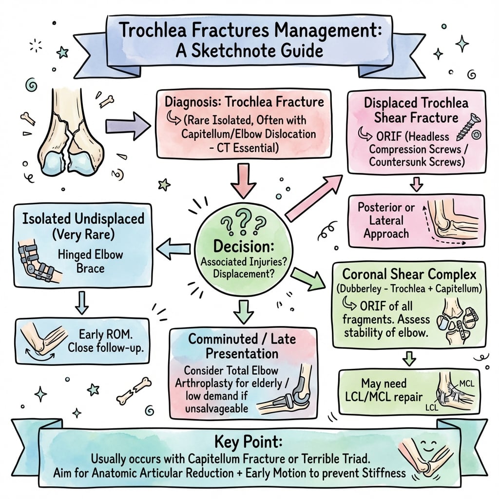
Decision Making:
-
Fragment Size:
- Small (less than 20%): Excision.
- Large (greater than 20%): Fixation.
-
Displacement:
- Non-displaced: Cast (verify with CT).
- Displaced: ORIF.
-
Bone Quality:
- Good: Headless Screws.
- Poor (Elderly): TEA (Total Elbow Arthroplasty).
Surgical Techniques
1. Medial Approach (Hotchkiss/Over-the-top):
- Incision over medial supracondylar ridge.
- Identify Ulnar Nerve. Release cubital tunnel.
- Elevate flexor-pronator mass anteriorly (or split it).
- Limitation: Hard to see the lateral extent of the trochlea.
2. Chevron Osteotomy (Medial Epicondyle):
- Pre-drill the medial epicondyle.
- Perform osteotomy.
- Reflect flexor mass distally.
- Benefit: Excellent view of the trochlea.
- Repair: Screw fixation of the epicondyle at the end.
3. Olecranon Osteotomy:
- Typically used for intercondylar fractures, but can be used here for posterior access.
Complications
- Nonunion / AVN:
- The fragment is devoid of soft tissue attachments.
- AVN creates a sequestrum which causes pain and locking.
- Treatment: Excision and TEA.
- Arthritis:
- Rapid onset if step-off remains.
- "The elbow tolerates congruity poorly."
- Ulnar Neuropathy:
- Due to surgical handling or scar tissue.
- Routine anterior transposition is debated but often done in complex cases.
- Heterotopic Ossification (HO):
- Medial side is lower risk than lateral/posterior, but still possible.
Postoperative Care
- Phase 1 (0-2 Weeks):
- Splint at 90 degrees.
- Elevation.
- Phase 2 (2-6 Weeks):
- Active Motion: Start AROM immediately if fixation is rigid.
- Gravity assisted flexion/extension.
- Avoid varus stress.
- Phase 3 (6+ Weeks):
- Strengthening.
- Wean splint.
Outcomes/Prognosis
Evidence is limited to retrospective series and one pooled meta-analysis (Fisher et al, JSES Int 2022, 45 studies / 899 patients). The figures below are the best-available pooled estimates for coronal-shear fractures (capitellum and/or trochlea), not isolated trochlear data, which remains case-report level.
- Functional result: Good with anatomic reduction and rigid fixation — mean MEPI ~91 and arc ~19 to 138 degrees in the Dubberley series; mean DASH ~24 in the Tanwar headless-screw series.
- Reoperation: ~14% pooled (contracture release, hardware removal, ulnar nerve procedures).
- Post-traumatic arthritis: ~21% pooled — driven by residual articular step-off.
- Heterotopic ossification: ~12% pooled. Routine pharmacological prophylaxis is NOT supported for isolated injuries; reserve for high-risk cases (associated dislocation/head injury, extensive dissection).
- Avascular necrosis / nonunion: ~7% pooled — higher with posterior comminution (Dubberley B) that strips the residual blood supply.
- ROM: A flexion contracture of 10 to 20 degrees is common and usually functional.
- Failures in the elderly: Comminution unreconstructable for stable fixation favours primary TEA (McKee RCT).
Controversies & Areas of Uncertainty
The isolated trochlear (Laugier) fracture is rare enough that almost every management decision rests on extrapolation from capitellar / coronal-shear series rather than dedicated evidence.
- Surgical approach: Anterolateral vs extended-lateral vs medial-column/epicondyle osteotomy. Pooled data hint at fewer complications with the anterolateral approach for Dubberley A, but the meta-analysis explicitly concludes evidence is insufficient to mandate one approach. A pure isolated-trochlear fragment is medial and may still need a medial exposure.
- Screw direction: Anterior-to-posterior (technically easier, countersunk under cartilage) vs posterior-to-anterior (preserves articular cartilage, mechanically stronger but harder for an anterior shear fragment). No comparative data exist.
- Excision vs fixation of small fragments: The "less than 20% excise" rule is a pragmatic teaching heuristic, not an evidence-derived threshold; the trochlea is load-bearing and intrinsically stabilising, so most authors favour fixation whenever technically feasible.
- HO prophylaxis: Routine indomethacin/radiotherapy is unproven for isolated coronal-shear injuries and carries its own risks (nonunion, GI); use is selective.
- Ulnar nerve handling: In-situ decompression vs routine anterior transposition during a medial approach remains debated, with no trochlear-specific data.
- Primary TEA threshold: RCT support exists for the elderly OTA-13C elbow, but the age/comminution threshold for choosing TEA over a salvageable ORIF is not defined for isolated articular shear patterns.
Evidence
Apparent capitellum fractures are more complex
- 21 articular distal humerus fractures; what looks like an isolated capitellum fracture often involves up to 5 components including the posterior trochlea.
- All fractures stabilised with implants buried beneath the articular surface healed, with no residual ulnohumeral instability.
- Average ulnohumeral arc 96 degrees (range 55 to 140); 10 of 21 required a second procedure, most often for contracture release.
Dubberley classification & outcome of capitellar/trochlear fractures
- 28 ORIF patients (mean age 43); fractures classified Types 1 to 3 by capitellar/trochlear involvement with modifier A/B for posterior comminution.
- More complex fractures needed more extensive surgery, had more complications/secondary procedures and poorer outcomes; overall mean MEPI 91 and arc 19 to 138 degrees.
- 2 comminuted fractures failed to unite and were converted to total elbow arthroplasty.
ORIF of coronal-plane capitellum/trochlea via anterolateral approach
- 10 patients with coronal-plane distal humerus fractures fixed with headless compression screws through an anterolateral approach.
- Mean DASH 24; union in all at a mean of 10 weeks (range 8 to 12).
- Low complication burden: one Broberg-Morrey grade 2 arthritis and one Brooker grade 1 heterotopic ossification.
Does surgical approach affect coronal-shear outcomes? (meta-analysis)
- Systematic review/meta-analysis of 45 studies, 899 patients (mean age 44.9); reoperation rate 13.8%.
- Pooled complications: post-traumatic arthritis 21.2%, heterotopic ossification 12.0%, nerve injury 7.8%, avascular necrosis 7.4%.
- For Dubberley A fractures, complication rate was higher with the extended-lateral (25.8%) than the anterolateral (16.7%) approach, but evidence is insufficient to mandate one approach.
TEA vs ORIF for displaced distal humerus fractures in the elderly (RCT)
- Multicentre RCT, 42 patients over 65 with OTA 13C fractures; TEA gave better MEPS at 3, 6, 12 and 24 months than ORIF (e.g. 86 vs 73 at 2 years).
- 5 of 21 (24%) randomised to ORIF were not amenable to stable fixation and converted to TEA intra-operatively.
- Reoperation rates (TEA 12% vs ORIF 27%) were not statistically different.
Salvage of unstable distal humerus nonunion
- 15 unstable distal humerus nonunions treated with rigid fixation, contracture release and autograft.
- 12 of 15 united (mean arc 95 degrees); 3 failed and were converted to total elbow arthroplasty.
- 6 of the 12 united cases needed further surgery for painful implants, ulnar neuropathy or contracture.
Laugier's isolated trochlear shear fracture
- First description of the isolated coronal shear fracture of the humeral trochlea, eponymous 'Laugier fracture'.
- Recognised the resulting ulnohumeral incongruity and instability.
Clinical Decision Scenarios
Use these scenarios to practise clinical reasoning and management decisions
"A 60-year-old female presents with a 'swollen elbow' after a fall. X-ray AP looks normal. She cannot flex past 90 degrees."
Diagnosis:
- Differential: Occult fracture (Radial head, Coronoid, or Trochlea). Ligamentous injury.
- Red Flag: Mechanical block suggests a loose body.
- Next Step: True Lateral X-ray (Look for Double Arc Sign) and CT Scan.
- Result: Likely an isolated Trochlear shear fracture (Laugier).
"Intra-op, you have fixed the trochlea fracture but the screw heads are prominent in the articular surface."
Options:
- Countersink: Ensure you have countersunk the hole before inserting the screw.
- Change Screw: Use a smaller headless screw (Acutrak Mini vs Micro).
- Direction: Insert from non-articular surface (posterior to anterior) if possible, but this is technically harder for coronal shear.
- Implication: Prominent hardware = Rapid chondrolysis and arthritis. Must be flush or buried.
"A 78-year-old osteoporotic woman has a comminuted coronal-shear distal humerus fracture involving the trochlea and capitellum (Dubberley 3B). At surgery the articular fragments crumble and you cannot achieve fixation stable enough for early motion."
Decision:
- Bail-out: Convert to primary semiconstrained total elbow arthroplasty rather than accept unstable fixation.
- Evidence: The McKee multicentre RCT showed superior Mayo Elbow Performance Scores with TEA versus ORIF in patients over 65, and 24% of ORIF-allocated patients had to be converted intra-operatively.
- Caveat: Reserve for low-demand patients; in a young high-demand patient, exhaust reconstruction (revision ORIF plus graft) first.
- Consent: Permanent lifting restriction (single-event ~5 kg, repetitive ~1-2 kg) and risks of bushing wear, infection and periprosthetic fracture.
MCQ Practice Points
Radiology
Q: What is the 'Double Arc Sign' on a lateral elbow radiograph pathognomonic for? A: Capitellum and Trochlea shear fractures (McKee Type IV / Dubberley).
Anatomy
Q: Which column of the distal humerus supports the Trochlea? A: The Medial Column.
Complications
Q: Which nerve is most at risk during fixation of a Laugier fracture? A: Ulnar Nerve (Posterior to medial epicondyle).
Treatment
Q: What is the preferred fixation method for a Type I coronal shear fracture? A: Headless Compression Screws (A-P direction).
Prognosis
Q: What factor most strongly predicts failure of fixation? A: Posterior Comminution (Dubberley B).
Guidelines, Registries & Global Practice
Global epidemiology
- Isolated coronal-shear fractures of the distal humerus are rare (apparent capitellum fractures are roughly 1% of all elbow fractures); the truly isolated trochlear (Laugier) variant is rarer still and reported only at case-series level worldwide.
- Bimodal: high-energy injuries in young adults (FOOSH with the elbow near extension) and low-energy fragility fractures in osteoporotic older women.
- There is no dedicated society guideline for this specific fracture; management is extrapolated from distal humerus articular-fracture principles common to AO, BOA and AAOS teaching.
Side-by-side principles (no fracture-specific national guideline exists)
How Major Schools Frame Coronal-Shear / Distal Humerus Articular Fractures
| Body | Emphasis | Practical message |
|---|---|---|
| AO Foundation | Anatomic articular reduction + absolute stability, early motion | Buried headless compression screws; reconstruct the articular block first |
| BOA / BOAST (UK principles) | Specialist upper-limb/major-trauma pathway, early definitive care | Refer complex intra-articular elbow injuries to a unit with elbow expertise and TEA capability |
| AAOS (US teaching) | CT-based planning, ORIF for reconstructable fractures | Primary TEA reserved for low-demand elderly with unreconstructable comminution |
| EFORT / European consensus | Joint-preserving fixation; arthroplasty as a considered salvage | Function-led rehab; outcome reporting via MEPS/DASH |
Registry note
- National joint registries (NJR England/Wales, AOANJRR Australia, SHAR Sweden, NZJR) capture total elbow arthroplasty implant survival and revision, which informs the salvage/elderly arm of this fracture — but they do not track ORIF of coronal-shear fractures, so primary-fixation outcomes rely on published series and the Fisher meta-analysis.
High- vs limited-resource practice
- Well-resourced: Routine pre-op CT with 3D reconstruction, headless compression screw systems, intra-operative fluoroscopy, and TEA available as backup.
- Limited-resource: CT may be unavailable, so a meticulous true-lateral radiograph and intra-operative assessment carry more weight; standard partially-threaded/mini-fragment screws may substitute for headless implants, and arthroplasty backup may be absent — pushing decisions toward fixation or, for tiny fragments, excision.
Trochlea Essentials
Clinical summary
Key Concepts
- •Coronal Shear Injury
- •Double Arc Sign
- •Medial Approach
- •Headless Screws
Classification (Dubberley)
- •Type 1: Capitellum
- •Type 2: Cap + Trochlea (Fused)
- •Type 3: Cap + Trochlea (Mallet)
- •Modifier B: Posterior Comminution
Imaging
- •Lateral View is key
- •CT Mandatory
- •Rule out terrible triad
- •Rule out capitellum fx
Complications
- •Ulnar Neuropathy
- •AVN (Thal)
- •Stiffness (Loss of extension)
- •Arthritis