Clinical Imaging
Imaging Gallery

Ulnar Club Hand (Ulnar Longitudinal Deficiency)
High Yield
At a Glance
Ulnar club hand (ulnar longitudinal deficiency) is a rare congenital anomaly (1:100,000 births) involving partial or complete ulnar absence—4x less common than radial club hand. The limb presents with radial deviation, ulnar digit absence (4th/5th fingers), and frequently associated syndactyly (50-60%) and radial head dislocation (40-50%). Unlike radial deficiency, systemic syndrome association is uncommon (~10-15%). The Bayne and Klug classification (Types I-IV) grades severity from hypoplastic ulna to complete absence. Treatment focuses on hand function rather than cosmesis: options include stretching/splinting, first web space deepening, radial osteotomy for severe deviation, and one-bone forearm creation for elbow instability.
ULNARCLUB
| U | U - Ulna absence (partial/complete) | C | C - Central deficiency pattern |
| L | L - Little finger often absent | L | L - Less common than radial deficiency |
| N | N - Nubbins common (finger remnants) | U | U - Ulnar nerve abnormalities |
| A | A - Associated anomalies (syndactyly, radial head) | B | B - Bones of hand affected (metacarpals absent) |
| R | R - Radial deviation posture |
| U | U - Ulna absence (partial/complete) | A | A - Associated anomalies (syndactyly, radial head) | L | L - Less common than radial deficiency |
| L | L - Little finger often absent | R | R - Radial deviation posture | U | U - Ulnar nerve abnormalities |
| N | N - Nubbins common (finger remnants) | C | C - Central deficiency pattern | B | B - Bones of hand affected (metacarpals absent) |
Hook:ULNAR CLUB
Introduction
Ulnar club hand represents a spectrum of congenital upper limb deficiencies involving partial or complete absence of the ulna. This condition is less common than radial club hand, occurring in approximately 1 in 100,000 live births. The deformity typically presents with radial deviation of the hand, absence of ulnar digits, and often associated elbow instability due to radial head dislocation.
RAREU
| R | R - Radial club hand is 4x MORE common |
| A | A - Associated with syndactyly (50-60%) |
| R | R - Radial head dislocation common |
| E | E - Elbow instability frequent |
| U | U - Ulnar-sided digits absent (4th/5th) |
| R | R - Radial club hand is 4x MORE common | E | E - Elbow instability frequent |
| A | A - Associated with syndactyly (50-60%) | U | U - Ulnar-sided digits absent (4th/5th) |
| R | R - Radial head dislocation common |
Hook:RARE-U
Epidemiology
Incidence: approximately 1 in 100,000 live births — roughly 3 to 4 times rarer than radial longitudinal deficiency (about 1 in 30,000). In population registry data (Hungarian Congenital Malformation Registry), all limb deficiency defects had a birth prevalence near 1 in 1,816, with the ulnar/fibular group being among the least frequent and least syndrome-associated.
Key Demographics:
- Male predominance (male:female roughly 1.5:1)
- Unilateral in the majority; bilateral in roughly a quarter of cases
- Predominantly sporadic; most cases are not inherited
- Frequently associated with musculoskeletal anomalies (notably ipsilateral hand/digit deficiency and proximal femoral or fibular deficiency) rather than visceral syndromes
Embryology and Pathogenesis
Critical Developmental Period: 4-8 weeks gestation
Zone of Polarizing Activity (ZPA): The limb bud's organizing center produces Sonic Hedgehog (SHH) protein, controlling ulnar-radial patterning. Disruption leads to ulnar deficiency.
Apical Ectodermal Ridge (AER): Maintains underlying mesenchyme proliferation. AER dysfunction causes longitudinal deficiencies.
Associated Conditions
Classification
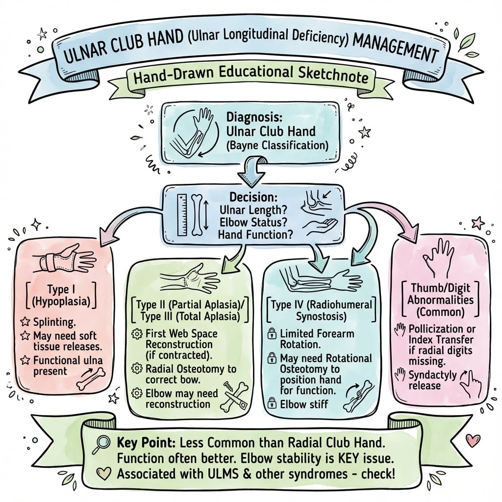
Bayne and Klug Classification (1987)
The most widely used classification system, based on radiographic appearance:
Bayne and Klug Classification of Ulnar Deficiency
| category | features | characteristics | elbow | hand | treatment | prognosis |
|---|---|---|---|---|---|---|
| Type I | Hypoplastic ulna | Ulna present but short, distal and proximal epiphyses present | Stable | Mild deviation, all digits usually present | Often observation, occasional soft tissue release | Excellent function |
| Type II | Partial absence (middle segment) | Proximal and distal ulnar segments present, middle absent | Variable stability | Moderate radial deviation, ulnar digits may be hypoplastic | One-bone forearm vs reconstruction | Good function with treatment |
| Type III | Complete absence (distal only) | Proximal ulna absent, distal ulnar remnant present | Unstable, radial head dislocation common | Severe radial deviation, often missing 4th/5th digits | One-bone forearm, radial head excision | Fair function, limited rotation |
| Type IV | Complete absence | Complete absence of ulna | Very unstable, severe radiohumeral dislocation | Severe deformity, multiple digit absence common | One-bone forearm mandatory, complex reconstruction | Limited function, stability priority |
Clinical Application: Type I rarely requires surgery. Types II-IV benefit from surgical intervention, with Type IV requiring most extensive reconstruction.
Cole and Manske Modification (1997)
Adds consideration of hand involvement:
- A: Normal first web space
- B: Mild first web space deficiency
- C: Moderate-to-severe first web space deficiency
- D: Absent thumb
This modification is critical for surgical planning as thumb function determines overall hand function.
Differential Diagnosis
The radially deviated, short forearm with absent ulnar-sided digits has several mimics. Distinguishing them changes management and counselling.
Differential diagnosis of the ulnar-deviated / ulnar-deficient limb
| condition | keyFeature | elbow | distinguisher | management |
|---|---|---|---|---|
| Ulnar longitudinal deficiency (ulnar club hand) | Radial deviation of hand; absent/hypoplastic ulnar (4th/5th) rays; ulna short or absent | Often unstable; radial head dislocation or radiohumeral synostosis | Thumb usually present and functional; low syndromic burden | Function-led; stabilise forearm/elbow only if grossly unstable |
| Radial longitudinal deficiency (radial club hand) | RADIAL deviation toward absent radius; thumb hypoplastic or absent | Often stiff; radial-sided wrist instability | Absent/deficient thumb; high syndromic burden (VACTERL, TAR, Fanconi, Holt-Oram) | Stretching, centralization/radialization, pollicization |
| Cleft hand (typical central deficiency) | Central (3rd ray) deficiency with V-shaped cleft; border rays present | Normal | Often bilateral, includes feet; thumb and small finger preserved | Cleft closure, syndactyly release, web reconstruction |
| Symbrachydactyly (ulnar/transverse type) | Short/absent fingers with nubbins and ectodermal pits; normal forearm bones | Normal | Soft-tissue (vascular) aetiology; bones of forearm intact; unilateral, sporadic | Soft-tissue/digit reconstruction, free toe transfer in selected cases |
| Constriction (amniotic) band sequence | Circumferential constriction rings, distal amputations, acrosyndactyly | Normal proximal bones | Bands at varying levels; asymmetric, sporadic; no longitudinal bone axis defect | Band release, syndactyly correction, reconstruction as needed |
Controversies & Areas of Uncertainty
The rarity of ulnar deficiency means most practice rests on small retrospective series and expert opinion; several questions remain genuinely unsettled.
- Value and timing of one-bone forearm. It reliably trades rotation for stability, but it is required far less often than older teaching implied — many type II-III limbs function well without it. Whether and when to convert an unstable two-element forearm to a single strut, and at what age, is not standardised.
- Management of the fibrocartilaginous anlage. Whether the anlage truly tethers growth and drives progressive radial bowing — and therefore whether prophylactic excision prevents deformity — is debated; excision risks wrist destabilisation and evidence of benefit is weak.
- Radiohumeral synostosis. Takedown to restore elbow motion is generally discouraged because of re-fusion and instability, but a minority advocate repositioning osteotomy in selected malpositioned limbs; the threshold is subjective.
- Radial head dislocation. Indications and timing for radial head excision (symptomatic, blocking motion) versus leaving it are not evidence-based and are extrapolated from other conditions.
- Outcome measurement. Few studies use validated, condition-specific paediatric upper-limb outcome tools, so comparisons across series and centres are unreliable; reported high satisfaction may reflect adaptation rather than reconstruction.
- Classification overlap. The mild end of the spectrum (Havenhill type 0, hand-only deficiency) overlaps clinically with ulnar-sided cleft hand and symbrachydactyly, and there is no consensus on where ulnar deficiency ends and these entities begin.
Clinical Presentation
Physical Examination
Functional Impact
Grasp Patterns:
- Power grip: Reduced with digit absence
- Precision grip: Depends on thumb function
- Key pinch: Limited if thumb hypoplastic
- Hook grip: Limited with 4th/5th digit absence
Activities of Daily Living:
- Bilateral involvement significantly impacts function
- Unilateral cases: Contralateral hand dominant for fine tasks
- Adaptive strategies develop early in childhood
- Early intervention improves adaptation
Investigations
Radiographic Evaluation
Laboratory Evaluation
Genetic Testing:
- Karyotype: If syndromic features present
- TBX3 gene: Ulnar-mammary syndrome suspected
- NIPBL gene: Cornelia de Lange features
- Fanconi anemia panel: If bilateral or family history
- Chromosomal microarray: Unexplained associated anomalies
Hematological:
- Complete blood count: Rule out Fanconi anemia (macrocytosis, cytopenias)
- Chromosome breakage test: Fanconi anemia screening
- Pre-operative: Standard pre-anesthetic workup
Cardiac Evaluation:
- Echocardiogram if syndromic association suspected
- ECG for Holt-Oram-like presentations
Non-Operative Management
Observation Criteria
Bayne Type I with minimal deformity:
- Functional hand with all digits present
- Stable elbow
- Minimal radial deviation
- No progressive deformity
- Good passive range of motion
Monitor for:
- Progressive radial deviation
- Elbow instability development
- Functional limitations emerging
- Pain (rare in children)
Splinting and Orthoses
Not typically effective for ulnar deficiency compared to radial deficiency:
- Radial deviation difficult to correct with splinting
- Bony deficiency limits orthotic correction
- May use temporarily post-operatively
- Dynamic splinting ineffective for this deformity
Occupational Therapy
Early Intervention (0-2 years):
- Promote bimanual activities
- Encourage grasp development in present digits
- Parent education on adaptation strategies
- Monitor developmental milestones
Ongoing Therapy (greater than 2 years):
- Adaptive equipment assessment
- Strengthening present muscles
- Range of motion maintenance
- Pre-operative preparation
- Post-operative rehabilitation
Functional Training:
- Compensatory techniques for absent digits
- Assistive device training if needed
- School activity modification
- Sports participation strategies
Surgical Management
ONEBONE
| O | O - Observation for Type I only | O | O - One-bone forearm (Types III-IV) |
| N | N - Nubbins excised if present | N | N - No rotation post-op (sacrifice made) |
| E | E - Elbow stabilization priority | E | E - Early surgery (1-2 years ideal) |
| B | B - Bone: radius fused to humerus |
| O | O - Observation for Type I only | B | B - Bone: radius fused to humerus | E | E - Early surgery (1-2 years ideal) |
| N | N - Nubbins excised if present | O | O - One-bone forearm (Types III-IV) | ||
| E | E - Elbow stabilization priority | N | N - No rotation post-op (sacrifice made) |
Hook:ONE BONE
Surgical Indications
Absolute Indications:
- Progressive elbow instability (Bayne Types III-IV)
- Severe radial deviation limiting function
- Radial head dislocation with pain or instability
- Cosmetically significant deformity in older child
Relative Indications:
- Bayne Type II with functional limitation
- Syndactyly release for border digits
- Nubbins causing functional or cosmetic concern
- Thumb reconstruction for severe hypoplasia
Timing of Surgery
Primary Reconstruction: 12-24 months
- Optimal time for one-bone forearm procedure
- Before habitual compensation patterns fixed
- Tissues adequate size for surgical technique
- Anesthetic risk acceptable
Syndactyly Release: 6-18 months
- Earlier if thumb-index syndactyly (release by 6 months)
- Border digit release improves appearance
- May stage with forearm reconstruction
Secondary Procedures: 4-8 years
- Radial head excision if painful
- Carpal stabilization if progressive deviation
- Thumb reconstruction if delayed
One-Bone Forearm Procedure
Indications: Bayne Types III and IV with unstable elbow
Principles:
- Create stable monorail between humerus and hand
- Sacrifice forearm rotation for stability
- Provide stable platform for hand function
- Position hand optimally for function
Technique:
- Approach: Posterior or lateral incision from elbow to wrist
- Radial head: Excise if dislocated and irreducible
- Radius preparation:
- Resect distal radius to appropriate length
- Preserve distal radial epiphysis if possible
- Create recipient site in distal humerus
- Fixation:
- Intramedullary rod or plate fixation
- Radius-to-humerus fusion
- Position in 20-30 degrees flexion, neutral pronation-supination
- Soft tissue: Muscle balancing around construct
- Wrist: May require additional stabilization
Post-operative Management:
- Long arm cast 8-12 weeks
- Serial radiographs to confirm union
- Gentle mobilization after union
- Occupational therapy for adaptation
Outcomes:
- Union rate greater than 90%
- Stable elbow in greater than 95%
- No forearm rotation (accepted trade-off)
- Improved hand positioning for function
- High patient/parent satisfaction
Radial Head Management
Excision Indications:
- Painful chronic dislocation
- Blocking elbow motion
- Cosmetically concerning prominence
- Failed closed reduction
Timing:
- Not in young children (growth concerns)
- Typically delayed until 4-6 years if symptomatic
- Often performed with one-bone forearm
Technique:
- Boyd approach to radial head
- Complete excision including neck
- Preserve annular ligament remnant if present
- Avoid ulnar nerve injury (if present)
Complications:
- Proximal radial migration
- Valgus deformity progression
- Posterolateral rotatory instability (rare)
Syndactyly Release
Timing: Early for thumb-index, 6-18 months for others
Technique:
- Standard syndactyly release principles
- Dorsal and volar zigzag incisions
- Full-thickness skin grafts to fill defects
- Staged if multiple web spaces
Considerations in Ulnar Deficiency:
- Often border digits (3-4 if present)
- Neurovascular anatomy may be aberrant
- Digital nerves may be shared
- Bony fusion common at phalangeal level
Centralization Procedures
Rarely performed in ulnar deficiency compared to radial deficiency:
- Radial deviation less severe than ulnar deviation in radial club hand
- Soft tissue on radial side adequate
- Focus on elbow stabilization instead
Indications:
- Severe progressive radial carpal deviation
- Failed non-operative management in Type II
- Adequate ulnar remnant for stabilization
Technique (if performed):
- Similar to radial deficiency centralization
- Carpus centered over radius
- Soft tissue rebalancing
- Temporary K-wire fixation
Management Algorithm
Complications
Surgical Complications
Complications of Ulnar Deficiency Surgery
| category | complication | incidence | prevention | management | outcome |
|---|---|---|---|---|---|
| Intra-operative | Neurovascular injury | Less than 5% | Careful dissection, identify aberrant anatomy | Immediate repair if recognized, vascular surgery consult | Good if repaired primarily |
| Early Post-op | Wound dehiscence | 5-10% | Tension-free closure, adequate soft tissue | Local wound care, possible revision | Heals with treatment |
| Early Post-op | Infection | Less than 5% | Pre-operative antibiotics, sterile technique | Antibiotics, possible I&D | Usually resolves with treatment |
| Late | Nonunion (one-bone forearm) | 5-10% | Rigid fixation, adequate immobilization | Revision fixation, bone graft | High success with revision |
| Late | Recurrent deformity | 10-20% | Adequate soft tissue release, balanced forces | Revision centralization, osteotomy | Variable, may require multiple revisions |
| Late | Stiffness | 20-30% | Early mobilization, therapy | Aggressive therapy, possible release | Improves with treatment |
| Long-term | Growth disturbance | Variable | Preserve epiphyses when possible | Monitor growth, osteotomy if needed | May require multiple procedures |
| Long-term | Degenerative arthritis | Unknown (long-term) | Anatomic reconstruction, stable joints | Activity modification, possible arthrodesis | May limit function in adulthood |
Specific Complications
One-Bone Forearm:
- Loss of rotation: Expected outcome, not truly a complication
- Malposition: If fused in excessive flexion/extension
- Proximal radial migration: If radial head not addressed
- Refracture: Through fusion site in active children
Syndactyly Release:
- Web creep: Proximal migration of web space
- Scar contracture: Limiting digital motion
- Nail deformity: If germinal matrix damaged
- Skin graft loss: Requiring revision grafting
Outcomes and Prognosis
Functional Outcomes
Unilateral Cases:
- Excellent adaptation with contralateral normal limb
- One-bone forearm provides stable platform
- Most activities of daily living achieved independently
- Sports participation usually possible with adaptation
Bilateral Cases:
- Greater functional impact
- Both limbs require optimization
- Staging of surgeries important
- May require more extensive adaptive equipment
Hand Function Determinants:
- Thumb presence and function (most critical)
- Number of digits present
- First web space adequacy
- Sensation in present digits
- Wrist stability
Long-Term Outcomes
Literature Review Findings:
- Stability: One-bone forearm maintains stability in greater than 90% long-term
- Function: Dependent on hand ray presence, not forearm length
- Satisfaction: High in properly selected and counseled families
- Revisions: 20-40% require secondary procedures
- Independence: Greater than 90% achieve age-appropriate ADL independence
Predictors of Poor Outcome
- Absent thumb (most significant predictor)
- Bilateral severe involvement
- Associated syndrome with global delays
- Inadequate soft tissue envelope
- Family non-compliance with therapy
- Late presentation (greater than 3 years)
Evidence Base
- Thumb/first-web abnormality present in 73% of ulnar-deficient hands
- Most surgery is for radial-hand (thumb/web) problems, not forearm realignment
- Four-tier (A-D) hand classification supplements forearm/elbow systems
- Ulnar deficiency can be isolated to the hand/carpus with a normal forearm (type 0)
- Carpal coalition (often capitohamate) and simple syndactyly frequently coexist
- Expands the Bayne spectrum at its mild end
- Limb deficiency birth prevalence about 1 in 1,816 in a national registry
- Ulnar/fibular defects show weak association with other-system anomalies
- Contrasts sharply with the high syndromic burden of radial defects
- Original Bayne-Klug paper graded RADIAL (not ulnar) deficiency
- Severity grading by radiologic degree of bone absence
- Outcome depends on soft-tissue release and rehabilitation compliance
- OMT is the current IFSSH-adopted classification of congenital upper-limb anomalies
- Ulnar deficiency is a malformation of the proximal-distal/radioulnar axis
- Bayne and Cole-Manske remain the operative-planning classifications
- Individualised, severity-based treatment
- Hand reconstruction prioritised over forearm length/rotation
- Stabilisation reserved for a grossly unstable forearm/elbow
Clinical Decision Scenarios
Use these scenarios to practise clinical reasoning and management decisions
"A 3-month-old infant is referred to your clinic with a right upper limb abnormality. On examination, the right forearm appears shortened with radial deviation of the hand. The 4th and 5th digits are absent, and there is syndactyly of the 2nd and 3rd digits. The parents ask about treatment options and prognosis."
"You are seeing a 15-month-old child in pre-operative clinic for planned one-bone forearm procedure for Bayne Type IV ulnar deficiency. The parents ask you to explain the surgery, why their child needs it, and what to expect. Walk me through your discussion."
"A 5-year-old presents with a short, radially deviated forearm and a fixed flexed elbow. Radiographs show complete ulnar absence with the radial head fused to the humerus. The thumb is present and the first web is normal. The parents want to know whether you can give the forearm rotation and straighten the elbow. How do you counsel and manage?"
Guidelines, Registries & Global Practice
Global Epidemiology
- Ulnar longitudinal deficiency is rare worldwide (approximately 1 in 100,000 live births), roughly 3 to 4 times less common than radial deficiency.
- Population registry data (Hungarian Congenital Malformation Registry, over 1.5 million births) place all limb deficiency defects near 1 in 1,816, with the ulnar/fibular group among the least frequent and least associated with other-system anomalies — supporting the low syndromic burden of ulnar deficiency.
- Upper-limb deficiencies markedly outnumber lower-limb deficiencies in registry data; male predominance and largely sporadic occurrence are consistent across populations.
Classification Frameworks (side by side)
Classification systems in use globally
| category | scope | use | strength |
|---|---|---|---|
| OMT (Oberg-Manske-Tonkin) | IFSSH-adopted overarching classification of all congenital upper-limb anomalies | Nosology, audit, registries; ulnar deficiency = malformation of the proximal-distal/radioulnar axis | Embryology/dysmorphology-based; international standard |
| Bayne (forearm/elbow) | Grades I-IV by degree of ulnar absence and elbow involvement | Surgical planning for the forearm and elbow | Simple, radiographic, widely taught |
| Cole-Manske (hand) | Types A-D by thumb and first-web involvement | Hand reconstruction planning; supplements Bayne | Captures the deficits that actually drive function |
Society Guidance and Practice Principles
- There is no high-level (RCT) guideline for ulnar deficiency given its rarity; management rests on expert consensus and case series. Major hand-surgery bodies (ASSH in the US, BSSH/BOA in the UK, FESSH in Europe, IFSSH globally) converge on the same principles rather than divergent protocols.
- Shared consensus across societies: function follows the hand (thumb and digits), not forearm length or rotation; the elbow/forearm are stabilised only when grossly unstable; multidisciplinary care (hand surgery, paediatric occupational therapy, genetics) is standard.
- Genetic screening: widely recommended where syndromic features are present (e.g. ulnar-mammary syndrome / TBX3, Cornelia de Lange / NIPBL); routine VACTERL and Fanconi work-up is less emphasised than in radial deficiency because of the lower syndromic association.
Registry and Resource-Setting Variation
- Dedicated congenital-limb registries are scarce; epidemiology derives largely from national malformation registries (e.g. EUROCAT in Europe, individual national registries) rather than implant/arthroplasty registries, which are not applicable to this paediatric reconstructive condition.
- High-resource settings: early multidisciplinary review, MRI/ultrasound for cartilaginous anatomy before ossification, staged reconstruction, and structured occupational therapy and adaptive-equipment provision.
- Limited-resource settings: later presentation is common; care prioritises function-critical, high-yield interventions (syndactyly release, first-web deepening, elbow/forearm stabilisation only when unstable) over cosmetic procedures, with greater reliance on adaptation and lower availability of advanced imaging and prosthetics. Prosthetic restoration has a limited role in ulnar deficiency in any setting, as digit prostheses for absent ulnar rays are largely cosmetic.
Exam Day Cheat Sheet
MCQ Practice Points
Clinical Pearl
Q: What is the Bayne classification for ulnar longitudinal deficiency (ulnar club hand)?
A: Bayne classification (4 types): Type I: Hypoplasia - ulna short but present; Type II: Partial aplasia - only proximal ulna present; Type III: Total aplasia - complete absence of ulna, radiohumeral synostosis common; Type IV: Radiohumeral synostosis with total ulnar aplasia. Additionally, the thumb and first web space are usually normal (unlike radial club hand). The more severe the ulnar deficiency, the greater the elbow involvement and the more likely associated digital anomalies (absent ulnar digits).
Clinical Pearl
Q: What digital anomalies are commonly associated with ulnar longitudinal deficiency?
A: Ulnar-sided digital deficiencies include: absent or hypoplastic 4th and 5th digits (ulnar rays), syndactyly of remaining digits, thumb abnormalities (less common than in radial deficiency). The first web space is usually adequate. Associated conditions include fibular hemimelia, scoliosis, proximal focal femoral deficiency. Unlike radial club hand, ulnar deficiency has fewer systemic associations - no VACTERL or thrombocytopenia-absent radius (TAR) syndrome. Function is often better preserved than radial deficiency.
Clinical Pearl
Q: How does ulnar club hand differ from radial club hand in terms of function and prognosis?
A: Ulnar club hand generally has better function because: 1) Thumb is usually present and functional (essential for grip); 2) Elbow motion is often better despite potential radiohumeral synostosis; 3) Wrist deviation is less severe; 4) Fewer systemic anomalies. Radial club hand has worse prognosis due to absent thumb, severe radial wrist deviation, elbow stiffness, and associated syndromes (VACTERL, TAR, Fanconi anemia, Holt-Oram). Ulnar deficiency is approximately 10 times rarer than radial deficiency.
Clinical Pearl
Q: What is the surgical management approach for ulnar club hand?
A: Surgery is less commonly required than for radial club hand. Options depend on type: Syndactyly release for fused digits; First web space deepening if contracted; Rotational osteotomy of radius for severe forearm pronation deformity; Radiohumeral synostosis release rarely indicated (often worsens stability); Ulnar anlage resection (fibrous band tethering radius) may improve forearm rotation. Function is often adequate without intervention. Goals are improving grip strength and cosmesis rather than correcting wrist alignment.
Clinical Pearl
Q: What is a fibrocartilaginous anlage and what is its clinical significance in ulnar club hand?
A: The anlage is a fibrocartilaginous remnant of the absent ulna that acts as a tether during growth, causing progressive radial bowing and forearm deformity. It connects the distal humerus to the carpus or ulnar digits. Clinical significance: 1) Progressive deformity with growth (worse during growth spurts); 2) Limitation of forearm rotation; 3) May require excision if causing progressive bowing. However, anlage excision risks destabilizing the wrist. Decision to excise depends on rate of deformity progression and functional limitations.
Clinical summary
One-Liner Definition
- •Ulnar club hand is a spectrum of congenital longitudinal upper limb deficiencies
- •Involves partial or complete absence of the ulna
- •Causes radial deviation of the hand
- •Often presents with absent ulnar digits and elbow instability
Classification - Bayne and Klug
- •Type I: Hypoplastic ulna (observe)
- •Type II: Partial absence, middle segment (variable treatment)
- •Type III: Complete absence except distal (one-bone forearm)
- •Type IV: Complete absence (one-bone forearm mandatory)
- •Cole-Manske adds A-D for thumb/first web involvement
Clinical Triad
- •Radial deviation of hand (opposite to radial club hand)
- •Absent 4th and 5th digits (common)
- •Radial head dislocation with elbow instability (40-50% of cases)
Key Examination Findings
- •Shortened forearm with radial hand deviation
- •Absent ulnar-sided digits with syndactyly (50-60%)
- •Palpable posterolateral radial head
- •Ulnar border soft tissue deficiency
- •Limited forearm rotation
- •Thumb hypoplasia (20-30%)
Investigations
- •Plain radiographs (forearm AP/lateral, hand AP, elbow AP/lateral)
- •MRI in infancy for soft tissue assessment
- •Genetic testing if syndromic features
- •Fanconi anemia screening if bilateral
Management Algorithm
- •Type I: Observe, rarely needs surgery
- •Type II: Variable (soft tissue release vs. one-bone forearm)
- •Type III-IV: One-bone forearm at 12-24 months
- •Syndactyly release 6-18 months
- •Thumb reconstruction if needed
- •Radial head excision if symptomatic
One-Bone Forearm Principles
- •Indications: Bayne III-IV with unstable elbow
- •Technique: Excise radial head, fuse radius to humerus with rod/plate
- •Position 20-30° flexion, neutral rotation
- •Trade-off: Gain stability, lose rotation (accepted)
- •Union rate greater than 90%
Surgical Timing
- •Syndactyly release: 6-18 months (thumb-index by 6 months)
- •One-bone forearm: 12-24 months
- •Radial head excision: 4-6 years if symptomatic
- •Thumb reconstruction: Variable based on severity
Prognosis Determinants
- •Thumb function (most critical)
- •Digit number/function
- •Wrist stability
- •Unilateral vs bilateral involvement
- •Associated anomalies and family support/therapy compliance
- •Greater than 90% achieve ADL independence with unilateral involvement
Common Viva Questions
- •Difference from radial club hand? (Less common, radial deviation, ulnar digits absent, less syndromic association)
- •Why sacrifice rotation? (Stability priority for hand function platform)
- •Alternatives to one-bone forearm? (None effective for Type III-IV)
- •What determines function? (Thumb presence)
Pearls and Pitfalls
- •PEARL: Thumb function determines overall outcome
- •PEARL: One-bone forearm very reliable for stability
- •PEARL: Early OT involvement critical
- •PITFALL: Attempting to preserve rotation in Type III-IV
- •PITFALL: Not screening for syndromes
- •PITFALL: Late presentation (greater than 3 years)
- •PITFALL: Inadequate family counseling about rotation loss
Classification & Global Practice
- •OMT (IFSSH) is the overarching classification; ulnar deficiency = proximal-distal axis malformation
- •Bayne (forearm/elbow, I-IV) + Cole-Manske (hand, A-D) for surgical planning
- •No RCT-level guideline (rare condition); consensus = function follows the hand
- •Multidisciplinary care (hand surgery, paediatric OT, genetics) is the global standard
Summary
Ulnar club hand (ulnar longitudinal deficiency) is a congenital spectrum disorder involving partial or complete absence of the ulna. It is less common than radial club hand (1 in 100,000 vs 1 in 30,000) and presents with characteristic radial deviation of the hand, often with absent 4th and 5th digits and elbow instability due to radial head dislocation.
The Bayne and Klug classification (Types I-IV based on degree of ulnar absence) guides management, with the Cole-Manske modification adding consideration of thumb and first web involvement. Type I (hypoplastic ulna) rarely requires surgery, while Types III-IV (complete or near-complete absence) typically require one-bone forearm reconstruction.
The one-bone forearm procedure (radius fused to humerus) sacrifices rotation to gain stability, providing a stable platform for hand function. Performed at 12-24 months of age, this procedure achieves union in greater than 90% and maintains long-term elbow stability in greater than 90% of cases. The trade-off of losing forearm rotation is accepted for functional stability.
Thumb function is the primary determinant of overall hand function, more so than forearm stability or length. Associated syndactyly (50-60% of cases) requires release at 6-18 months. Radial head excision may be needed if painful or blocking motion.
Prognosis is generally excellent for unilateral involvement, with greater than 90% achieving age-appropriate independence in activities of daily living. Multidisciplinary team management including pediatric hand surgeon, occupational therapist, and genetic counseling is essential for optimal outcomes.