Ulnocarpal Abutment | TFCC Degenerative Tears | Positive Ulnar Variance
- Positive Ulnar Variance: Ulna over 2mm longer than radius = increased ulnocarpal loading
- TFCC Central Tear: Palmer 2C most common - degenerative perforation from chronic impaction
- Ulnar Shortening Osteotomy (USO): Gold standard - shortens ulna 2-4mm to unload ulnocarpal joint
- Wafer Procedure: Arthroscopic distal ulna resection - the operation removes only 2-4mm, so it is contraindicated above 4mm positive variance (Feldon); most series confine it to 2-3mm
- Palmer 2E: End-stage with arthritis - requires salvage (Darrach, Sauvé-Kapandji, ulnar head replacement)
- “Positive ulnar variance = ulna longer = increased ulnocarpal impaction
- “Each millimetre of positive variance markedly increases ulnocarpal load; shortening reverses this
- “Wafer only for variance under about 4mm with stable DRUJ - USO for greater variance
- “DRUJ stability must be preserved with any ulnar procedure
Overview and Epidemiology
Ulnar impaction syndrome, or ulnocarpal abutment, is a degenerative condition caused by excessive load transmission between the distal ulna and the ulnar carpus, the lunate and triquetrum. It is a common cause of ulnar-sided wrist pain, and understanding the relationship between ulnar variance, degenerative TFCC tears and the treatment options is essential for hand surgery practice.
Who. Patients are aged 40-60, reflecting a degenerative process in which chronic overload leads to progressive degeneration. The sexes are equally affected, manual labourers and overhead workers are typical, and repetitive pronation-supination activities add to the risk. Bilateral variance is common, present in 50%.
Why. Any process that increases ulnar variance predisposes to ulnar impaction. The variance may be constitutional or acquired, and which it is divides the condition in two.
Idiopathic versus Secondary Ulnar Impaction
Ulnar impaction is either idiopathic, a constitutional or dynamic positive variance with no history of fracture or physeal arrest, or secondary, a positive variance manufactured by shortening or malposition of the radius. The distinction changes the operation: when the radius is the primary problem, correcting the radius may be preferable to simply shortening the ulna.
Idiopathic impaction is the classic degenerative picture, and the group in which ulnar shortening osteotomy has been most robustly validated. Baek et al reduced a mean variance of +4.6 mm to -0.7 mm with excellent scores.
Secondary impaction. These mechanisms create relative ulnar overlength:
- Distal radius fracture malunion, with radial shortening with or without dorsal tilt: the commonest acquired cause
- Premature distal radial physeal arrest in the skeletally immature, whether post-traumatic, infective, or Madelung deformity
- Radial head excision or Essex-Lopresti injury, where proximal migration of the radius unloads the elbow onto the wrist (see the dedicated Essex-Lopresti topic)
- Galeazzi or forearm malunion shortening the radius
Correct the radius when the radius is the problem. With a significant distal radius malunion, meaning large radial shortening, marked dorsal tilt or DRUJ incongruity, a radial corrective osteotomy restores anatomy and DRUJ congruity. An isolated ulnar shortening only neutralises the variance and leaves the radial deformity uncorrected, so assess the magnitude of the deformity and DRUJ articular congruity before choosing.
Isolated shortening after malunion still works. A systematic review of isolated USO performed for distal radius malunion (Laane et al) reported a 33% overall complication rate dominated by implant irritation, with removal in about 13% and nonunion only about 3%, and improved functional and patient-rated scores in most patients. It is a simpler, reliable option for lesser deformity; prefer a buried or low-profile implant to limit hardware irritation.
Pathophysiology and Biomechanics
Load sharing. In a neutral wrist with neutral ulnar variance, approximately 60% of axial load passes through the radius and 40% through the ulna and TFCC. Every millimetre of positive variance increases ulnocarpal load: with 2.5mm of positive variance, ulnocarpal load rises to over 60%, and studies show TFCC load increases by over 40%.
Why it matters. This excess load causes TFCC degeneration and chondromalacia. It also explains why shortening the ulna by 2-4mm dramatically relieves symptoms, and why biomechanical correction is the goal of surgery.
The cascade. Positive variance sets off a biomechanical cascade:
- Positive ulnar variance, constitutional or acquired
- Increased ulnocarpal loading, especially at the lunate and triquetrum
- TFCC central wear and perforation (Palmer 2C)
- Lunate and ulnar head chondromalacia (Palmer 2B-2C)
- Lunotriquetral ligament tear (Palmer 2D)
- Ulnocarpal arthritis (Palmer 2E, end-stage)
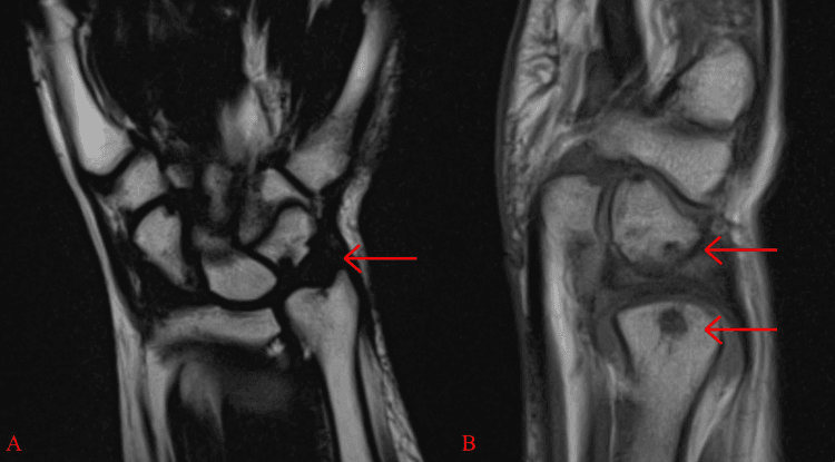
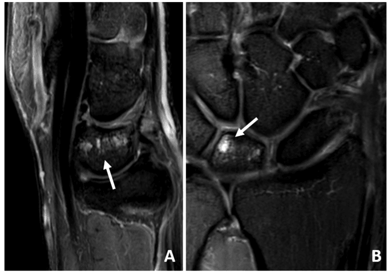
Not only the lunate. The impaction can centre on the triquetrum rather than only on the lunate. Positive variance then accompanies opposing subcortical cystic degeneration in the distal ulna and proximal triquetrum.
Classification Systems
Palmer's class 2 describes the degenerative TFCC tears of chronic ulnar impaction, distinct from the class 1 traumatic tears. The stage guides the treatment algorithm, and Palmer 2C is the commonest presentation requiring surgical intervention. Variance is the second axis, and ulnar impaction usually requires over 2mm of positive variance.
- TFCC Status
- Attritional thinning
- Associated Pathology
- No perforation, minimal chondromalacia
- Treatment
- Conservative, debridement
- TFCC Status
- Thinning + chondromalacia
- Associated Pathology
- Lunate or ulnar head cartilage damage
- Treatment
- USO or wafer
- TFCC Status
- Central perforation
- Associated Pathology
- TFCC tear + chondromalacia
- Treatment
- USO (gold standard)
- TFCC Status
- 2C + LT tear
- Associated Pathology
- Lunotriquetral ligament disruption
- Treatment
- USO + LT repair
- TFCC Status
- 2D + arthritis
- Associated Pathology
- Ulnocarpal arthrosis
- Treatment
- Darrach, SK, or replacement
The Ulnar-Sided Impaction and Impingement Spectrum
Ulnocarpal abutment sits within a spectrum of ulnar-sided impaction and impingement disorders. Examiners expect you to separate them, because each has a different key measurement and a different operation.
- Mechanism
- Long ulna abuts lunate/triquetrum with central TFCC wear
- Key measurement
- Positive ulnar variance (over 2 mm)
- Operation
- Ulnar shortening osteotomy or wafer (this topic)
- Mechanism
- Excessively long ulnar styloid abuts the triquetrum
- Key measurement
- Raised styloid process index (styloid length / ulnar head width, normal about 0.2-0.3); variance near-neutral
- Operation
- Ulnar styloid resection
- Mechanism
- SHORT distal ulna converges on the radius (post-Darrach, physeal arrest) - the opposite of impaction
- Key measurement
- Negative variance / short ulna, radioulnar convergence
- Operation
- Stabilisation/reconstruction, not shortening
- Mechanism
- Type II lunate with a medial facet articulates with the proximal hamate pole
- Key measurement
- Presence of a lunate medial (hamate) facet on imaging
- Operation
- Arthroscopic debridement of the proximal hamate pole
The most examinable error is applying an ulnar-shortening operation to ulnar impingement syndrome (a short ulna) or to stylocarpal impaction with near-neutral variance. In stylocarpal impaction it is the long styloid, not overall ulnar length, that abuts the triquetrum, so styloid resection is the answer; in a genuinely short converging ulna, further shortening worsens instability. Do not reflexively shorten the ulna for every ulnar-sided abutment: confirm variance AND the styloid process index before committing.
Clinical Assessment
History. The pain is dorsal-ulnar, worse with ulnar deviation, grip and pronation, and better with rest and avoiding provocative activities. Insidious onset over months to years is typical, and there may be occasional clicking with forearm rotation.
Examination. Swelling is usually minimal and the range of motion normal or slightly reduced. The hallmark is ulnar-sided tenderness over the ulnocarpal joint, just distal to the ulnar styloid, and grip is weak because it hurts. Assess DRUJ stability with the piano key sign, then run the provocative tests.
- Technique
- Axial load + ulnar deviation + pronation
- Positive Finding
- Reproduces ulnar pain
- Sensitivity
- 70-80%
- Technique
- Forearm pronation-supination with ulnar deviation
- Positive Finding
- Pain over TFCC
- Sensitivity
- 60-70%
- Technique
- Push up from chair using hands
- Positive Finding
- Ulnar-sided pain
- Sensitivity
- 50-60%
Differential diagnosis. Ulnar-sided wrist pain has several mimics.
- Key feature
- Load-related dorsal-ulnar pain, worse pronation/grip
- Variance
- Positive (over 2mm)
- Discriminator
- Lunate/ulnar-head sclerosis and cysts; central (2C) TFCC tear
- Key feature
- Trauma history, DRUJ instability
- Variance
- Often neutral/negative
- Discriminator
- Foveal tenderness, positive fovea sign, repairable peripheral tear
- Key feature
- Ulnar pain, painful clunk
- Variance
- Variable
- Discriminator
- Positive ballottement/shear test, LT widening
- Key feature
- Pain along ECU groove
- Variance
- Normal
- Discriminator
- Snapping with supination, tenderness over tendon not joint
- Key feature
- Volar-ulnar pain
- Variance
- Normal
- Discriminator
- Tender pisiform, pain on pisiform grind
- Key feature
- Pain on forearm rotation
- Variance
- Any
- Discriminator
- Positive piano-key, joint-space loss on radiograph
The decisive discriminator for UIS is positive ulnar variance plus subchondral sclerosis/cysts in the lunate and ulnar head. A traumatic peripheral (1B) tear with DRUJ instability is repaired and is NOT treated by shortening alone. Do not confuse it with the degenerative 2C tear: confirm variance and TFCC tear pattern before committing to USO.
Investigations
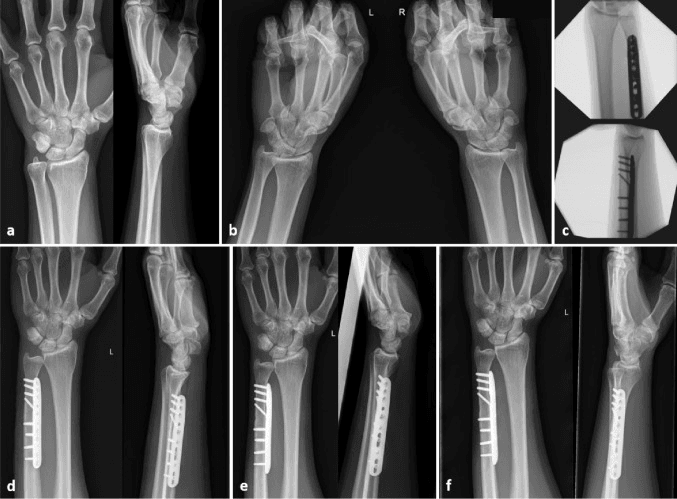
Plain radiographs. PA and lateral wrist radiographs come first. Measure the ulnar variance on the PA view and look for lunate sclerosis (increased density on the PA view), subchondral cysts in the ulnar head, and ulnocarpal joint narrowing, which marks end-stage Palmer 2E. A clenched-fist view may increase the variance and demonstrate pathology.
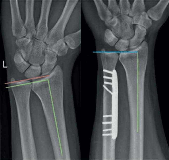
Measuring variance. Take the PA view with the forearm in neutral rotation, the shoulder abducted 90 degrees and the elbow flexed 90 degrees. Draw a perpendicular line from the ulnar articular surface to the radial articular surface. The variance is positive when the ulna extends distal to the radius and negative when it lies proximal to it.
Dynamic variance. Forearm rotation changes the reading. Pronation increases positive variance by 1-2mm and supination decreases it, which is why neutral rotation is critical; grip force also increases dynamic loading. Because rotation and grip both change the measurement, pre- and postoperative films must be taken in identical positions to be compared.
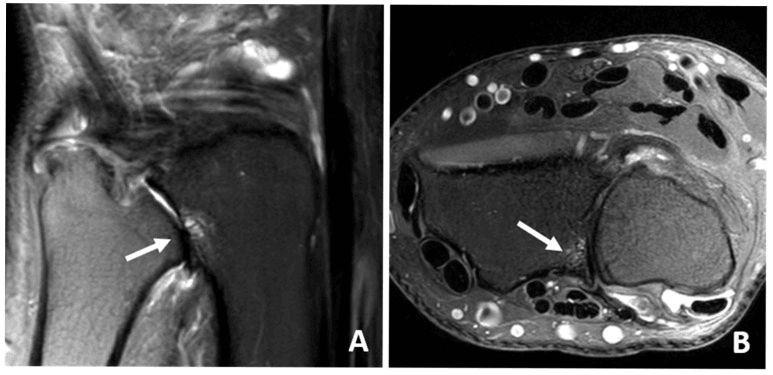
MRI. T1, T2 and proton density sequences show the TFCC central tear, lunate chondromalacia, bone marrow oedema and the state of the LT ligament. An MR arthrogram increases sensitivity for TFCC tears but is usually not necessary. The findings:
- TFCC central perforation: high signal on T2
- Lunate bone marrow oedema: low T1, high T2 signal
- Chondromalacia: cartilage thinning or defects
- LT ligament tear: discontinuity or high signal
Reading the MRI against the film. Lunate marrow oedema in the ulnocarpal loading zone is a supportive sign when correlated with positive or dynamic variance and TFCC degeneration. Neutral films can look normal while MRI shows a central triangular-disc perforation, so dynamic variance and tissue injury must be considered when the PA film appears normal.
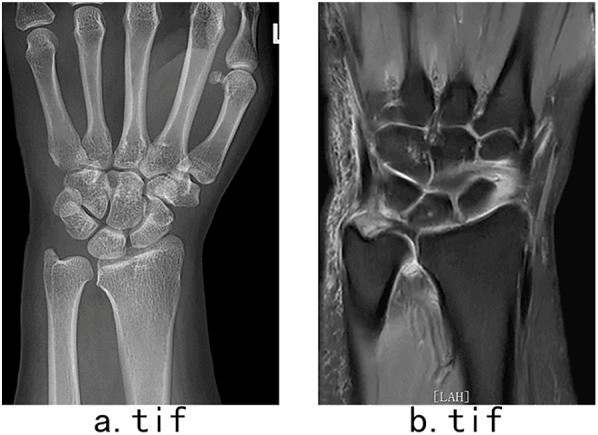
Arthroscopy. Wrist arthroscopy is the diagnostic and therapeutic gold standard. It directly visualises the TFCC tear and the extent of chondromalacia and allows treatment (debridement, wafer), and radiocarpal and midcarpal portals assess all the pathology.
Management Algorithm
Conservative treatment. A trial of 3-6 months is the reasonable initial approach for early disease: Palmer 2A, mild symptoms, or a patient who prefers a non-operative trial. It has three parts:
- Activity modification: avoid provocative activities, reduce repetitive pronation, grip and ulnar deviation, and modify the workplace ergonomically
- Splinting: a forearm-based splint immobilising the wrist in neutral, worn during activities and at night, may decrease symptoms 30-50%
- NSAIDs and injection: NSAIDs for inflammation; a corticosteroid injection into the ulnocarpal joint may provide temporary relief (weeks to months), with a limit of 2-3 injections
Of these patients, 30-40% improve. Palmer 2C and beyond usually require surgery, and for Palmer 2A, arthroscopic TFCC debridement alone is an option.
Unloading: shortening or wafer. Both operations aim to unload the ulnocarpal joint, and DRUJ stability must be preserved with either. Ulnar shortening osteotomy is the gold standard: indicated for Palmer 2C-2D with positive variance over 2-3mm after failed conservative management, it shortens the ulna by 2-4mm to neutral or 1mm negative variance. It is more powerful, but it needs 3-4 months of healing and therapy and carries the hardware burden.
The limit of the wafer. The arthroscopic wafer procedure suits Palmer 2B-2C with mild positive variance (under 2-3mm) in a patient who wants a faster recovery. The wafer resects only the distal 2-4mm of the ulnar head, so Feldon's original contraindication is positive variance over 4mm, and because the resection must also leave the styloid and its ligaments intact, most surgeons confine it to 2-3mm. Every comparative trial of wafer against USO capped entry at 4mm, so nothing is known about the wafer above that.
Weighing the wafer. Its advantages are a faster recovery, no hardware, no nonunion risk and an outpatient procedure. Against it, the shortening possible is limited (a maximum of 2-3mm), it is less powerful than USO, and it cannot address LT tears or severe pathology. It is effective for mild disease, while USO is preferred for significant variance.
Salvage for Palmer 2E. Ulnocarpal arthritis, a failed USO or wafer, and end-stage disease call for salvage. Severe DRUJ degeneration, with cartilage thinning, joint-space loss and subchondral cysts, likewise moves treatment from isolated unloading toward DRUJ salvage. The choice depends on age, activity and DRUJ status; distal ulna hemiresection (the Bowers procedure) is a rare alternative.
- Technique
- Excise the distal 1-2cm of ulna
- Suited to
- Low-demand, elderly patients; avoid in young, high-demand patients
- Advantages
- Simple; reliably relieves pain
- Drawbacks
- DRUJ instability (especially supination), ulnar stump pain, convergence
- Technique
- Fuse the DRUJ and create a pseudarthrosis proximally in the ulna
- Suited to
- Young patients needing stability
- Advantages
- Maintains DRUJ stability and good rotation; preserves an ulnar buttress; good pain relief with preserved function
- Drawbacks
- Proximal stump instability; technically demanding
- Technique
- Implant arthroplasty replacing the ulnar head
- Suited to
- DRUJ arthritis, failed Darrach
- Advantages
- Preserves stability and rotation; good short-to-medium-term results
- Drawbacks
- Implant wear, loosening, cost; long-term results unknown
The decision in one table.
- Variance
- Mild positive
- Treatment
- Conservative 3-6 months, NSAIDs, splint
- Key Pearl
- Many respond to activity modification
- Variance
- 2-3mm positive
- Treatment
- USO or wafer procedure
- Key Pearl
- Wafer faster recovery but less powerful
- Variance
- Over 3mm positive
- Treatment
- Ulnar shortening osteotomy
- Key Pearl
- Gold standard, 85-90% success
- Variance
- Any variance + arthritis
- Treatment
- Darrach, Sauvé-Kapandji, or ulnar head replacement
- Key Pearl
- Salvage procedures for end-stage
Surgical Technique
Planning the shortening. Measure the exact variance before surgery and plan the amount to remove. Too little fails to relieve symptoms; too much, over 4mm, risks DRUJ instability.
The osteotomy. A mid-diaphyseal or metaphyseal cut may take one of three configurations:
- Transverse: a perpendicular cut, easier but with delayed union
- Oblique: an angled cut, with a larger surface area for faster healing
- Step-cut: the most stable, but technically harder
Ulnar shortening osteotomy, step by step.
- Positioning and approach. Supine on an arm table with a tourniquet. A direct lateral or dorsal approach to the mid-distal ulna through an 8-10cm incision; identify and protect the dorsal sensory branch of the ulnar nerve.
- Exposure. Elevate ECU and FCU from the ulna by subperiosteal dissection, exposing 6-8cm of the shaft and protecting the interosseous membrane.
- Osteotomy. Mark the osteotomy site, then measure and mark the bone to remove (2-4mm, from the preoperative plan) before cutting, to ensure accuracy. Make the transverse or oblique cut with a saw, remove the segment and compress the osteotomy.
- Plate fixation. Apply a 6-8 hole compression plate with 3-4 screws each side of the osteotomy. Achieve rigid fixation with maximum interfragmentary compression, which decreases nonunion; a locking or non-locking plate is acceptable, and a low-profile plate minimises prominence.
- DRUJ check and closure. Release the tourniquet and check DRUJ stability with forearm rotation, which should be smooth and stable. If the DRUJ is unstable, the shortening is excessive and should be revised. Close in layers and splint in neutral.
Guides and parallel cuts. A patient-specific guide translates the planned shortening and obliquity to the ulna, controls the saw cuts and plate holes, and permits compression to the intended postoperative variance. Parallel guided cuts create a minimal gap that accepts lag-screw compression; nonparallel freehand cuts leave a long reduction gap that the lag screw cannot compress, and union then comes slowly.
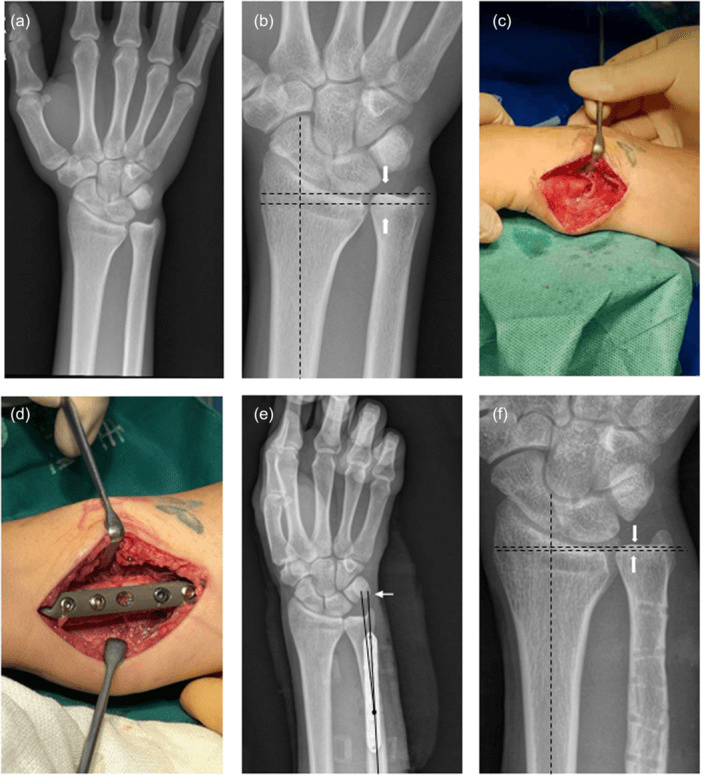
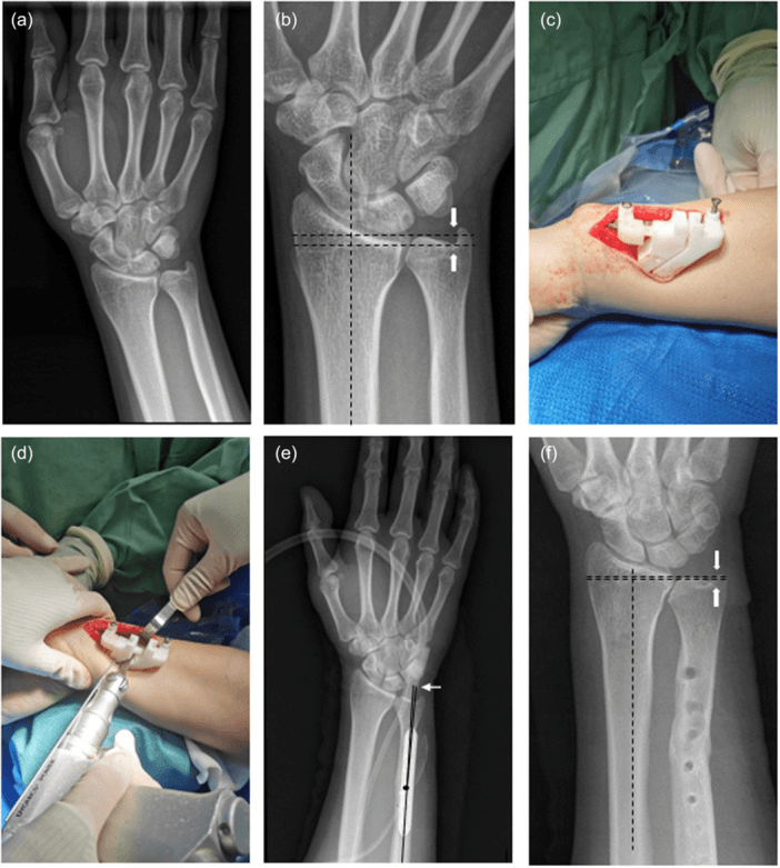
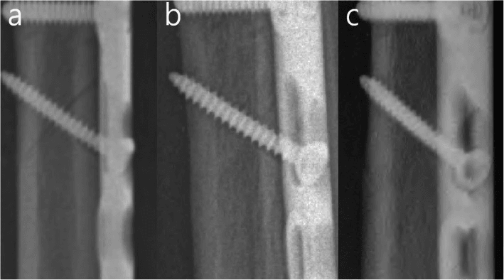
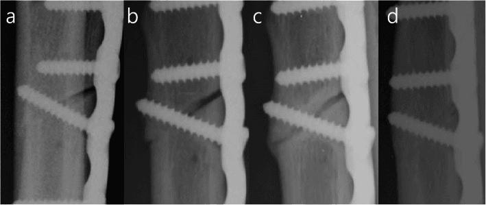
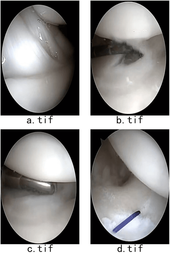
Arthroscopic wafer, step by step. The wafer is a technically demanding arthroscopic procedure, and adequate visualisation is critical.
- Set-up. Wrist arthroscopy tower with 10-15 lbs of traction; radiocarpal 3-4 and 6R portals. Inspect systematically: radiocarpal joint, TFCC, lunate, scaphoid.
- TFCC assessment. Probe the central TFCC, identify the degenerative perforation and unstable flaps, and grade the Palmer class arthroscopically.
- TFCC debridement. Debride the degenerative flap with a shaver to a stable rim, using the radiofrequency probe for haemostasis. Preserve the peripheral 1-2mm of the TFCC, which is vascular and stabilising.
- Ulnar dome resection. Pass the arthroscopic burr through the radiocarpal joint and burr 2-3mm from the distal ulnar dome (articular surface); more causes instability. Smooth the contour to prevent catching, and use the probe to verify adequate decompression and the space created.
- Final check. Visualise the decompression and a smooth ulnar surface, secure haemostasis, close the portals and apply a soft dressing.
Complications
- Early Complications
- Infection, nerve injury (dorsal sensory)
- Late Complications
- Nonunion (3-6%), hardware prominence
- Management
- ORIF with bone graft, hardware removal
- Early Complications
- Portal site infection, synovitis
- Late Complications
- Recurrent impaction (if inadequate resection)
- Management
- Revision to USO
- Early Complications
- Haematoma, nerve injury
- Late Complications
- DRUJ instability, convergence, ulnar stump pain
- Management
- Revision to SK or ulnar head replacement
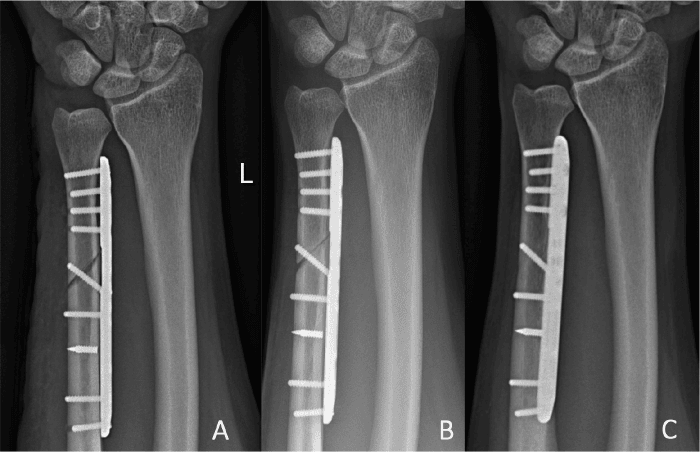
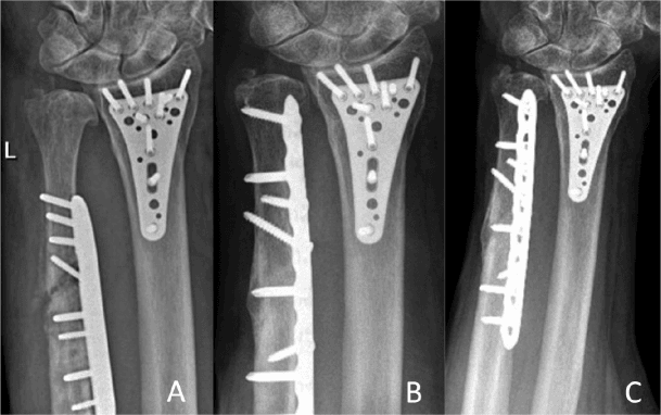
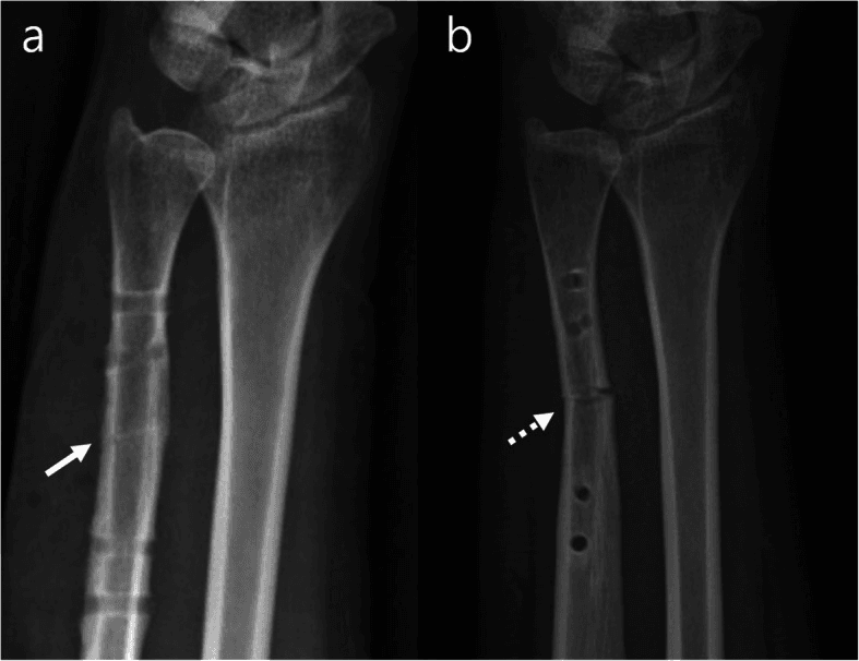
Hardware irritation. Hardware prominence or irritation (10-15%) is the most common complication of USO and dominates pooled series, where the plate needs removal in roughly 1 in 8. Low-profile or buried implants help prevent it. Remove the plate only once union is confirmed and remodelled: a persistent osteotomy trace can precede refracture after removal, so hardware should not be removed solely for irritation until cortical remodelling and consolidation are convincing.
Nonunion. Nonunion is uncommon, about 3-6%, and smoking, diabetes and inadequate fixation are the risk factors. Prevent it with rigid fixation, interfragmentary compression and smoking cessation; treat it with ORIF, bone graft and compression plating.
Other complications of USO. DRUJ stiffness occurs in 5%, and the dorsal sensory branch of the ulnar nerve can be injured. DRUJ instability follows excessive shortening (over 4mm) and is worse in supination, with dorsal subluxation; splint it if mild and perform a Sauvé-Kapandji procedure if severe.
Persistent pain after USO. Persistent pain requires direct assessment of the osteotomy rather than an assumption that the symptoms are TFCC in origin. The causes:
- Hardware prominence, the most common: remove the plate after union
- Inadequate shortening: consider revision
- Unrecognised pathology (LT tear, arthritis): MRI and arthroscopy
Postoperative Care
After shortening. The osteotomy is protected until it consolidates; a fall before union can loosen the plate. Full activities follow by 3-4 months if healed:
- Weeks 0-2: sugar-tong splint or cast; finger and shoulder range of motion; no forearm rotation; wound check at 10-14 days
- Weeks 2-6: removable splint; gentle wrist range of motion from week 3-4; no strengthening; radiograph at 6 weeks to assess union
- Weeks 6-12: progressive strengthening if union is progressing, with therapy for range of motion and grip
After a wafer. With no osteotomy to heal, rehabilitation is much faster:
- Weeks 0-2: soft dressing and removable splint; finger range of motion immediately; wrist range of motion from 1-2 weeks
- Weeks 2-6: progressive strengthening, with return to normal activities by 6-8 weeks
- Wafer
- 2-3 weeks
- USO
- 4-6 weeks
- Wafer
- 6-8 weeks
- USO
- 10-12 weeks
- Wafer
- 12 weeks
- USO
- 16 weeks
Outcomes and Prognosis
- Pain Relief
- 30-40% improvement
- Function Preservation
- Maintained
- Durability
- Temporary, often progresses
- Pain Relief
- 85-90% good-excellent
- Function Preservation
- Grip 85-90% normal
- Durability
- Durable 10+ years
- Pain Relief
- 70-80% good
- Function Preservation
- Grip 80-85% normal
- Durability
- Good 5-7 years
- Pain Relief
- 80-90% pain relief
- Function Preservation
- Instability issues
- Durability
- Variable
The USO figure is good-to-excellent pain relief with functional improvement; the wafer figure applies to appropriate indications.
Prognostic factors.
- Better Outcome
- 2B-2C
- Worse Outcome
- 2E (arthritis)
- Better Outcome
- 2-4mm (correctable)
- Worse Outcome
- Over 6mm
- Better Outcome
- Under 60
- Worse Outcome
- Over 65
- Better Outcome
- Good therapy adherence
- Worse Outcome
- Non-compliant
Natural history. Untreated, the disease progresses from Palmer 2A to 2E over 5-15 years. Most symptomatic patients eventually require surgery, and conservative management only temporises.
Guidelines, Registries & Global Practice
Global epidemiology: Ulnar impaction syndrome is one of the commonest causes of chronic ulnar-sided wrist pain worldwide. Positive ulnar variance is present in roughly 10-15% of the general population, but only a subset becomes symptomatic. Manual workers (construction, agriculture, mining), gymnasts and athletes loading the wrist in pronation are over-represented. There is no dedicated international registry; high-quality evidence comes from cohort series and a small number of randomised trials and meta-analyses.
Guideline and society positions (side by side):
- Position on UIS
- No disease-specific guideline; consensus favours staged care — conservative trial, then USO for significant variance, arthroscopic wafer for mild variance with stable DRUJ
- Position on UIS
- Emphasise MRI/arthroscopy confirmation and Palmer staging; USO regarded as the workhorse for positive-variance impaction
- Position on UIS
- Recommend correcting variance to neutral or slight negative; preserve DRUJ stability and foveal TFCC attachment
- Position on UIS
- Provides technical standards for diaphyseal USO — rigid compression plating, oblique or step-cut osteotomy to reduce nonunion
There is broad international agreement on the principle (unload the ulnocarpal joint by correcting positive variance) and the staged algorithm; the main variation is the threshold and enthusiasm for arthroscopic wafer versus formal USO.
No arthroplasty-style registry tracks UIS. The best pooled evidence is the meta-analysis of USO versus arthroscopic wafer (no difference in patient-rated outcomes; grip favours wafer, variance correction favours USO) and a systematic review of isolated USO performed for distal radius malunion reporting a 33% overall complication rate dominated by implant irritation (removal in ~13%) with nonunion only ~3% — attach that qualifier whenever the figure is quoted, because a previously fractured and often previously operated wrist is not the same risk as an idiopathic impaction.
- Well-resourced settings: routine MRI and wrist arthroscopy; arthroscopic wafer and ulnar head arthroplasty available; low-profile/locking USO plates standard.
- Limited-resource settings: diagnosis is often clinical plus plain radiographs (ulnar variance, lunate sclerosis, cysts); USO with conventional compression plating remains the mainstay because it needs no arthroscopy tower; salvage defaults to Darrach or Sauvé-Kapandji rather than implant arthroplasty.
Counsel for nonunion (uncommon but increased by smoking and diabetes), hardware prominence frequently requiring later removal, DRUJ stiffness, dorsal sensory ulnar nerve injury and a 3-4 month recovery. Document exact pre-operative variance and plan the precise amount of shortening; follow with serial radiographs to confirm union. Smoking cessation is the single most useful modifiable factor for bone healing.
Related pages: Ulnar-Sided Wrist Pain is the parent presentation and the differential this diagnosis must be extracted from - impaction is one cause among many and the others are excluded before an osteotomy is contemplated; TFCC Injuries carries the Palmer classification in full and, more importantly, the foveal attachment that this page's Nishiwaki card shows must be intact for shortening to stabilise the DRUJ - a complete foveal avulsion needs repair and cannot be shortened into stability; Lunotriquetral Instability is the Palmer 2D lesion and coexists often enough that it must be assessed arthroscopically at the same sitting; DRUJ Arthritis is both a contraindication to the wafer and the endpoint (Palmer 2E) that pushes management to salvage - Darrach, Sauvé-Kapandji or ulnar head replacement; Distal Radius Fractures are the commonest cause of SECONDARY impaction through radial shortening and malunion, and the population in which the 33% complication figure was measured; Kienbock's Disease is the key differential for lunate signal change - note that it associates with NEGATIVE ulnar variance, the opposite of this condition, which is the discriminator examiners use; Madelung Deformity produces positive variance by a growth mechanism and presents younger; and Scapholunate Dissociation and Hamate Fractures complete the differential for mechanical wrist pain with a normal initial radiograph.
Controversies and Areas of Uncertainty
The wafer-versus-shortening threshold. The variance cut-off for choosing the wafer over USO is not firmly evidence-based. Trials and meta-analysis pooling subtle-variance patients (under 4mm) show equivalent patient-rated outcomes, with the wafer offering faster recovery and fewer complications, and most surgeons reserve it for stable-DRUJ, low-variance disease.
Osteotomy configuration. Oblique and step-cut osteotomies increase the union surface and allow interfragmentary compression, but high-quality comparative data are limited. The dominant complication is implant irritation requiring removal, not nonunion, and that has reframed the debate toward low-profile fixation.
Does shortening help an unstable DRUJ? Shortening re-tensions the TFCC and can stabilise the DRUJ, but cadaveric data show this works only if the radioulnar ligament remains attached to the fovea. A complete foveal avulsion is not corrected by shortening and requires separate foveal repair.
Salvage choice in Palmer 2E. For end-stage ulnocarpal or DRUJ arthritis there is no consensus winner. Selection is driven by age, demand and resources rather than by robust comparative trials.
MCQ Practice Points
Q: What amount of positive ulnar variance typically causes ulnar impaction syndrome? A: Over 2mm positive ulnar variance significantly increases ulnocarpal loading and predisposes to UIS. Normal population: 60-70% neutral variance, 10-15% positive variance (usually under 2mm).
Q: What defines Palmer 2C TFCC tear? A: Central TFCC perforation (degenerative tear) PLUS chondromalacia of lunate or ulnar head. Most common stage requiring surgical intervention.
Q: What is the variance threshold for choosing USO over wafer procedure? A: Over 2-3mm positive variance warrants USO. Wafer procedure can only address 2-3mm maximum shortening; greater variance requires formal ulnar shortening osteotomy.
Q: What is the success rate of ulnar shortening osteotomy for UIS? A: 85-90% good to excellent outcomes for pain relief and functional improvement in cohort series. The commonest complication is hardware irritation (plate removal in roughly 13%); nonunion is uncommon (about 3-6%).
Q: How does ulnar shortening decrease ulnocarpal loading? A: Positive ulnar variance shifts axial load onto the ulnocarpal joint and TFCC; each millimetre of positive variance increases ulnocarpal load substantially. Shortening to neutral (or slight negative) redistributes load back through the radius and re-tensions the TFCC, which can also improve DRUJ stability when the foveal attachment is intact.
Q: What are salvage options for Palmer 2E (ulnocarpal arthritis)? A: Darrach procedure (distal ulna excision), Sauvé-Kapandji (DRUJ fusion with proximal pseudarthrosis), or ulnar head arthroplasty. Selection based on age and activity level.
Exam Viva Scenarios
Practise clinical reasoning and management decisions out loud
“A 52-year-old carpenter presents with 12 months of ulnar-sided wrist pain, worse with gripping and pronation. X-rays show 3mm positive ulnar variance with mild lunate sclerosis. MRI shows Palmer 2C TFCC central tear and lunate chondromalacia. Conservative management with splint has failed over 3 months.”
“A 45-year-old woman has ulnar-sided wrist pain with 1.5mm positive ulnar variance. MRI shows Palmer 2B-2C tear. She is active and wants fastest return to work. What do you recommend?”
“A patient returns 6 months after USO with persistent ulnar pain. X-rays show the osteotomy has healed, and variance is now neutral. What is your approach?”
Key Pathophysiology
- Positive ulnar variance = ulna longer than radius by over 2mm
- Excessive ulnocarpal loading causes TFCC degeneration
- Each 1mm positive variance increases TFCC load significantly
- Biomechanical problem requiring mechanical solution
Palmer Classification (Class 2)
- 2A: TFCC wear, no perforation
- 2B: Wear + chondromalacia
- 2C: Central perforation + chondromalacia (most common surgical)
- 2D: 2C + LT tear
- 2E: 2D + ulnocarpal arthritis (end-stage)
Clinical Diagnosis
- Ulnar-sided wrist pain, worse with pronation and grip
- Ulnar impaction test: axial load + ulnar deviation reproduces pain
- X-ray: measure ulnar variance (PA view, neutral rotation)
- MRI: TFCC tear, chondromalacia, bone marrow edema
Treatment Algorithm
- Conservative: activity modification, splint, injection (30-40% success)
- Wafer: arthroscopic resection for variance under 2-3mm (70-80% success)
- USO: gold standard for variance over 2-3mm (85-90% success)
- Salvage: Darrach, SK, or ulnar head replacement for 2E
USO Technique
- Shorten ulna 2-4mm to neutral or 1mm negative variance
- Rigid plate fixation with compression (prevent nonunion)
- Check DRUJ stability intraoperatively
- Recovery 3-4 months, nonunion risk 3-6%
Evidence Base
Baek et al — USO for Idiopathic UIS
- 31 wrists (29 patients) with idiopathic UIS treated by USO
- Mean ulnar variance reduced from +4.6mm to -0.7mm
- Modified Gartland-Werley score improved from 69.5 to 92.5; 29 of 31 good or excellent
- Dorsal DRUJ subluxation reduced and carpal cysts resolved after shortening
Feldon, Terrono & Belsky — Original Wafer Description
- 13 wafer resections in 12 patients; distal 2-4mm of ulnar head removed, styloid and ligaments preserved
- All good-to-excellent at minimum 1-year follow-up
- Explicitly contraindicated if positive ulnar variance exceeds 4mm
- Contraindicated with DRUJ instability or DRUJ degenerative arthritis
Afifi et al — AWP vs USO Randomised Trial
- 60 patients with idiopathic UIS randomised to arthroscopic wafer (AWP) or USO
- No significant difference in DASH, Modified Mayo Wrist Score, VAS, ROM or grip at 2 years
- All USO patients united by 12 weeks
- AWP had earlier return to work, fewer complications and fewer secondary procedures
Oh, Choi et al — AWP vs USO Comparative Cohort
- 42 patients aged 45+ with Palmer 2C/2D and under 4mm positive variance, stable DRUJ
- Equivalent clinical outcomes at 6, 12 and 24 months
- AWP superior at 3 months for grip, Mayo Wrist Score and DASH
- Complication rate 34.8% for USO (implant irritation, DRUJ arthritis, refracture) vs 10.5% for AWP
Yu et al — AWP vs USO Systematic Review & Meta-analysis
- 7 studies, 133 USO and 118 AWP patients pooled
- No difference in Mayo Wrist Score, DASH, VAS, revision rate or time to return to work
- Grip strength favoured AWP (SMD -0.73)
- Correction of positive variance favoured USO; overall evidence quality moderate
Nishiwaki et al — Ulnar Shortening and DRUJ Stability
- Cadaveric study of DRUJ stiffness with progressive ulnar shortening
- 6mm shortening increased DRUJ stiffness 26-44% across rotation positions
- Stabilising effect depends on radioulnar ligament remaining attached to the fovea
- Complete foveal RUL avulsion abolishes the stabilising benefit of shortening
Laane et al — Complications of Isolated USO
- 12 cohorts, 185 patients undergoing isolated USO after distal radius malunion
- Overall complication rate 33% (95% CI 16-51%)
- Implant irritation most common (22%); removal required in 13%
- Nonunion uncommon at 3%; buried/low-profile implants may reduce irritation
Palmer — TFCC Lesion Classification
- Original classification dividing TFCC lesions into Class 1 (traumatic) and Class 2 (degenerative)
- Class 2 degenerative tears (2A-2E) reflect progressive ulnocarpal impaction
- 2C (central perforation + chondromalacia) is the common surgical lesion
- Framework still underpins treatment algorithms for ulnar-sided wrist pain