LTIL Injury | VISI Deformity | LT Arthrodesis
- An isolated LTIL tear does not automatically create static VISI; secondary extrinsic ligament failure is usually required
- VISI means volar flexion of the lunate on a true lateral view; use scapholunate and capitolunate relationships with the whole carpal alignment
- Arthroscopy directly grades intercarpal instability and evaluates cartilage and associated lesions
- LT arthrodesis has variable union, pain and satisfaction and is not a universal gold standard
- Timing, reducibility, tissue, ulnar variance, cartilage and associated injury guide repair, reconstruction, shortening, fusion or salvage
- “VISI is the volar-flexed intercalated-segment pattern; do not diagnose it from one poorly positioned lateral radiograph
- “Dynamic LT instability may have normal static radiographs
- “Ballottement pain or laxity is supportive but not specific; compare the opposite side and examine TFCC, ECU and ulnocarpal loading
- “No single operation is preferred for every chronic LT instability phenotype
Overview and Epidemiology
Lunotriquetral (LT) instability is a spectrum of injury to the lunotriquetral interosseous ligament (LTIL) and the secondary carpal instability that may follow it, presenting as ulnar-sided wrist pain. An LTIL tear may be occult or dynamic; a fixed VISI generally indicates broader failure of the secondary restraints.
Describing the injury. The LTIL label alone does not decide treatment, so describe each of these:
- Symptoms and dynamic laxity
- The LT interval and associated TFCC and ulnocarpal pathology
- Static carpal alignment and reducibility
- Cartilage and the secondary extrinsic restraints
Who gets it. LT instability is uncommon and frequently missed among ulnar-sided wrist disorders, and it is one of the harder diagnoses because radiographs are often normal. It may follow trauma or occur with perilunate and other carpal injuries. No robust age, sex or incidence denominator supports a fixed patient profile.
Anatomy and Pathophysiology
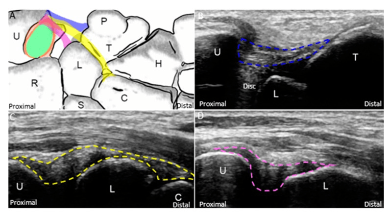
The ligament. The LTIL has dorsal, proximal membranous and volar components connecting the lunate and triquetrum, and the volar component is substantial. The complex works with the dorsal radiocarpal, dorsal intercarpal and ulnocarpal restraints, so an LTIL injury may permit translation or rotation without a fixed radiographic deformity.
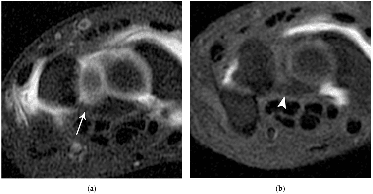
Carpal kinematics. The proximal row moves as an intercalated segment under competing scaphoid and triquetral forces. After an isolated intrinsic injury the secondary ligaments may preserve alignment; when they fail as well, the lunate may assume persistent volar flexion. That is VISI (volar intercalated segment instability), volar flexion of the lunate relative to the radius and capitate, the counterpart of DISI, in which the lunate extends dorsally after scapholunate injury.
From dynamic to static. When only the LTIL is torn, the triquetrum can be ballotted on the lunate and pain or crepitus reproduced, but the proximal row still tracks together and the lateral radiograph is normal. Reagan, Linscheid and Dobyns described this "LT sprain" as a discrete, often-unrecognised cause of ulnar wrist pain. Cadaver and clinical work shows that sectioning the LTIL alone yields only dynamic instability.
The secondary stabilisers. An isolated LTIL tear does not usually produce a fixed VISI. That usually requires attrition or rupture of the extrinsic secondary stabilisers as well, principally:
- The dorsal radiocarpal (dorsal radiotriquetral) ligament
- The palmar ulnocarpal ligaments (ulnolunate/ulnotriquetral arcuate limb)
Only when these give way does the lunate lose its dorsal and volar tethers and drop into a fixed volar-flexed posture, which Reagan termed progression to LT dissociation. A static VISI is therefore a more advanced injury than an isolated LTIL tear: the extrinsic envelope has failed too, so a simple LTIL repair is unlikely to restore alignment.
The reverse-perilunate spectrum. The same secondary-constraint concept explains an ulnar-to-radial progression of ligament failure that starts at the LT joint. The full perilunate and greater-arc pattern and Mayfield staging are covered in the perilunate-dislocations topic.
Degenerative tears. Many "LT tears" are actually degenerative, driven by positive ulnar variance and ulnocarpal impaction; what that means for treatment is covered under Management.
In the viva: a positive ballottement with a normal lateral film is dynamic instability, where repair or reconstruction of the LTIL is most logical and best-timed early. A fixed volar lunate tilt is a more extensive injury with secondary-constraint failure, favouring realignment or arthrodesis over isolated LTIL repair.
Classification Systems
LT injuries are classified by severity, timing and associated injury.
By severity. The severity pattern guides the treatment approach:
- Predynamic / dynamic. Pain, clicking or arthroscopic laxity, with static PA and lateral radiographs that may remain normal. Treatment depends on symptoms, associated pathology and repairability; the evidence is heterogeneous.
- Static VISI. Fixed volar lunate alignment on a true lateral view, which usually implies secondary ligament failure. Assess reducibility and cartilage before reconstruction, fusion or salvage, and aim to restore whole-carpus alignment.
- Arthritic / combined. LT cartilage loss or broader carpal collapse. Treat the whole wrist pathology rather than the LTIL label; there is no verified universal success rate.
By timing. A recent injury with mobile, reducible alignment and viable tissue may permit direct repair, and any associated perilunate or extrinsic-ligament injury must also be restored. In a chronic, deficient injury, scar, attenuation, malalignment or cartilage loss may require reconstruction, shortening, fusion or salvage. Direct repair is most feasible before retraction and degeneration, but no universal six-week cut-off is validated to select the operation.
By associated injury. An isolated LT injury gets LT-specific treatment. With a perilunate dislocation the LT tear is part of a greater arc injury and every component must be addressed; with other carpal injuries several ligaments are involved and the reconstruction must be comprehensive. Associated injuries affect management.
Arthroscopic grading (Geissler)
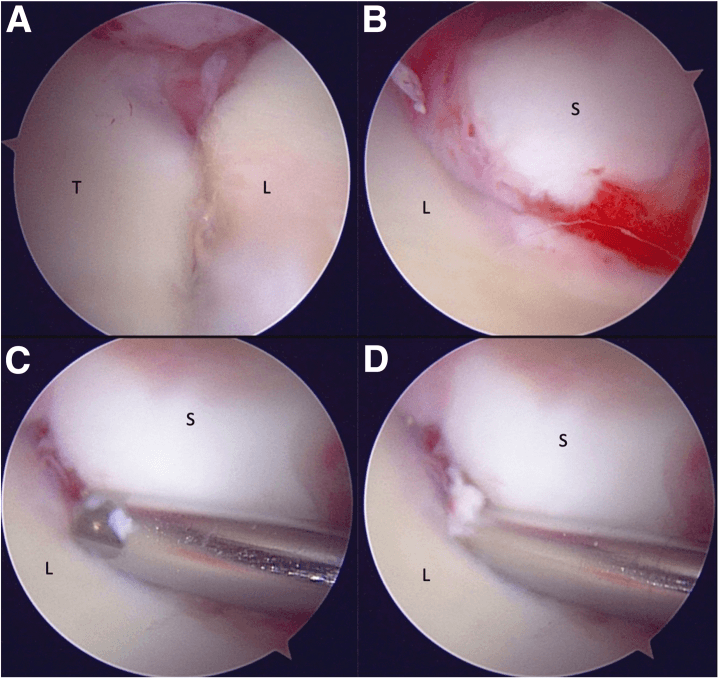
The Geissler classification is the standard arthroscopic scale for intrinsic (interosseous) carpal ligament injury. It is most familiar from scapholunate injury but is equally used to stage the LT interval, inspected from both the radiocarpal and the midcarpal portals.
- Radiocarpal view
- Attenuation or haemorrhage of LTIL; no step-off
- Midcarpal view / probe test
- Carpal alignment congruent; no gap
- Typical management concept
- Immobilisation / debridement; often stable
- Radiocarpal view
- Attenuation/haemorrhage of LTIL
- Midcarpal view / probe test
- Incongruency/step-off; gap smaller than a probe tip
- Typical management concept
- Debridement +/- pinning; early instability
- Radiocarpal view
- Step-off/gap at LT interval
- Midcarpal view / probe test
- Probe passes between lunate and triquetrum
- Typical management concept
- Reduction and pinning / repair
- Radiocarpal view
- Gross step-off/gap
- Midcarpal view / probe test
- Arthroscope passes freely across the interval (drive-through sign); gross instability
- Typical management concept
- Repair or reconstruction; consider arthrodesis if chronic
Higher grades correlate with more complete ligament disruption and a greater likelihood of requiring stabilisation rather than debridement alone. The grade complements, rather than replaces, the dynamic-versus-static distinction on plain radiographs.
The Geissler grade is defined largely by the midcarpal appearance. Inspecting the LT joint from the radiocarpal portal alone under-grades the injury, so always assess from the midcarpal portal.
Clinical Assessment
History. The patient reports ulnar wrist pain, clicking or clunking with movement, weakness of grip, or a feeling of the wrist giving way. Mechanisms include:
- A fall on the outstretched hand with ulnar deviation, or other fall or torsional loading
- Direct trauma to the ulnar side of the wrist
- High-energy trauma with perilunate dislocation
- Repetitive ulnocarpal loading
Examination. Look for swelling on the ulnar side of the wrist and, if the instability is static, a VISI deformity with prominence of the ulnar head. The LT interval, just ulnar to the lunate, is tender, and there may be crepitus at the LT joint. Wrist motion may be limited, with pain on ulnar deviation and clicking through the arc.
The LT ballottement (Reagan's) test is the key clinical test:
- Stabilise the lunate with one hand
- Translate the triquetrum dorsally and palmarly with the other
- Pain, crepitus or increased motion is positive
- Compare with the contralateral side
Ballottement pain or laxity is supportive but not specific, because it overlaps with other ulnar-sided disorders. Examine the TFCC, ECU and ulnocarpal loading, and evaluate ulnar variance and the other intercarpal ligaments, rather than attributing all ulnar pain to the LT interval.
Other tests. The LT compression and ulnar snuffbox tests add to ballottement:
- LT compression test. With the wrist in ulnar deviation, apply an axial load through the ring and small finger metacarpals; pain at the LT interval is positive.
- Ulnar snuffbox test. Palpate the ulnar snuffbox, between the triquetrum and the ulnar styloid; tenderness indicates LT injury.
Differential diagnosis. Ulnar-sided wrist pain has many causes, and these are the key mimics to exclude.
- Key Distinguishing Feature
- Positive ballottement; VISI on lateral (if static)
- Best Test
- Arthroscopy / dynamic stress views
- Key Distinguishing Feature
- Pain on ulnocarpal stress/grind; foveal tenderness
- Best Test
- MRI / arthroscopy
- Key Distinguishing Feature
- Positive ulnar variance; lunate/triquetrum chondral lesions
- Best Test
- PA grip radiograph + MRI
- Key Distinguishing Feature
- Piano-key sign; pain on forearm rotation
- Best Test
- Clinical + CT in pro/supination
- Key Distinguishing Feature
- Pain over pisiform; positive grind
- Best Test
- 30-degree supinated oblique X-ray
- Key Distinguishing Feature
- Snapping ECU with supination/ulnar deviation
- Best Test
- Dynamic ultrasound / MRI
- Key Distinguishing Feature
- Point tenderness over hook; grip pain
- Best Test
- Carpal tunnel view / CT
Investigations
Radiographs. Dynamic LT instability may have normal static radiographs. The views and what each shows:
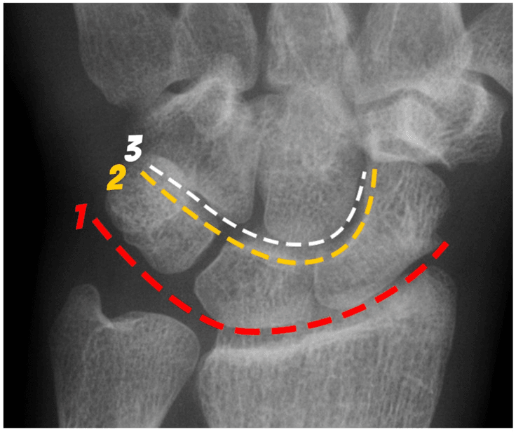
- PA. The LT interval may be widened and carpal height may be reduced in VISI; measure ulnar variance. Gilula's arcs screen proximal- and distal-row congruity.
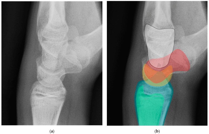
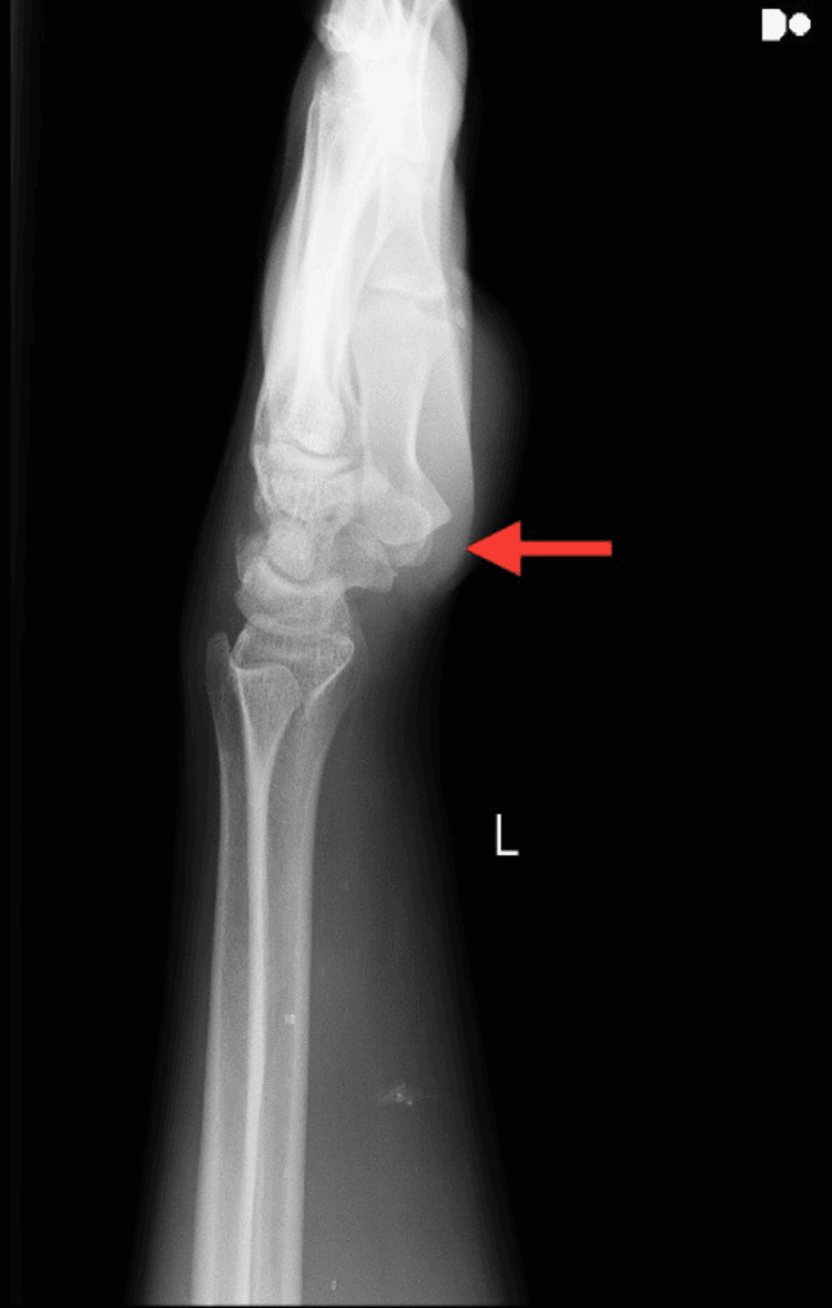
- Lateral. The critical view, essential for diagnosing VISI, with the lunate flexed volarly.
- Clenched-fist view. May show dynamic instability as widening of the LT interval.
Reading the lateral. Use a true lateral, with the radius, lunate, capitate and third metacarpal aligned, and do not diagnose VISI from one poorly positioned film. The scapholunate ligament is intact, but volar flexion of the lunate decreases the scapholunate angle and alters the capitolunate relationship. A capitolunate angle below 0° (normal 0-15°) is quoted for VISI, but do not use it as an isolated rule: read it with the scapholunate relationship and the whole carpal alignment.
CT and MRI. CT assesses for fractures and is not routine for LT instability. MRI assesses LTIL integrity and may show the tear, but is not always necessary if the clinical diagnosis is clear.
Arthroscopy. Arthroscopy is the reference assessment and the most reliable test. It visualises the LTIL directly, grades intercarpal instability, shows the location and severity of the tear, evaluates cartilage and associated lesions, and allows repair or debridement. Central LTIL perforations (Palmer 1B type, degenerative) may be incidental and asymptomatic, so correlate the arthroscopic findings with clinical instability rather than treating every tear seen.
Management Algorithm
Non-operative treatment. Suited to mild symptoms, dynamic instability only, patient preference or medical contraindications to surgery. It consists of activity modification, a wrist splint, NSAIDs and physiotherapy. Success is limited, and the instability may progress to static VISI.
Indications for surgery. Static VISI deformity, failed non-operative treatment and significant functional impairment are absolute indications. Dynamic instability with persistent symptoms and patient preference for definitive treatment are relative ones.
Choosing the operation. Timing, reducibility, tissue, ulnar variance, cartilage and associated injury guide the choice between repair, reconstruction, ulnar shortening, fusion and salvage. No single operation is preferred for every chronic LT instability phenotype. Direct repair is offered for acute injuries (less than 6 weeks) and arthrodesis for chronic ones, but that boundary is not validated as universal; tissue, reducibility, associated ligaments and cartilage decide.
- Acute and repairable. Direct repair with suture anchors through a dorsal or palmar approach, which gives better outcomes than repair of a chronic tear.
- Chronic or static VISI. Reconstruction, selected arthrodesis or salvage, according to reducibility and cartilage.
- LT joint arthritis. LT fusion addresses both the instability and the pain. Severe arthritis or a failed fusion calls for salvage: proximal row carpectomy (PRC) or wrist fusion.
Arthrodesis versus reconstruction. The traditional teaching is that LT arthrodesis is the "gold standard" for chronic instability, because the LT joint contributes less than 5% of wrist motion and the fusion is reported to relieve pain reliably. That is challenged by series reporting nonunion rates up to ~45% (Vandesande 2001), and published union and satisfaction are variable. Arthrodesis may stabilise selected chronic reducible instability, but not every chronic tear needs fusion: assess the cartilage before choosing it, and correct the global carpal alignment, not just the LT gap.
The alternatives. Soft-tissue reconstruction (e.g. ECU strip, Pillukat 2015) and capsulodesis are increasingly favoured in some centres to preserve motion and proprioception. No randomised data exist to settle the choice.
Treat the ulnar variance. In a degenerative tear driven by positive ulnar variance and ulnocarpal impaction, ulnar-shortening osteotomy, not LT surgery, may be the definitive treatment. Failing to assess variance is a classic error.
Surgical Technique
LT arthrodesis
Indications. Chronic LT instability (over 6 weeks), static VISI deformity, or failed repair or reconstruction.
Approach. A dorsal ulnar incision between the 4th and 5th extensor compartments. Identify and protect the DRUJ and the ECU tendon, and expose the LT joint through a dorsal capsulotomy.
Technique. The steps of the fusion:
- Remove the articular cartilage from the lunate and triquetrum with curettes or a burr
- Pack autograft or allograft bone graft
- Fix with a headless cannulated compression screw, or a plate and screws
- Confirm reduction and hardware position fluoroscopically
Direct repair
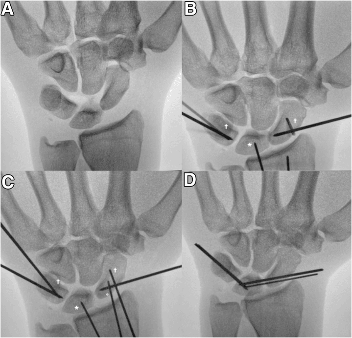
Direct repair suits an acute injury (less than 6 weeks) with good tissue quality and no fixed deformity. Through a dorsal or palmar approach, expose the LTIL and identify where it has torn. Repair it with suture anchors in the lunate and triquetrum, which may be augmented with K-wires, then immobilise in a cast (see Postoperative Care).
Ligament reconstruction
Reconstruction is used for chronic instability, failed repair, or young, high-demand patients as an alternative to arthrodesis. A tendon graft (FCR or ECU) is passed through bone tunnels and tensioned appropriately. Outcomes are less predictable than arthrodesis, with a higher failure rate.
Complications
- Incidence
- 10-15%
- Risk Factors
- Smoking, poor fixation, inadequate graft
- Prevention/Management
- Rigid fixation, bone graft, smoking cessation
- Incidence
- 10-15%
- Risk Factors
- Incomplete fusion, arthritis
- Prevention/Management
- Complete cartilage removal, adequate graft
- Incidence
- 5-10%
- Risk Factors
- Prominent screws
- Prevention/Management
- Countersink screws, remove if symptomatic
- Incidence
- 5-10%
- Risk Factors
- Inadequate fixation
- Prevention/Management
- Rigid fixation, compression
Nonunion is managed by revision fusion with bone graft. The table's 10-15% is lower than published arthrodesis series reporting nonunion up to ~45% (see Management).
Persistent pain may come from incomplete fusion, arthritis or other pathology. Assess for other causes and consider revision.
Postoperative Care
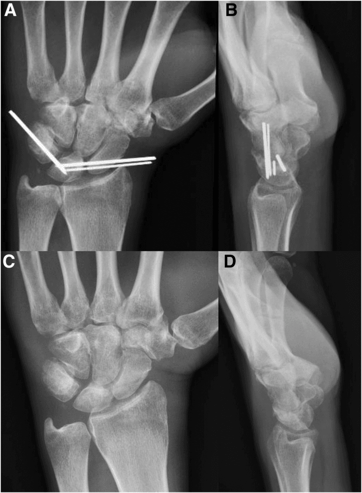
Immobilisation. A short arm cast for 6-8 weeks, non-weight-bearing on the hand, with finger range of motion from the start and wrist range of motion after cast removal. Union typically takes 8-12 weeks.
- Weeks 0-6. Short arm cast, finger range-of-motion exercises, elevation to reduce swelling
- Weeks 6-8. Cast removal, wrist range-of-motion exercises, progressive strengthening
- Weeks 8-12. Full range of motion, progressive activity, return to sport and activity
Hardware removal. Consider it if the hardware is prominent or symptomatic, usually after union is confirmed (3-6 months).
Outcomes and Prognosis
The figures below sit beside a heterogeneous literature: published arthrodesis union and satisfaction vary between series, and there is no verified universal success rate.
LT arthrodesis. Success (union and pain relief) is quoted at 85-90%, with 80-85% returning to their pre-injury level and minimal motion loss.
Direct repair. If acute (less than 6 weeks), 70-80% have good results and pain relief, 70-75% return to their pre-injury level, and full motion is preserved. Results are less predictable if the tear is chronic.
LT joint arthritis. LT fusion, or salvage where arthritis is severe or fusion has failed, has a success rate of 80-85%.
Return to activity. Return takes 3-6 months after surgery, with 80-85% reaching their pre-injury level; the rate depends on the treatment method, timing and rehabilitation compliance.
Long-term arthritis. After arthrodesis, 5-10% develop adjacent joint arthritis; without treatment, 20-30% develop arthritis. Chronic instability and associated injuries are the risk factors.
Guidelines, Registries & Global Practice
LT instability is a relatively uncommon, often under-diagnosed cause of ulnar-sided wrist pain worldwide. There are no large dedicated registries or formal society guideline statements specific to LT instability - management is guided by hand-surgery consensus and case-series evidence. The cross-cutting controversy globally is whether chronic LT instability is best treated by LT arthrodesis, soft-tissue/tendon-graft reconstruction, or by addressing an underlying positive ulnar variance with ulnar-shortening osteotomy.
Global Epidemiology
- Relative frequency: Far less common than scapholunate injury; estimated at a minority of intrinsic carpal-ligament injuries presenting with ulnar wrist pain.
- Two distinct populations: (1) younger patients with a traumatic hyperextension/twisting mechanism or perilunate-spectrum injury; (2) older patients with degenerative tears related to ulnar-positive variance and ulnocarpal impaction (Shin AY et al, JAAOS 2000).
- Frequent comorbidity: TFCC tears and positive ulnar variance commonly coexist and must be assessed - they change the operation.
Side-by-Side Practice (no single society guideline)
- Diagnostic Emphasis
- Ballottement + arthroscopy; assess ulnar variance
- Preferred Surgical Strategy
- Acute repair; chronic = reconstruction or LT fusion; ulnar shortening if ulna positive
- Diagnostic Emphasis
- MRI + arthroscopy staging; proprioceptive focus
- Preferred Surgical Strategy
- Capsulodesis / tendon reconstruction favoured; fusion reserved for failures
- Diagnostic Emphasis
- Arthroscopic grading, exclude TFCC pathology
- Preferred Surgical Strategy
- Address ulnar impaction first; selective reconstruction or fusion
Registry and Evidence Notes
- No implant registry captures LT-specific procedures (these are bone-and-soft-tissue, not arthroplasty), so evidence rests on small retrospective series and systematic reviews (Athlani L et al, Hand Surg Rehabil 2023).
- Arthrodesis outcomes are not uniform: union/failure rates vary widely between centres - from union in all patients with rigid Herbert-screw + K-wire fixation (Nelson DL et al, 1993) to ~45% nonunion in another series (Vandesande W et al, 2001). This heterogeneity is itself an exam discussion point.
High- vs Limited-Resource Settings
- High-resource: Wrist arthroscopy for diagnosis and staging, MRI, intra-operative fluoroscopy, and CT to confirm union are routine.
- Limited-resource: Diagnosis relies on clinical ballottement testing, plain radiographs (including stress/clenched-fist views) and, where available, arthrography. Immobilisation and ulnar-shortening or fusion with simpler fixation are pragmatic; advanced arthroscopic reconstruction may be unavailable.
LT instability is a common viva topic globally. Know that VISI = Volar Intercalated Segment Instability (lunate flexes volarly, LT-side injury), that the LT ballottement (Reagan) test is the key bedside test, and that treatment is matched to chronicity. Be ready to argue BOTH sides of the arthrodesis-versus-reconstruction debate, to mention ulnar-shortening when the ulna is positive, and to quote that arthrodesis nonunion rates can reach ~40% in published series.
MCQ Practice Points
Q: What is VISI and how does it differ from DISI? A: VISI (Volar Intercalated Segment Instability) = lunate flexes volarly (LT injury) - Opposite of DISI (Dorsal Intercalated Segment Instability) = lunate extends dorsally (SL injury). Capitolunate angle less than 0 degrees in VISI (normal 0-15 degrees).
Q: Why is LT arthrodesis the gold standard for chronic LT instability? A: High union rate (85-90%), minimal motion loss (LT contributes less than 5% to wrist motion), reliable pain relief, predictable outcomes - Preferred over ligament reconstruction for chronic instability. LT arthrodesis is treatment of choice.
Q: How do you perform the LT ballottement test? A: Stabilise lunate with one hand, translate triquetrum dorsally and palmarly with other hand - Positive = pain, crepitus, or increased motion. Compare to contralateral side. Also known as Reagan's test.
Q: When is direct repair preferred over LT arthrodesis? A: Acute injuries (less than 6 weeks) with good tissue quality - Direct repair achieves 70-80% good results if acute, but only 50-60% if chronic. LT arthrodesis preferred for chronic injuries (over 6 weeks).
Q: Why does LT arthrodesis cause minimal functional impairment? A: LT joint contributes less than 5% to total wrist motion - Fusion of LT joint causes minimal motion loss functionally. This supports LT arthrodesis as treatment of choice for chronic instability.
Exam Viva Scenarios
Practise clinical reasoning and management decisions out loud
“A 35-year-old woman presents with 3 months of ulnar wrist pain and clicking. Clinical examination shows positive LT ballottement test. Lateral X-ray shows VISI deformity with lunate flexed volarly. The capitolunate angle is negative 10 degrees.”
“A 28-year-old athlete presents 2 weeks after fall on outstretched hand with ulnar deviation. He has ulnar wrist pain and clicking. Clinical examination shows positive LT ballottement test. X-rays show no fixed deformity (dynamic instability).”
“A 52-year-old manual worker has 8 months of activity-related ulnar wrist pain, worse with gripping and forearm pronation. Ballottement is mildly positive and the ulnocarpal stress test reproduces pain. Radiographs show no VISI but a positive ulnar variance of plus 3 mm with subchondral changes in the lunate and triquetrum. MRI shows a central LTIL perforation and lunate chondromalacia.”
Key Concepts
- VISI = Volar Intercalated Segment Instability (lunate flexes volarly, LT injury)
- DISI = Dorsal Intercalated Segment Instability (lunate extends dorsally, SL injury)
- LTIL = Lunotriquetral Interosseous Ligament
- LT contributes less than 5% to wrist motion
Clinical Features
- Ulnar wrist pain
- Clicking or clunking with movement
- VISI deformity (if static)
- LT ballottement test positive (Reagan's test)
Treatment
- Acute (less than 6 weeks): Direct repair with suture anchors (70-80% good results)
- Chronic (over 6 weeks): LT arthrodesis (85-90% good results, gold standard)
- Static VISI: LT arthrodesis (preferred)
- With arthritis: LT fusion or salvage procedures
LT Arthrodesis Technique
- Dorsal ulnar approach between 4th and 5th extensor compartments
- Remove articular cartilage from lunate and triquetrum
- Pack bone graft (autograft or allograft)
- Fix with compression screw (headless cannulated) or plate/screws
- Cast 6-8 weeks, then ROM exercises
Complications
- Nonunion: 10-15% (prevent with rigid fixation, bone graft)
- Persistent pain: 10-15% (assess for other causes)
- Hardware issues: 5-10% (remove if symptomatic)
- Loss of correction: 5-10% (prevent with rigid fixation)
Evidence Base
Lunotriquetral Sprains - Original Description (Reagan)
- Defined LT sprain as discrete cause of ulnar wrist pain
- Described the LT ballottement manipulation (crepitus, laxity)
- Progression to VISI = LT dissociation
- Treatment depends on chronicity and severity
VISI / DISI - Carpal Instability Classification (Linscheid)
- Defined VISI and DISI patterns
- VISI = volar lunate tilt; DISI = dorsal lunate tilt
- Established lateral radiograph and intercalated-segment concept
LT Instability - Diagnosis and Treatment (Shin/AAOS review)
- Spectrum: partial tear to dislocation, dynamic to static
- Positive ulnar variance and attrition are key associations
- Ulnar shortening is a recognised option when ulna is positive
- Treatment matched to instability degree and chronicity
Lunotriquetral Arthrodesis - Technique and Union (Nelson)
- Herbert screw + K-wire superior to K-wires alone
- Immobilise over 6 weeks until union documented (at least 8 weeks)
- Union in all when both conditions met
- Plain films under-read the fusion - use fluoro/CT
LT Arthrodesis - High Failure Rate (Cautionary Series)
- Union in only 16/29 (45% nonunion)
- 17/29 remained painful; many needed reoperation
- Only 5/29 fully satisfied
- Challenges arthrodesis as automatic gold standard
Normal Carpal Kinematics (Kobayashi)
- Lunate rotates least of the proximal row; scaphoid most
- Lunate and triquetrum move closely together
- Translation minimal across all carpal bones
- Biomechanical basis for tolerability of LT fusion
Intercarpal Arthrodesis - Systematic Review
- Limited fusions predictable but reduce wrist ROM
- Nonunion is the principal complication
- Surface prep + bone graft + rigid fixation are critical
- LT fusion is a lesser-used, lesion-specific arthrodesis