Guyon's Canal Compression | Ulnar Nerve at Wrist | Zone-Specific Deficits
- GUYON'S CANAL BOUNDARIES: floor = pisiform/hamate/pisohamate ligament; roof = palmaris brevis and palmar carpal ligament
- ZONE 2 = PURE MOTOR DEFICIT: the most common presentation (50%) - weakness with NORMAL sensation
- GANGLION CYST IS THE COMMONEST CAUSE: 85-90% of Zone 2 compression comes from a ganglion arising from the pisotriquetral joint
- HOOK OF HAMATE FRACTURE: a classic cause in cyclists and golfers - palpate for tenderness
- NO FROMENT'S SIGN: adductor pollicis is supplied BEFORE Guyon's canal, so a positive Froment's indicates a more proximal ulnar lesion
- “Zone 2 compression = interosseous weakness WITHOUT sensory loss (DDx from CuTS)
- “Ganglion cyst most common space-occupying lesion in Guyon's
- “Hook of hamate fracture = missed on standard radiographs, need carpal tunnel view or CT
- “Guyon's vs cubital tunnel: Guyon's has normal Froment's sign (adductor spared)
Overview
Ulnar tunnel syndrome (Guyon's canal syndrome) is compression of the ulnar nerve or its branches at the wrist, within the fibro-osseous tunnel formed by the pisiform, the hook of hamate and the overlying soft tissues. It is rare compared with carpal tunnel and cubital tunnel syndromes.
How it presents. Unlike cubital tunnel syndrome there are no elbow symptoms, and the deficit depends on where in the canal the nerve is caught: it may be motor, sensory or mixed. The zone of compression is therefore the organising idea for the whole topic, from examination to imaging to the operation.
What causes it. A ganglion cyst arising from the pisotriquetral joint is the most common cause, followed by the hook of hamate fracture in cyclists and golfers. Ulnar artery thrombosis is also among the common causes.
Anatomy and Pathophysiology
The canal. Guyon's canal (the ulnar tunnel) is a fibro-osseous tunnel about 4 cm long through which the ulnar nerve enters the hand. Its walls:
- Roof: palmaris brevis, the palmar (volar) carpal ligament, and skin and subcutaneous tissue
- Floor: the pisiform (ulnar), the hook of hamate (radial) and the pisohamate ligament, with the transverse carpal ligament deeper
- Ulnar border: the pisiform and the flexor carpi ulnaris tendon
- Radial border: the hook of hamate and the flexor digitorum superficialis tendons
Contents. The ulnar nerve, the ulnar artery and the ulnar veins. The artery lies radial to the nerve.
The bifurcation. The nerve divides within the canal. The deep motor branch curves around the hook of hamate into the palm; the superficial sensory branch continues distally to supply the hypothenar eminence and the ulnar one and a half digits. Because the division happens inside the canal, compression can catch the trunk, the motor branch alone or the sensory branch alone, and that is what produces the zone-specific presentations.
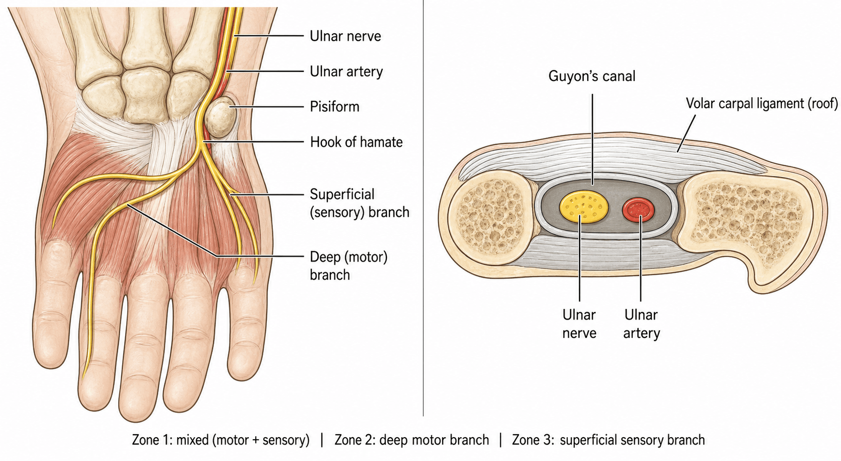
What the deep branch supplies. A lesion of the trunk or of the deep branch (Zones 1 and 2) weakens the ulnar intrinsics:
- Interossei, palmar and dorsal: the patient cannot spread or adduct the fingers; it is the palmar interossei that adduct
- Lumbricals 3 and 4: contributes to clawing
- Abductor digiti minimi: hypothenar wasting
- Flexor digiti minimi brevis: weak small finger flexion
- Opponens digiti minimi: weak small finger opposition
- Flexor pollicis brevis (deep head): usually minimal impact
What is spared. Four ulnar-supplied structures take their supply from branches that leave the nerve proximal to the canal, and finding them normal is what places the lesion at the wrist:
- Adductor pollicis: its branch leaves the nerve before Guyon's canal
- Flexor carpi ulnaris: motor branch proximal, in the forearm
- FDP to the ring and small fingers: motor branch proximal, in the forearm
- Dorsal ulnar cutaneous nerve: branches 5-8 cm proximal to the wrist
Classification
Shea and McClain (1969). The classification divides Guyon's canal into three zones by the branching of the nerve. The zone determines the clinical presentation, and with it the imaging and the surgical approach.
- Anatomical Location
- Proximal to bifurcation
- Motor Deficit
- All ulnar intrinsics weak
- Sensory Deficit
- Hypothenar + ulnar 1.5 digits numb
- Common Causes
- Ganglion, ulnar artery aneurysm, fracture
- Anatomical Location
- Deep motor branch
- Motor Deficit
- All ulnar intrinsics weak
- Sensory Deficit
- NO sensory loss (normal sensation)
- Common Causes
- Ganglion from pisotriquetral joint (85-90%)
- Anatomical Location
- Superficial sensory branch
- Motor Deficit
- NO motor deficit (full strength)
- Sensory Deficit
- Hypothenar + ulnar 1.5 digits numb
- Common Causes
- Ulnar artery thrombosis, direct trauma
Zone 2 is the most common, at 50% of cases, because pisotriquetral ganglion cysts preferentially compress the deep motor branch as it curves around the hook of hamate.
Interosseous weakness with NORMAL sensation = Zone 2 compression until proven otherwise. This presentation is pathognomonic, and it separates Zone 2 from cubital tunnel syndrome. Always look for a ganglion cyst on imaging.
By cause. The causes fall into four groups, and the proportions are worth knowing:
- Space-occupying lesions, 60%: ganglion cyst (85-90% of these), lipoma (including spindle cell lipoma), neurofibroma (a rare nerve sheath tumour), ulnar artery aneurysm, and rarely a carpal bone tumour
- Bony or traumatic, 30%: hook of hamate fracture, pisotriquetral arthritis, distal radius malunion
- Vascular, 5%: hypothenar hammer syndrome and ulnar artery thrombosis
- Chronic pressure, 5%: cycling (handlebar palsy), wheelchair use, occupational tool use
Ganglion cysts. They arise from the pisotriquetral joint and preferentially compress the deep motor branch (Zone 2). They may be small and difficult to palpate, so MRI or ultrasound makes the diagnosis.
Hook of hamate fracture. It occurs in cyclists, golfers and baseball players, from a direct blow to the hypothenar eminence. Nonunion is common, and the fibrous callus compresses the nerve.
Pisotriquetral arthritis. It follows chronic wrist trauma, and osteophytes compress the nerve.
Vascular lesions. Hypothenar hammer syndrome (see Complications and Special Situations) is thrombosis of the ulnar artery from chronic repetitive trauma such as hammering; it may compress the nerve or cause ischaemia, and Allen's test is abnormal. The ulnar artery may also thrombose from an aneurysm or atherosclerosis.
Chronic pressure and the idiopathic case. Handlebar pressure in cyclists, wheelchair use and tool use put chronic pressure on the hypothenar eminence. When no cause is found the syndrome is idiopathic, a diagnosis of exclusion, and it often responds to conservative treatment.
Clinical Presentation and Diagnosis
History. The questions that localise and explain the lesion:
- Onset, acute or gradual
- Hand dominance and occupation
- Trauma: a fall on the hand, a hook of hamate fracture
- Cycling, tool use or wheelchair use
- A palpable mass in the hypothenar region
- Weakness or numbness, which points to the zone
Inspection. Look for hypothenar wasting (Zones 1 and 2), interosseous wasting on the dorsum of the hand, a claw deformity of the ring and small fingers, and a visible or palpable mass.
Palpation. Tenderness over the hook of hamate suggests a fracture. A ganglion may be palpable, but may be subtle. Test for Tinel's sign over Guyon's canal, and perform Allen's test for ulnar artery patency.
Motor testing. Test interosseous function by finger spread and adduction (Wartenberg's sign), and hypothenar strength by abduction and opposition of the small finger. Froment's sign should be negative, and flexor carpi ulnaris and FDP to the ring and small fingers should be normal; these are the findings that localise the lesion to the wrist.
Sensory testing. Test the hypothenar eminence (superficial branch) and the palmar surface of the ulnar one and a half digits (the small finger and the ulnar side of the ring finger). Dorsal ulnar cutaneous sensation should be normal.
The Riche-Cannieu anastomosis is a common communication in the palm between the deep motor branch of the ulnar nerve and the recurrent (thenar) motor branch of the median nerve. Because it shifts motor supply across the usual median/ulnar boundary in the hand, the motor findings of a distal (Guyon's) ulnar lesion can be atypical and may not match the sensory pattern. Where ulnar-territory intrinsics receive median fibres through the anastomosis, intrinsic weakness can be milder than expected despite genuine compression; in the "all-ulnar hand" the thenar muscles are ulnar-innervated, so a Guyon's lesion can add unexpected thumb (thenar) weakness. A coexisting forearm Martin-Gruber anastomosis (median fibres crossing into the ulnar nerve, producing an "all-median hand") can further mask intrinsic loss. The practical message: when the motor deficit does not fit the sensory deficit or the imaging, suspect a crossover anastomosis and interpret nerve conduction studies with this anatomy in mind.
A more distal ulnar nerve lesion produces a MORE marked ulnar claw than a proximal (elbow) lesion — the "ulnar paradox." At the wrist (Guyon's canal) the FDP to the ring and little fingers is preserved (it is innervated in the proximal forearm), so distal interphalangeal flexion of these fingers is intact and unopposed; combined with loss of the lumbricals and interossei this exaggerates the metacarpophalangeal hyperextension and interphalangeal flexion posture of the fourth and fifth fingers. A proximal lesion at the elbow also paralyses FDP to the ring and little fingers, so the distal flexion pull is weaker and the claw is paradoxically LESS obvious. The corollary catches candidates out: as a proximal lesion recovers (or as the level descends), the claw can worsen before it improves. The clawing deformity is therefore a clue to the LEVEL of the lesion, not simply its severity.
Localising the lesion. The question in every ulnar neuropathy is the level: wrist, forearm or elbow. A Guyon's lesion spares everything supplied above the wrist, has no elbow signs, and can produce a pure motor or a pure sensory deficit depending on the zone. Its common causes are a ganglion or a hamate fracture, so imaging is often diagnostic; the common causes of cubital tunnel syndrome are chronic pressure and anatomical. Sensory loss in cubital tunnel syndrome covers the hypothenar area and the ulnar 1.5 digits.
- Guyon's Canal (Wrist)
- Negative (adductor spared)
- Cubital Tunnel (Elbow)
- Positive (adductor weak)
- Forearm (Martin-Gruber)
- Positive
- Guyon's Canal (Wrist)
- Absent (normal FCU)
- Cubital Tunnel (Elbow)
- Present if severe
- Forearm (Martin-Gruber)
- Present
- Guyon's Canal (Wrist)
- Absent (normal FDP)
- Cubital Tunnel (Elbow)
- Present if severe
- Forearm (Martin-Gruber)
- Present
- Guyon's Canal (Wrist)
- Normal (nerve branches proximal)
- Cubital Tunnel (Elbow)
- Abnormal if affected
- Forearm (Martin-Gruber)
- Abnormal
- Guyon's Canal (Wrist)
- None
- Cubital Tunnel (Elbow)
- Elbow pain, +Tinel's at elbow
- Forearm (Martin-Gruber)
- None
- Guyon's Canal (Wrist)
- Wrist segment slowing
- Cubital Tunnel (Elbow)
- Elbow segment slowing
- Forearm (Martin-Gruber)
- Forearm abnormality
Froment's sign should be NEGATIVE in pure Guyon's canal syndrome. If it is positive (the thumb IP joint flexes during key pinch), the compression is proximal to the wrist: at the elbow or above, or in the forearm.
Systematic exam: (1) Check Froment's (adductor), (2) Check FCU/FDP strength (forearm branches), (3) Check dorsal hand sensation (dorsal ulnar cutaneous). All normal except intrinsics + hypothenar sensation = Guyon's canal.
Three patterns in practice. Three illustrative cases, one for each zone.
- The cyclist with a pure motor deficit. A 35-year-old cyclist with a 3-month history of weak grip and difficulty with fine motor tasks has interosseous wasting and Wartenberg's sign, normal sensation in the hand and no Froment's sign. This is Zone 2 compression, likely a ganglion, and MRI shows a pisotriquetral ganglion.
- The baseball catcher with hypothenar pain. A 22-year-old catcher has hypothenar pain after a foul tip, tenderness over the hook of hamate, interosseous weakness and hypothenar numbness. This is a hook of hamate fracture compressing the nerve in Zone 1, and CT shows the fracture with displacement.
- The carpenter with pure sensory loss. A 50-year-old carpenter has hypothenar numbness without weakness and an abnormal Allen's test with no ulnar artery flow. This is Zone 3, from ulnar artery thrombosis, and Doppler ultrasound shows the occlusion.
Investigations
The work-up has two jobs: confirm the level of the neuropathy, and find a treatable cause.
- Indication
- All patients
- Key Findings
- Hook of hamate fracture, arthritis
- Indication
- Suspected hamate fracture
- Key Findings
- Gold standard for bony pathology
- Indication
- Soft tissue mass suspected
- Key Findings
- Ganglion cyst, nerve pathology
- Indication
- All surgical candidates
- Key Findings
- Confirms level, severity, prognosis
Radiographs. PA, lateral and oblique views of the wrist, with a carpal tunnel view, which is the best view of the hook of hamate; a supinated oblique is an alternative hamate view. Look for a hook of hamate fracture or nonunion, pisotriquetral arthritis and carpal bone masses. Standard views have a sensitivity of only 50% for hamate fracture.
CT is the gold standard for hook of hamate fracture. Thin slices through the carpus identify fracture, nonunion and displacement, and can show bony impingement on the nerve. It is indicated for a suspected hamate fracture with negative radiographs, for planning excision, and to assess pisotriquetral arthritis.
MRI is best for soft-tissue masses: a ganglion cyst (high T2 signal), ulnar artery aneurysm or thrombosis, a nerve sheath tumour or a lipoma. Its sensitivity for ganglion cyst is greater than 90%. Use a dedicated wrist coil, T1, T2 and fat-suppressed sequences, and coronal and axial planes through Guyon's canal.
Ultrasound gives a dynamic, real-time view of the nerve and any mass, with Doppler for the ulnar artery, and can guide aspiration of a ganglion. It involves no radiation, is quick and inexpensive, and is good for ganglion cysts and vascular lesions, but it is operator-dependent.
Nerve conduction studies. Motor studies stimulate the ulnar nerve at the wrist and record from the hypothenar eminence (ADM), comparing the wrist-to-hypothenar latency with the normal side. Distal latency is prolonged in Guyon's compression, and amplitude may be reduced if severe. Sensory studies of the superficial branch are abnormal in Zones 1 and 3 and normal in Zone 2, and the dorsal ulnar cutaneous nerve should be normal, which localises the lesion to the wrist.
EMG shows denervation in the ulnar intrinsics in Zones 1 and 2, with first dorsal interosseous and ADM the most sensitive muscles. Adductor pollicis should be normal.
Vascular tests. Allen's test assesses ulnar artery patency, and angiography is used if hypothenar hammer syndrome is suspected.
NCS is essential to: (1) Confirm ulnar neuropathy (vs cervical radiculopathy or other), (2) Localise to wrist (vs elbow or forearm), (3) Assess severity (demyelination vs axonal loss). Do NOT operate without NCS confirmation showing wrist-level slowing, and image to identify a treatable cause.
Management
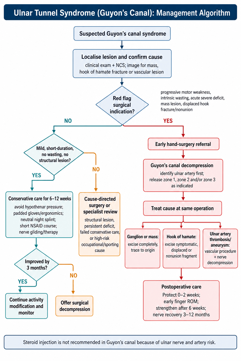
Who is treated conservatively. Non-operative treatment suits:
- Mild symptoms of short duration (less than 3 months)
- No muscle wasting
- Idiopathic compression, with no mass lesion on imaging
- An occupational cause with modifiable activity
The conservative programme.
- Activity modification: avoid repetitive hypothenar pressure, with padded gloves for cyclists, ergonomic tool handles and wheelchair cushion padding
- Splinting: a wrist splint in neutral, as a night splint for 6-8 weeks, to reduce pressure in Guyon's canal; the evidence is limited but the risk is low
- NSAIDs: a short course of 2-4 weeks for their anti-inflammatory effect, with minimal evidence in nerve compression
- Therapy: nerve-gliding exercises, then strengthening as symptoms improve; there is no evidence of benefit, but it is commonly prescribed
What it achieves. 30-40% improve with conservative treatment. Results are best in idiopathic and occupational cases and poor when a ganglion or other structural lesion is present.
Steroid injection NOT recommended for Guyon's canal syndrome. Unlike carpal tunnel, injection into confined space near ulnar artery is risky. May damage nerve or artery. Not supported by evidence.
Surgical indications. Any of these moves the patient to decompression:
- A ganglion cyst or other mass lesion on imaging
- Progressive motor weakness
- Muscle atrophy
- No improvement after 3 months of conservative treatment
- Acute onset with a severe deficit
- Hook of hamate fracture nonunion
The hook of hamate fracture. Fractures are typed as I (tip avulsion), II (base fracture) or III (comminuted). Nonunion is common because the hook has a poor blood supply.
- Acute (less than 4 weeks), non-displaced: a trial of immobilisation for 4-6 weeks in a short arm cast with an ulnar gutter. Success is only 30-50%
- Acute, displaced: primary excision is recommended. Union is rare even with prolonged immobilisation, and early excision prevents nerve compression
- Chronic nonunion or delayed presentation: excision of the hook is the treatment of choice if symptomatic; an asymptomatic, incidental nonunion can be observed
Excision is treatment of choice, NOT fixation. Hook has poor blood supply, high nonunion rate even with fixation. Excision has excellent outcomes and faster recovery. ORIF reserved for rare cases of acute base fractures in elite athletes.
Surgical Technique
Set-up. Supine with the arm on a hand table, an upper-arm tourniquet and the hand supinated. Loupe magnification is recommended.
Incision. Longitudinal, along the ulnar border of the hypothenar eminence, starting 1 cm proximal to the wrist crease and extending 3-4 cm distally into the palm, radial to the FCU tendon and ulnar to the thenar crease.
Decompression, proximal to distal.
- Incise skin and subcutaneous fat, and identify and protect the palmar cutaneous branch of the ulnar nerve, a small sensory branch
- Expose palmaris brevis and incise it longitudinally
- Identify the ulnar artery first: it lies radial to the nerve
- Identify the ulnar nerve, ulnar to the artery, and follow it distally
- Zone 1: release the palmar carpal ligament proximal to the wrist crease
- Identify the bifurcation into motor and sensory branches
- Zone 2: follow the deep motor branch as it curves around the hook of hamate, and release all fibrous tissue compressing it
- Zone 3: follow the superficial sensory branch distally and release any constricting tissue
Release the zones as needed, tracing the nerve through each.
Dealing with the cause. The cause is treated at the same operation:
- Ganglion cyst: excise completely, tracing it to its origin in the pisotriquetral joint
- Hook of hamate fracture: excise the hook fragment rather than fixing it
- Ulnar artery thrombosis: ligate or resect the thrombosed segment
- Ulnar artery aneurysm: excision with vascular reconstruction, or ligation
- Pisohamate ligament: release if it compresses the nerve
Closure. Meticulous haemostasis matters beside the ulnar artery. Close the skin with interrupted 4-0 nylon, apply a soft bulky dressing, and, optionally, a wrist splint in neutral for 1-2 weeks.
Ulnar artery lies RADIAL to ulnar nerve in Guyon's canal. Always identify artery first before aggressive decompression. Injury causes hypothenar ischaemia and difficult repair. Use loupe magnification.
Hook of hamate excision. The indications are a symptomatic nonunion, a displaced acute fracture, and a chronic fracture with ulnar neuropathy. Through the same incision, identify and protect the ulnar nerve and artery and expose the hook, then remove the fragment with a small osteotome or rongeur. Remove every fragment and smooth the edges with a rasp: a sharp edge left behind risks flexor tendon rupture.
After excision. A soft dressing and early motion, no forceful gripping for 6 weeks, and return to sport at 8-12 weeks. Pain relief is achieved in 90-95%, grip strength returns to 85-90% of normal, and nerve symptoms, if present, resolve in 80-90%.
Complications and Special Situations
Ulnar artery injury. The most serious complication, at 1-2%. Prevention is identifying the artery before decompressing the nerve, loupe magnification and no monopolar cautery near the artery. Repair it primarily with 8-0 or 9-0 suture, which may need microvascular expertise; ligation is a last resort because of the risk of hypothenar ischaemia.
Nerve branch injury. The superficial sensory branch is most at risk. Identify every branch before dividing anything, protect the palmar cutaneous branch in the subcutaneous tissue, and dissect the bifurcation carefully. A recognised injury is repaired primarily and a symptomatic neuroma is excised later, but the sensory deficit may be permanent.
Early complications (0-6 weeks).
- Haematoma, 2-3%: a risk beside the ulnar artery. Prevent it with meticulous haemostasis and consider a drain; evacuate it if symptomatic and apply a compressive dressing
- Infection, less than 1%: rare with clean technique. Treat with antibiotics, and washout if severe; a deep infection puts nerve recovery at risk
- Wound dehiscence: the incision lies in a mobile area of the palm. Prevention is careful closure and 1-2 weeks of immobilisation; treat with local wound care, and re-closure may be needed
Late complications (after 6 weeks).
- Incidence
- 10-20%
- Prevention
- Early surgery (less than 6 months symptoms)
- Treatment
- Tendon transfers if severe
- Incidence
- 5-10%
- Prevention
- Complete excision to joint origin
- Treatment
- Revision excision
- Incidence
- 5%
- Prevention
- Protect palmar cutaneous nerve
- Treatment
- Scar massage, desensitisation
- Incidence
- 10-15%
- Prevention
- Less aggressive retinaculum release
- Treatment
- Therapy, usually resolves by 3 months
Ulnar artery injury or ligation can cause hypothenar ischaemia, especially if palmar arch incomplete. Always assess Allen's test preoperatively. If abnormal, ulnar artery must be preserved. Consider vascular surgery consultation if injured.
Hypothenar hammer syndrome. The ulnar artery thrombosis or aneurysm it produces may compress the nerve in Zone 1 or 3. The patient presents with cold intolerance, pain and numbness. Doppler ultrasound or angiography makes the diagnosis, and treatment is vascular reconstruction or ligation with nerve decompression.
Ulnar artery aneurysm. A pulsatile mass in the hypothenar eminence and Guyon's canal that compresses the nerve in Zone 1, and may thrombose and cause acute ischaemia. Ultrasound with Doppler makes the diagnosis.
Pisotriquetral arthritis. Degenerative arthritis of the pisotriquetral joint gives chronic wrist pain with an ulnar neuropathy, and radiographs show the arthritis. Treatment is pisiform excision with nerve decompression.
Pisiform excision is treatment for chronic pisotriquetral arthritis causing ulnar neuropathy. Remove entire pisiform (origin of FCU). Decompress nerve simultaneously. Good outcomes for pain and neuropathy. FCU function preserved (dynamic muscle origin).
Combined carpal and Guyon's tunnel compression. The "double crush" phenomenon: the median nerve compressed in the carpal tunnel and the ulnar nerve in Guyon's canal, giving combined median and ulnar deficits. Consider it when a patient having carpal tunnel release has hypothenar symptoms; both tunnels are released, and this can be done simultaneously.
Revision surgery. Revision is considered for persistent or recurrent symptoms after 6 months or more, an incomplete first decompression, a recurrent ganglion, or the nerve caught in scar tissue.
- Before operating: repeat NCS to confirm persistent compression, MRI for a recurrent mass or nerve scarring, and exclude a proximal lesion (cubital tunnel)
- At operation: an extended incision with careful dissection through scar, complete decompression of all three zones, neurolysis if the nerve is encased in scar, excision of a recurrent ganglion to its joint origin, and nerve wrapping (fat, vein) if the scarring is extensive
- Salvage: tendon transfers for a permanent motor deficit, nerve grafting for a nerve defect, neuroma excision for intractable pain
What revision achieves. It is less predictable than a primary release, at 60-70% success, so set realistic expectations. If a second revision is being considered, consider an alternative diagnosis.
Postoperative Care
Rehabilitation.
- Weeks 0-2, protection: bulky soft dressing, finger range of motion immediately, gentle wrist mobilisation, sutures out at 10-14 days
- Weeks 2-6, active motion: remove the dressing or splint, active wrist and finger range of motion, light activities of daily living, no forceful gripping
- Weeks 6-12, strengthening: progressive grip strengthening, return to light-duty work, therapy for stiffness if needed, and sport-specific training begins
- Months 3-6, full recovery: return to full activities, sport-specific training, and a final assessment of nerve recovery
Return to activity. Office work at 2 weeks, light manual work at 4-6 weeks, heavy manual work at 8-12 weeks, and cycling or golf at 8-12 weeks after hamate excision.
Monitoring. Review clinically at 2, 6 and 12 weeks, assessing motor recovery by intrinsic strength; sensory recovery typically precedes motor. Obtain a final NCS at 6 months if symptoms persist.
Outcomes
Decompression succeeds in 90%. How completely the hand recovers depends on the factors below.
- Good Prognosis
- Less than 6 months
- Poor Prognosis
- Greater than 12 months
- Good Prognosis
- No atrophy
- Poor Prognosis
- Severe wasting
- Good Prognosis
- Ganglion cyst (removable)
- Poor Prognosis
- Chronic arthritis or fibrosis
- Good Prognosis
- Younger (less than 40)
- Poor Prognosis
- Older (greater than 60)
- Good Prognosis
- Demyelination only
- Poor Prognosis
- Axonal loss
Sensory recovery. Sensation recovers faster than motor function, over 3-6 months. Protective sensation returns first, at 1-2 months; dysaesthesias may persist for 3-6 months; and numbness resolves in 95%, by 6 months.
Motor recovery. Improvement begins at 2-4 months and reaches its maximum at 6-12 months. Intrinsic strength improves but may not return to baseline if there was chronic or severe atrophy. Clawing resolves in 90%, and grip strength improves by 30-50%.
Set realistic expectations: If severe atrophy present preoperatively, complete strength recovery unlikely. Goal is to halt progression and improve function, not restore normal strength. Emphasise to patients preoperatively.
Guidelines, Registries & Global Practice
Global Epidemiology
Ulnar tunnel syndrome is an uncommon, well-described entity that is substantially rarer than both carpal tunnel and cubital tunnel syndrome, and it is frequently mis-diagnosed because its symptoms overlap with carpal tunnel syndrome, cubital tunnel syndrome, thoracic outlet syndrome, C8-T1 radiculopathy and peripheral neuropathy (Chen & Tsai, J Hand Surg Am 2014, DOI; Bachoura & Jacoby, Orthop Clin North Am 2012, DOI). The hook of hamate fracture - a classic structural cause - is itself uncommon with an unclear true prevalence and is often diagnosed late (Tian & Goldfarb, Hand Clin 2021, DOI). Vascular causes such as hypothenar hammer syndrome arise in people who repeatedly use the hypothenar eminence as a hammer (Schröttle et al, Vasa 2015, DOI).
- Stance Relevant to Ulnar Tunnel Syndrome
- Electrodiagnostic confirmation and targeted imaging (MRI/US for mass, CT for hook) before decompression; surgery for confirmed compressive or structural lesion
- Evidence Level
- Expert consensus / Level V
- Stance Relevant to Ulnar Tunnel Syndrome
- Localise lesion clinically and electrophysiologically; exclude proximal (elbow) ulnar neuropathy first; decompression for structural cause or failed conservative care
- Evidence Level
- Expert consensus / Level V
- Stance Relevant to Ulnar Tunnel Syndrome
- Zone-based (Shea-McClain) assessment guides exploration; address underlying cause (ganglion excision, hook excision, vascular repair)
- Evidence Level
- Expert consensus / Level V
- Stance Relevant to Ulnar Tunnel Syndrome
- No high-level RCT evidence; management driven by cause-specific case series and anatomical principles
- Evidence Level
- Level IV-V
- Contribution
- Defining three-zone classification (PMID 5805411)
- Limitation
- Small original series; descriptive
- Contribution
- Outcomes for ganglion excision, hook excision, vascular repair
- Limitation
- Level IV; no comparative RCTs
- Contribution
- Ulnar tunnel syndrome is not tracked in arthroplasty-style registries (e.g. AOANJRR/NJR cover joint replacement, not peripheral nerve decompression)
- Limitation
- Epidemiology relies on hospital and surgeon series
Practice Variation & Global Notes
- Diagnostic work-up is broadly consistent worldwide: clinical localisation, nerve conduction studies/EMG, then targeted imaging (MRI/ultrasound for soft-tissue masses, CT for the hook of hamate). The dorsal ulnar cutaneous nerve and Froment's sign are universally used to separate wrist-level from elbow-level lesions.
- Surgical thresholds vary by structural cause and patient demand: an identified ganglion, hook of hamate fracture, or vascular lesion lowers the threshold for surgery, whereas idiopathic mild cases are managed conservatively first.
- Hook of hamate management converges on excision across regions for symptomatic fractures and nonunions, particularly in athletes (golf, racquet sports, baseball) and cyclists.
- Vascular causes require Allen's test and duplex ultrasound first-line with angiography as reference standard; smoking cessation is a universal adjunct.
- Cycling (handlebar pressure), wheelchair use and repetitive tool use are recognised occupational/recreational drivers worldwide.
- Hook of hamate injuries cluster in golf, racquet sports and baseball internationally; document occupational exposure for compensation pathways.
Exam Viva Point - Global Practice: Management worldwide rests on cause-specific Level IV evidence and anatomical principles rather than randomised trials. Nerve conduction studies are standard of care before decompression in all major systems. Hook of hamate injuries are seen in baseball, golf and cyclists; the dorsal ulnar cutaneous nerve and Froment's sign are the universal clinical tools for localising the lesion to the wrist rather than the elbow.
Shea & McClain: Ulnar-Nerve Compression Syndromes at and Below the Wrist (Landmark Classification)
Hook of Hamate Excision in Athletes (Symptomatic Partial Union / Nonunion)
Ulnar Tunnel Syndrome: Contemporary Review of Anatomy, Diagnosis and Treatment
Zone-Based Approach to Ulnar Tunnel Syndrome
Hook of Hamate Fractures: Diagnosis and Excision
Hypothenar Hammer Syndrome (Vascular Cause of Ulnar Tunnel Pathology)
MCQ Practice Points
Q: What are the boundaries of Guyon's canal?
A: Floor: Pisiform, hook of hamate, pisohamate ligament, hypothenar muscles. Roof: Volar carpal ligament (not the transverse carpal ligament) and palmaris brevis. Contents: Ulnar nerve and ulnar artery. The artery lies radial to the nerve. Canal is approximately 4 cm long from proximal wrist to palm.
Q: Describe the Shea-McClain classification of ulnar tunnel syndrome.
A: Zone 1 (proximal): Before bifurcation - mixed motor and sensory deficit. Zone 2 (deep): Deep motor branch only - pure motor deficit (intrinsic weakness, no sensory loss) - accounts for 50% of cases. Zone 3 (superficial): Superficial sensory branch - pure sensory deficit (ulnar 1.5 digits). Ganglion from pisotriquetral joint is most common Zone 2 cause.
Q: How do you differentiate Guyon's canal syndrome from cubital tunnel syndrome?
A: Dorsal ulnar cutaneous nerve: NORMAL in Guyon's (branches proximal to wrist), ABNORMAL in cubital tunnel. Froment's sign: Often NEGATIVE in pure Zone 2 Guyon's (FPL compensation intact), typically POSITIVE in cubital tunnel. FCU weakness: Absent in Guyon's, may be present in cubital tunnel. NCS localization: Wrist segment in Guyon's, elbow segment in cubital tunnel.
Q: What is the most common cause of Zone 2 (pure motor) ulnar tunnel syndrome?
A: Ganglion cyst arising from the pisotriquetral joint accounts for 85-90% of Zone 2 lesions. Other causes include hook of hamate fracture (especially in racquet sports, baseball, golf), ulnar artery aneurysm/thrombosis, lipoma, and direct trauma from cycling ("handlebar palsy"). MRI or ultrasound is diagnostic.
Q: What clinical finding is pathognomonic for Zone 2 ulnar tunnel syndrome?
A: Intrinsic hand weakness WITHOUT sensory loss. Patient has weak finger abduction/adduction, weak thumb adduction, interosseous atrophy, and potential clawing (4th/5th fingers), but normal sensation in the ulnar 1.5 digits. This pattern cannot occur with cubital tunnel syndrome (sensory fibers would also be affected at elbow level).
Clinical Decision Scenarios
Practise clinical reasoning and management decisions out loud
“A 40-year-old cyclist presents with a 6-month history of weak grip and inability to spread fingers. Examination shows interosseous wasting, positive Wartenberg's sign, but normal sensation throughout the hand including hypothenar and ring/small fingers. Froment's sign is negative. What is your diagnosis and management?”
“A 24-year-old baseball player presents 3 months after a foul tip injury to his catching hand. He has hypothenar pain, weak grip, and numbness in the small finger. Examination shows tenderness over the hook of hamate and intrinsic weakness. What is your diagnosis and how would you manage this?”
“A patient presents with ulnar-sided hand numbness and weak grip. How do you differentiate between Guyon's canal syndrome and cubital tunnel syndrome on clinical examination?”
Guyon's Canal Anatomy (Memorize!)
- Floor: pisiform, hook of hamate, pisohamate ligament
- Roof: palmaris brevis muscle, palmar carpal ligament
- Contents: ulnar nerve and artery (the artery lies RADIAL to the nerve)
- Nerve bifurcates into deep motor and superficial sensory branches
- Length: approximately 4 cm from wrist to palm
Shea-McClain Classification (Essential)
- Zone 1: proximal to the bifurcation → combined motor and sensory deficit
- Zone 2: deep motor branch → pure MOTOR deficit (50% of cases)
- Zone 3: superficial sensory branch → pure SENSORY deficit
- Zone 2 is pathognomonic: weakness WITHOUT sensory loss
- Ganglion cyst: the cause in 85-90% of Zone 2 cases, arising from the pisotriquetral joint
Guyon's vs Cubital Tunnel (High Yield)
- Froment's sign: NEGATIVE in Guyon's, POSITIVE in cubital tunnel
- Dorsal ulnar cutaneous: NORMAL in Guyon's, ABNORMAL in cubital tunnel
- FCU/FDP strength: NORMAL in Guyon's, WEAK in cubital tunnel if severe
- Elbow symptoms: ABSENT in Guyon's, PRESENT in cubital tunnel
- Pure motor or pure sensory deficit: possible in Guyon's (zone-dependent), NOT in cubital tunnel
- Nerve conduction localisation: slowing across the wrist versus across the elbow
Common Causes
- Ganglion cyst (85-90% of masses, from the pisotriquetral joint)
- Ulnar artery thrombosis or aneurysm (hypothenar hammer syndrome)
- Neurofibroma (rare nerve sheath tumour)
- Spindle cell lipoma or other mass
- Hook of hamate fracture (cyclists, baseball catchers)
- Occupational pressure (handlebar, wheelchair)
- Thrombosed ulnar artery
Investigations
- Radiographs: PA, lateral, and a carpal tunnel view for the hamate
- CT: the most reliable investigation for hook of hamate fracture
- MRI: best for ganglion cyst and soft-tissue masses
- Ultrasound: dynamic assessment, with Doppler for the ulnar artery
- NCS/EMG: confirms ulnar neuropathy and localises it to the wrist segment
- Obtain nerve conduction studies before surgery - they confirm both diagnosis and location
Surgical Technique Pearls
- Incision: along the ulnar border of the hypothenar eminence, from 1 cm proximal to the wrist crease and 3-4 cm distally
- Identify the ulnar artery FIRST - it lies radial to the nerve and must be protected
- Decompress all three zones completely
- Ganglion: excise back to its pisotriquetral joint origin or it recurs
- Hook of hamate: excise the fragment rather than fixing it, and smooth the edges
- Outcomes: 90% success, with motor recovery over 6-12 months and sensory recovery over 3-6 months
Common Viva Questions
- Describe Guyon's canal anatomy - boundaries, contents, bifurcation
- What are the three zones? Shea-McClain zones 1, 2 and 3, with their motor and sensory deficits
- How do you differentiate it from cubital tunnel? Froment's, dorsal sensation, FCU and FDP
- What is the commonest cause of Zone 2 compression? Ganglion cyst from the pisotriquetral joint
- How is a hook of hamate fracture treated? Excision rather than fixation, because of the poor blood supply
- Why is Froment's negative? Adductor pollicis branches proximal to Guyon's canal
Exam Day Traps
- DON'T assume Froment's is positive in all ulnar neuropathy - a negative sign means a wrist lesion
- DON'T forget to check dorsal hand sensation - normal points to Guyon's, abnormal to a proximal lesion
- DON'T operate without nerve conduction studies - they confirm level, severity, and axonal versus demyelinating loss
- DON'T injure the ulnar artery - it lies radial to the nerve, and injury causes hypothenar ischaemia
- DON'T fix a hook of hamate fracture - excision is the treatment of choice and fixation fails
- DON'T excise the ganglion incompletely - trace it to the pisotriquetral joint or it recurs
Evidence Base
Key Evidence Summary
- Landmark paper defining three anatomical zones
- Basis for all subsequent classification systems
- Guides surgical approach and prognostication
- Multiple case series: 78-91% of Zone 2 lesions
- Arise from pisotriquetral joint
- Complete excision to origin prevents recurrence
- Level IV evidence supports excision over ORIF
- Excision: 95% success, return to sport 10 weeks
- ORIF: 67% nonunion, conversion to excision common
References
-
Shea JD, McClain EJ. Ulnar-nerve compression syndromes at and below the wrist. J Bone Joint Surg Am. 1969;51(6):1095-1103.
-
Murata K, Tamai M, Gupta A. Anatomic study of variations of hypothenar muscles and arborization patterns of the ulnar nerve in the hand. J Hand Surg Am. 2004;29(3):500-509.
-
Kleinert JM, Mehta S. Radial, ulnar, and median nerve entrapment at the wrist and forearm. J Hand Surg Am. 1996;21(4):532-559.
-
Bozkurt MC, Tagil SM, Ozçakar L, Ersoy M, Tekdemir I. Anatomical variations as potential risk factors for ulnar tunnel syndrome: a cadaveric study. Clin Anat. 2005;18(4):274-280.
-
Gross MS, Gelberman RH. The anatomy of the distal ulnar tunnel. Clin Orthop Relat Res. 1985;(196):238-247.
-
Depukat P, Mizia E, Kuniewicz M, Bonczar M, Mazur M, Długosz-Chrzanowska E. Anatomy of Guyon's canal - a systematic review. Folia Med Cracov. 2014;54(2):81-86.
-
Bachoura A, Jacoby SM. Ulnar tunnel syndrome. Orthop Clin North Am. 2012;43(4):467-474.
-
Aguiar PH, Pereira CU, Lobo IJ, Matushita H, Cardoso AC, Martins RS. Ulnar nerve entrapment at Guyon's canal: report of 28 cases. Arq Neuropsiquiatr. 2001;59(3-B):728-732.
-
Netscher D, Cohen V. Ulnar nerve entrapment at the wrist. Hand Clin. 2002;18(2):329-338.
-
Friedman RJ, Cochran TP. A clinical and electrophysiological investigation of anterior transposition for ulnar neuropathy at the elbow. Arch Orthop Trauma Surg. 1987;106(6):375-380.
-
Sarris IK, Papadimitriou NG, Sotereanos DG. Ulnar nerve compression at the wrist: Guyon's canal syndrome. Techniques in Hand & Upper Extremity Surgery. 2002;6(3):109-114.
-
Antoniadis G, Scheglmann K. Posterior interosseous nerve entrapment: etiology, diagnosis, and treatment. Clin Orthop Relat Res. 2008;466(5):1225-1232.
-
Harvie P, Patel N, Ostlere SJ. Prevalence and epidemiological variation of CT-detected hamate hook fractures. J Hand Surg Eur Vol. 2011;36(9):737-739.
-
Hirano K, Inoue G. Classification and treatment of hamate fractures. Hand Surg. 2005;10(2-3):151-157.
-
Cohen SB, Mont MA, Campbell KR, Vogelstein BN, Loewy JW. Upper extremity physical factors affecting tennis serve velocity. Am J Sports Med. 1994;22(6):746-750.