Wells Score for Deep Vein Thrombosis
- Trusting a negative D-dimer in the 'likely' group. A negative D-dimer only excludes DVT when Wells is under 2 — in the DVT-likely (≥2) group, skip the D-dimer and image directly.
- Ordering a D-dimer after surgery. Post-operative (and trauma/cancer/pregnancy) D-dimer is predictably raised and uninformative — let clinical suspicion drive early duplex ultrasound.
- Using the fixed 500 µg/L cut-off in older patients. In over-50s use the age-adjusted threshold (age × 10 µg/L FEU) to cut false positives without losing sensitivity.
- Forgetting the −2. A convincing alternative diagnosis (ruptured Baker cyst, cellulitis, muscle tear) subtracts 2 and can move a patient from likely to unlikely — use it honestly.
- Over-reassurance from a negative proximal scan. Ultrasound is over 95% for proximal DVT but lower for calf thrombus — in a high-risk patient with persistent symptoms, repeat at 5–7 days.
- Confusing the DVT and PE Wells scores. They are different rules — don't apply the DVT items to a suspected PE.
The Wells score components
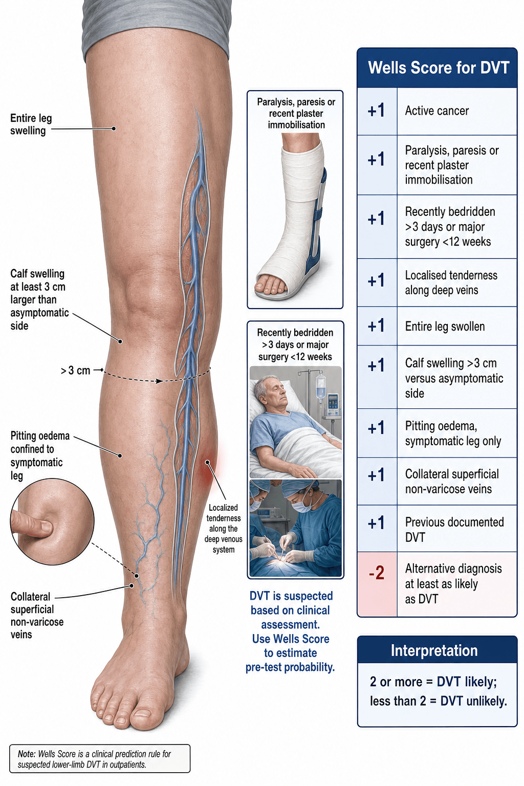
Each variable is scored as present or absent. The total ranges from −2 (a clear alternative diagnosis and no risk factors) to +8.5 (every variable present). The modern two-tier system splits at a threshold of 2.
- Points
- +1
- Clinical detail
- Includes solid tumours and haematological malignancies
- Points
- +1
- Clinical detail
- Any neuromuscular deficit reducing calf-pump function, or rigid immobilisation
- Points
- +1
- Clinical detail
- Applies to most post-operative orthopaedic patients (arthroplasty, fracture fixation)
- Points
- +1
- Clinical detail
- Palpate the popliteal fossa and posterior calf along the deep veins (not shin/knee)
- Points
- +1
- Clinical detail
- The whole limb from thigh to foot, not just ankle or calf
- Points
- +1
- Clinical detail
- Measure 10 cm below the tibial tuberosity bilaterally; over 3 cm asymmetry is positive
- Points
- +1
- Clinical detail
- Bilateral oedema suggests systemic causes; only unilateral counts
- Points
- +1
- Clinical detail
- Newly prominent superficial veins suggesting deep venous obstruction
- Points
- +1
- Clinical detail
- Confirmed by ultrasound or venography, not just clinical suspicion
- Points
- −2
- Clinical detail
- A convincing alternative: cellulitis, ruptured Baker cyst, muscle tear, superficial thrombophlebitis
CAPS-LEO-CVThe nine plus-one variables
Hook:CAPS-LEO-CV: nine variables each +1. Subtract 2 for a convincing alternative diagnosis. Under 2 = unlikely, 2 or more = likely.
A convincing alternative diagnosis (ruptured Baker cyst on ultrasound, cellulitis with a clear entry point, a muscle tear with a history of injury) subtracts 2 — and this single variable can shift a patient from DVT-likely to DVT-unlikely. Use it honestly, not as a shortcut to avoid imaging.
Score interpretation & diagnostic algorithm
The modern two-tier interpretation replaced the older three-tier (low/moderate/high) system — simpler and equally safe.
- Category
- DVT unlikely
- DVT prevalence (approx.)
- ~5–12%
- Recommended action
- D-dimer first; if positive → duplex ultrasound; if negative → DVT effectively excluded
- Category
- DVT likely
- DVT prevalence (approx.)
- ~25–40%
- Recommended action
- Skip D-dimer; proceed directly to duplex ultrasound of the affected leg
- 1Score the nine items (− 2 if an alternative is as likely)Each clinical/risk item scores +1; subtract 2 for a convincing alternative diagnosis.
- 2Under 2 → D-dimerA high-sensitivity D-dimer; a NEGATIVE result safely excludes DVT without imaging. Use the age-adjusted cut-off (age × 10 µg/L) in patients over 50.
- 32 or more → duplex ultrasoundSkip the D-dimer (it cannot rule out DVT in the likely group) and image the leg directly.
- 4Post-operative / high suspicion → imageAfter surgery the D-dimer is predictably raised and uninformative; if clinical suspicion is high, scan regardless. A negative proximal scan in a high-risk patient → repeat at 5–7 days.
A negative D-dimer in a DVT-likely patient (Wells 2 or more) does NOT exclude DVT. D-dimer has high sensitivity but poor specificity — a negative result is reassuring only in the DVT-unlikely group. In a post-operative orthopaedic patient the D-dimer is routinely elevated and essentially uninformative; if clinical suspicion is high, image regardless of the D-dimer.
The algorithm decides whether to image; the examiner then wants what you do with a confirmed DVT:
- Anticoagulation: a DOAC is first-line — apixaban or rivaroxaban start without a heparin lead-in, whereas dabigatran/edoxaban need ~5 days of LMWH first; LMWH-to-warfarin is the older standard.
- Special groups: pregnancy → LMWH (DOACs and warfarin contraindicated); cancer-associated VTE → a DOAC (edoxaban/apixaban/rivaroxaban) or LMWH, with caution/LMWH preference for luminal GI/GU tumours (bleeding).
- Duration: provoked by a transient major risk factor (e.g. recent surgery) → 3 months; unprovoked, persistent risk factor, or recurrent → extended/indefinite, weighing bleeding risk.
- Isolated distal (calf) DVT: either anticoagulate, or serial surveillance ultrasound (repeat in ~1 to 2 weeks) with anticoagulation if it extends — decided by symptom severity, extension risk and bleeding risk.
- IVC filter only when anticoagulation is absolutely contraindicated (active bleeding) or VTE recurs despite adequate anticoagulation — retrievable, removed when able.
- Phlegmasia cerulea dolens (massive iliofemoral DVT with limb ischaemia) is an emergency → catheter-directed thrombolysis or thrombectomy.
Orthopaedic-specific considerations
VTE is a leading cause of readmission and death after lower-limb arthroplasty and major trauma. The Wells score is relevant but has important limitations in the surgical patient.
- Post-operative D-dimer is reliably elevated for weeks-to-months after hip/knee arthroplasty — a positive result is expected and non-diagnostic; rely on the Wells score and ultrasound.
- Most post-arthroplasty patients already score 1–2 from recent major surgery before any leg signs — the majority are DVT-likely, so keep a low threshold for duplex ultrasound.
- Pharmacological prophylaxis (LMWH, DOACs, fondaparinux) reduces but does not eliminate DVT risk — the Wells score applies regardless.
- Distal (calf) DVT is more common than proximal post-operatively — the ultrasound must include the calf veins; distal DVT is managed by surveillance or anticoagulation depending on extent/symptoms.
- PE may present without leg symptoms — in the post-operative patient with tachycardia, dyspnoea, pleuritic chest pain or hypotension, investigate for PE (using the separate Wells PE score) even if the leg is normal.
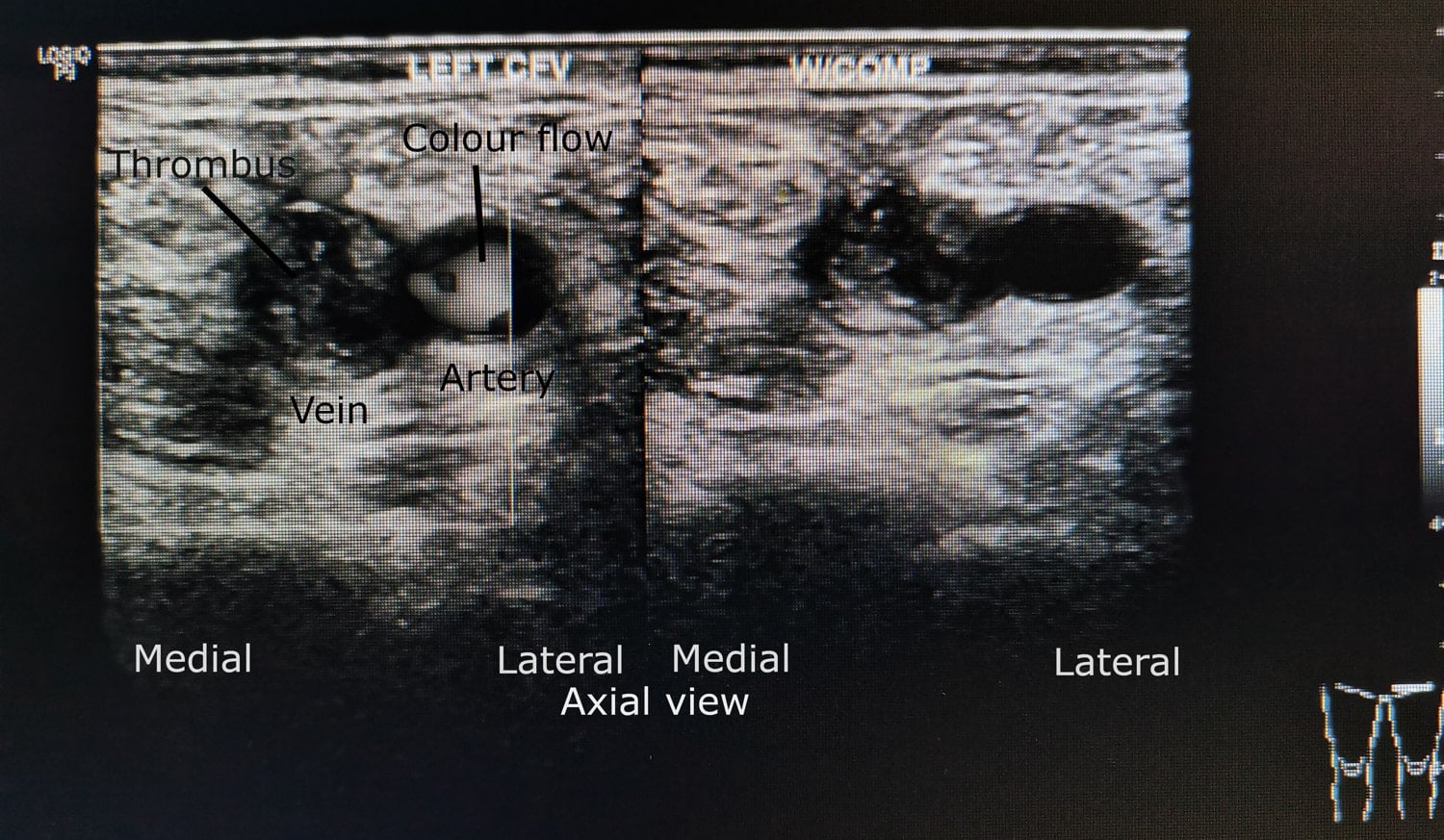
- Duplex ultrasound sensitivity exceeds 95% for proximal DVT but is lower (~70–80%) for calf thrombus — a negative scan in a high-risk patient with persistent symptoms warrants a repeat at 5–7 days.
The topic keeps pointing to "the separate Wells PE score" — here it is, because DVT and PE are one disease and PE is the post-operative killer that can present with a normal leg:
- Wells PE score (7 items): clinical signs of DVT (+3); PE the most likely diagnosis / an alternative less likely (+3); heart rate over 100 (+1.5); immobilisation at least 3 days or surgery in the prior 4 weeks (+1.5); previous DVT/PE (+1.5); haemoptysis (+1); active malignancy (+1).
- Two-tier: 4 or less = PE unlikely → D-dimer (a negative result excludes); over 4 = PE likely → CTPA (the first-line test; V/Q scan if contrast is contraindicated).
- PERC (PE rule-out criteria): in a patient already low-probability by gestalt, if all eight criteria are negative (age under 50, heart rate under 100, SaO2 at least 95%, no haemoptysis, no oestrogen use, no prior VTE, no unilateral leg swelling, no recent surgery/trauma), PE can be excluded without even a D-dimer.
- So the post-op patient with dyspnoea/tachycardia/pleuritic pain → apply the PE Wells (not the DVT one) and go to CTPA if likely.
D-dimer: role, limitations & assays
- Sensitivity for DVT
- Over 97%
- Specificity
- ~35–45%
- Notes
- Standard of care for VTE exclusion; use the age-adjusted cut-off in over-50s (age × 10 µg/L)
- Sensitivity for DVT
- ~95%
- Specificity
- ~40%
- Notes
- Older assays; acceptable but less commonly used
- Sensitivity for DVT
- ~85–90%
- Specificity
- ~50%
- Notes
- Fast in the ED; may miss some DVTs — confirm with a lab assay if concern persists
SNAPD-dimer clinical rules
Hook:SNAP: Sensitive-not-specific, Negative only excludes in low-risk, Age-adjusted in over-50s, Post-surgical = unhelpful.
In patients aged 50 and older, use age × 10 µg/L (FEU) as the threshold rather than the fixed 500 µg/L — this cuts false positives in older patients without sacrificing sensitivity. A 75-year-old, for example, has a cut-off of 750 µg/L.
Limitations & modern context
- Derived/validated in medical and ED populations, not specifically in early post-surgical orthopaedic patients — its performance in the first ~2 weeks post-op is less rigorously studied.
- Inter-observer variability affects tenderness assessment, calf-circumference technique, and the "alternative diagnosis" judgement; standardised measurement (10 cm below the tibial tuberosity) improves reproducibility.
- Clinical gestalt can match the score in experienced hands, but the score gives a structured, defensible documentation framework.
- The two-tier system has replaced the three-tier in most guidelines (NICE, ACCP/CHEST, ASH); the older low/moderate/high classification is still occasionally examined.
- Compression ultrasound is first-line imaging; CT/MR venography are alternatives when ultrasound is equivocal or limited (obesity, cast); contrast venography is historical.
- A negative complete compression ultrasound plus a negative D-dimer in a low-risk patient safely excludes DVT; in a high-risk patient, a negative proximal scan should be repeated at 5–7 days to catch an extending calf DVT.
Viva practice
Exam viva
Practise clinical reasoning and management decisions out loud
“A 68-year-old woman is 10 days post left total knee arthroplasty. She reports new left calf swelling and mild tenderness in the popliteal fossa. Her entire left leg is visibly swollen, with 2 cm of asymmetric calf swelling measured 10 cm below the tibial tuberosity. She is otherwise afebrile and haemodynamically stable. How would you investigate?”
“A 55-year-old man with no past medical history presents to the emergency department with a 2-day history of right calf pain and swelling after a long-haul flight. Examination reveals tenderness in the posterior calf, 2 cm asymmetric calf swelling, and no pitting oedema. He is otherwise well. His Wells score is 1 (tenderness +1; no other variables; no alternative diagnosis). How do you proceed, and how would an age-adjusted D-dimer influence your decision?”
Exam cheat sheet
DASHThe two-tier action rule
Hook:DASH: D-dimer if low, Always image if high, Surgery falsifies D-dimer, Hold anticoagulation only when both are negative.
- Nine +1 items, −2 for a convincing alternative; two-tier split at 2 (under 2 unlikely, 2 or more likely).
- Under 2 → D-dimer first (negative excludes without imaging); 2 or more → duplex ultrasound directly.
- A negative D-dimer does NOT exclude DVT in the likely group.
- Post-operative D-dimer is predictably raised and unhelpful — let suspicion drive imaging; most post-arthroplasty patients are already DVT-likely.
- Age-adjusted D-dimer (age × 10 µg/L) in over-50s.
- Ultrasound over 95% for proximal DVT, lower for calf — repeat at 5–7 days if high-risk and initially negative.
The nine plus-one variables
- Active cancer (+1)
- Paralysis, paresis, or plaster immobilisation of the lower limb (+1)
- Bedridden over 3 days or major surgery within 12 weeks (+1)
- Localised tenderness along the deep venous system (+1)
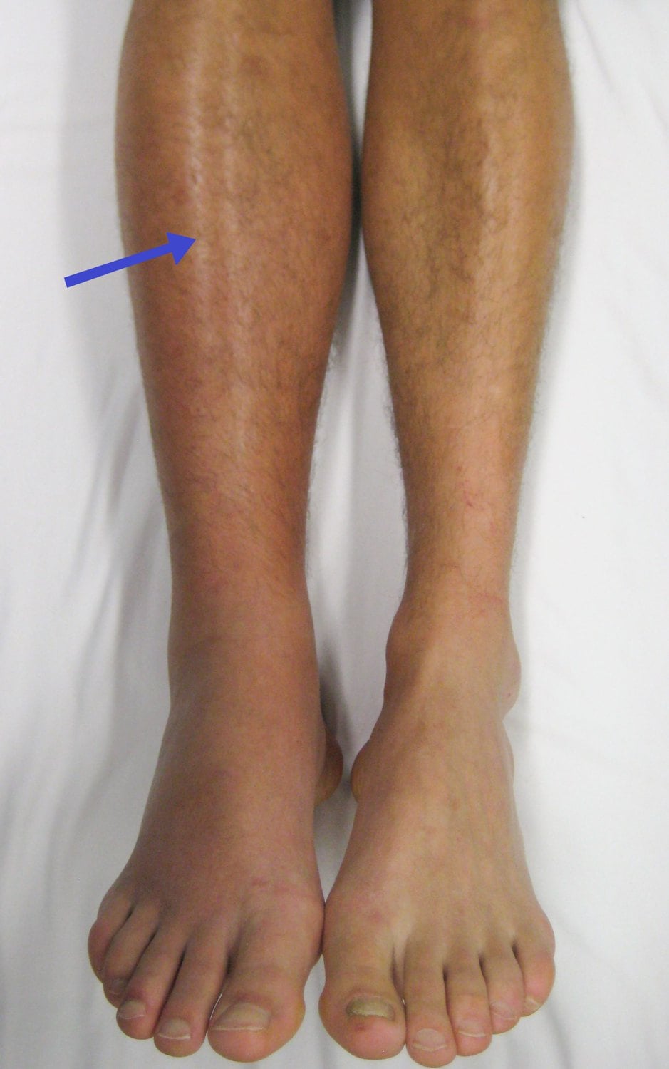
- Entire leg swollen (+1)
- Calf swelling over 3 cm vs the asymptomatic side, at 10 cm below the tibial tuberosity (+1)
- Pitting oedema confined to the symptomatic leg (+1)
- Collateral non-varicose superficial veins (+1)
- Previously documented DVT (+1); Alternative diagnosis at least as likely (−2)
Interpretation and action
- Under 2: DVT unlikely — D-dimer first; ultrasound only if positive
- 2 or more: DVT likely — skip D-dimer, proceed directly to duplex ultrasound
- Negative D-dimer + Wells under 2 safely excludes DVT
- Post-surgical patients: D-dimer unreliable — low threshold for ultrasound
Key ortho exam points
- Post-arthroplasty patients score from major surgery before leg signs — most are DVT-likely
- D-dimer predictably raised after surgery, trauma, malignancy, pregnancy
- Age-adjusted D-dimer: age × 10 µg/L FEU in over-50s
- Ultrasound over 95% for proximal DVT, lower for calf; repeat at 5–7 days if high-risk and negative
- Distal (calf) DVT is the commonest post-operative DVT — scan must include the calf veins
Evidence Base
Value of assessment of pretest probability of deep-vein thrombosis in clinical management
- Prospective management study (593 outpatients) using a clinical model to stratify DVT pretest probability into low, moderate and high.
- DVT prevalence tracked the score: ~3% (low), ~17% (moderate), ~75% (high).
- Combining the score with proximal compression ultrasound was safe and reduced the need for serial ultrasonography and venography.
Evaluation of D-dimer in the diagnosis of suspected deep-vein thrombosis
- RCT (1096 outpatients) of a D-dimer + clinical-probability strategy versus ultrasound alone.
- A clinically DVT-unlikely patient with a negative D-dimer could safely skip ultrasound (0.4% subsequent VTE), and 39% of the D-dimer group avoided imaging.
- Established the modern two-tier (likely/unlikely) Wells pathway combined with D-dimer.
According to PubMed, the original probability model and the 3%/17%/75% prevalence gradient come from Wells et al. 1997 (DOI), and the two-tier (likely/unlikely) + D-dimer pathway that allows imaging to be safely omitted from Wells et al. 2003 (DOI). The age-adjusted D-dimer cut-off, the post-operative D-dimer caveat and the ultrasound performance figures are standard, well-established VTE-diagnosis teaching.