Where Leukocyte Scintigraphy Still Adds Value
Leukocyte positive and marrow negative: infection likely
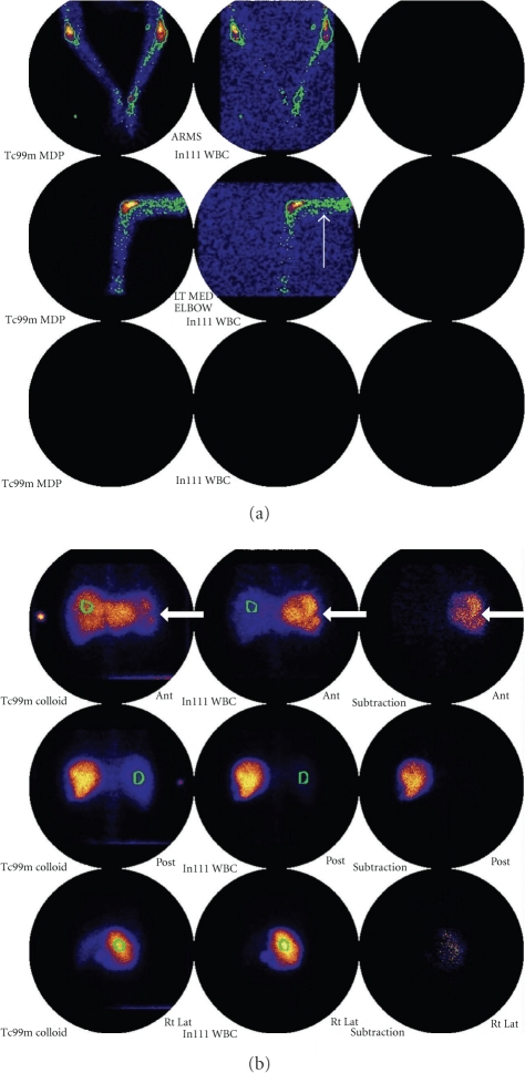
Leukocyte positive and marrow matched: marrow expansion or postoperative change
Leukocyte negative: infection less likely at the imaged site
Key: The marrow comparison is what turns leukocyte imaging into a high-specificity test for PJI.
- The best-established indication is chronic or prosthetic joint infection, not routine acute osteomyelitis.
- Interpretation hinges on mismatch: leukocyte uptake without matching marrow uptake suggests infection.
- MRI is superior for anatomy, marrow oedema, abscess extent, and vertebral infection.
- In the spine, labelled leukocyte imaging performs poorly and can even appear photopenic rather than hot.
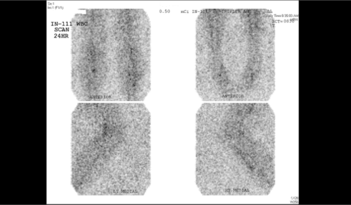
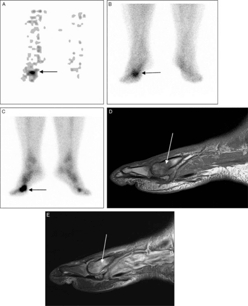
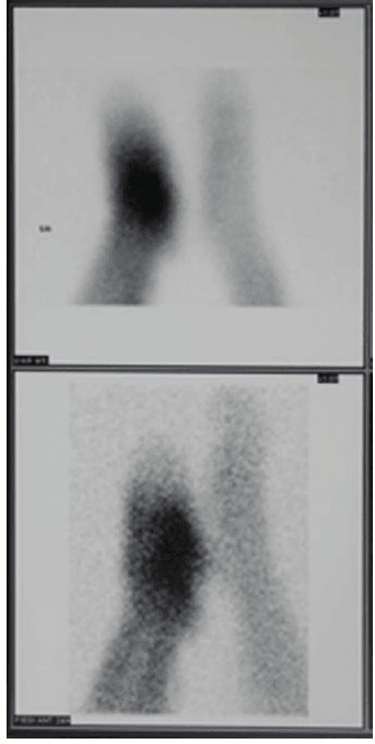
- SPECT/CT improves localisation, particularly in the diabetic foot and around complex hardware.
- “If the stem is failed arthroplasty with equivocal aspiration, think combined leukocyte and marrow imaging.
- “A positive bone scan alone is not enough after arthroplasty because postoperative turnover stays high.
- “Do not recommend labelled white cell scanning as first-line imaging for vertebral osteomyelitis.
- “Tc-99m HMPAO gives better image quality, whereas In-111 provides more stable delayed imaging.
Labelled white cell scanning is a problem-solving test. Order it when specificity matters more than raw sensitivity, especially around arthroplasty or chronic hardware. If the main question is early marrow infection or vertebral osteomyelitis, MRI is usually the better study.
SCANSCAN Indications
Hook:SCAN when the question is chronic, hardware-related, or postsurgical infection specificity.
MATCHMATCH Interpretation
Hook:MATCH the leukocyte scan against marrow before calling infection.
Overview
Labelled leukocyte imaging is based on ex vivo labelling of the patient's white blood cells, which are then reinjected and tracked to sites of active neutrophil accumulation. In orthopaedics, that mechanism is valuable when anatomical imaging is either equivocal or limited by metal, and when the clinician needs a more specific answer than a three-phase bone scan can provide.
The practical hierarchy is simple. MRI remains the best first-line test for most acute osteomyelitis, septic arthritis extent, and spinal infection. Labelled white cell imaging becomes attractive when the question is chronic infection activity, prosthetic joint infection, or diabetic foot osteomyelitis in a patient where marrow, postoperative change, Charcot change, or hardware makes MRI and bone scan less decisive.
Clinical Imaging
Imaging Atlas
Systematic Approach
| Step | Question | Reason |
|---|---|---|
| 1. Pick the right patient | Is this a chronic, hardware-related, or post-arthroplasty question? | This is where specificity matters most |
| 2. Choose the tracer | Do you need same-day Tc-99m HMPAO or delayed In-111 imaging? | Balances workflow against delayed stability |
| 3. Add marrow imaging | Will marrow expansion confound the leukocyte signal? | Critical in prosthetic joint infection |
| 4. Localise uptake | Is planar imaging enough, or do you need SPECT/CT? | Improves anatomic localisation |
| 5. Check the alternative | Would MRI answer the question better? | Avoids mis-ordering the wrong modality |
| Agent | Advantage | Limitation |
|---|---|---|
| In-111 oxine | Stable delayed imaging and established literature | Higher radiation burden and slower workflow |
| Tc-99m HMPAO | Better count statistics and same-day imaging | Less stable label than In-111 |
| Sulfur colloid marrow scan | Maps marrow distribution | Adds a second acquisition but markedly improves specificity |
Clinical Applications
| Problem | Why WBC imaging helps | Pitfall avoided |
|---|---|---|
| Postoperative bone turnover | Marrow comparison improves specificity | Bone scan false positives |
| Metal artefact on MRI | Nuclear imaging is less affected by hardware | Non-diagnostic MRI |
| Equivocal aspiration | Provides another line of evidence | Over-calling aseptic loosening |
Differential Interpretation & Mimics
The hardest part of a leukocyte study is not detecting uptake but deciding what the uptake means. Use the marrow scan, the time-course of the images, and the clinical question to separate true infection from physiological or sterile causes.
| Pattern | Most likely cause | Discriminator |
|---|---|---|
| Leukocyte positive, marrow negative (mismatch) | Infection | The classic high-specificity signature; uptake usually intensifies on delayed images |
| Leukocyte positive, marrow positive (matched) | Marrow expansion or displaced marrow after arthroplasty | Matched marrow uptake at the same site argues strongly against infection |
| Leukocyte positive, decreasing over time | Healing surgical wound or sterile inflammation | Stable or fading focus over early-to-delayed imaging favours non-infection |
| Photopenic vertebral lesion | Vertebral osteomyelitis (false negative for WBC) | Spine is unreliable for leukocyte imaging; switch to MRI or FDG-PET |
| Diffuse marrow or splenic uptake | Reticuloendothelial physiology or recent transfusion | Symmetrical, non-focal; not a focal infective pattern |
Controversies & Areas of Uncertainty
- FDG-PET versus leukocyte/marrow for PJI. After two decades there is still no consensus on FDG for periprosthetic joint infection because it cannot reliably separate infection from physiological or aseptic inflammation and lacks standardised uptake criteria; combined leukocyte/marrow imaging retains the better specificity track record (Palestro, Semin Nucl Med 2022).
- Shoulder and upper-limb arthroplasty. Evidence is limited and suggests leukocyte/marrow SPECT/CT and FDG-PET/CT are specific but not sensitive, so a negative study is less reassuring than around hip or knee prostheses.
- In-111 versus Tc-99m HMPAO. Both perform well; Tc-99m HMPAO gives superior count statistics and same-day imaging, while In-111 offers more stable delayed images. In the diabetic foot, the Tc-99m HMPAO meta-analytic specificity (92 percent) outperformed In-111 (75 percent).
- Is the marrow scan always necessary? Around hardware and in the postoperative skeleton, omitting marrow correlation undermines specificity; in a virgin appendicular skeleton without marrow-altering disease, the incremental value is smaller.
- Workflow burden. Ex-vivo labelling is labour-intensive, requires blood handling, and is not available everywhere, which increasingly pushes practice toward FDG-PET/CT where access and expertise allow.
Guidelines, Registries & Global Practice
Global epidemiology
-
Prosthetic joint infection complicates roughly 1 to 3 percent of primary lower-limb arthroplasties and is one of the leading causes of revision worldwide; the diagnostic challenge is separating it from aseptic loosening, the commonest cause of failure.
-
Diabetic-foot osteomyelitis is a high-volume global problem; imaging is invoked when probe-to-bone testing, plain films, and inflammatory markers leave the diagnosis open.
Society Positions on Imaging Bone & Joint Infection Body Emphasis Where leukocyte imaging sits EANM (procedural guideline) Standardised WBC acquisition with early/delayed imaging plus marrow correlation and SPECT/CT Endorsed best-practice technique for neutrophil-mediated infection MSIS / ICM (PJI consensus) Diagnosis is multi-criteria (aspiration, synovial markers, histology); imaging is adjunctive Nuclear imaging reserved for equivocal cases, not a stand-alone criterion IDSA / IWGDF (diabetic foot) MRI first-line for bone infection; bone biopsy is reference standard Labelled WBC (ideally Tc-99m HMPAO SPECT/CT) when MRI is contraindicated or equivocal IDSA (vertebral osteomyelitis) MRI first-line; FDG-PET as alternative Labelled WBC NOT recommended for the spine
Registry and access notes
- Arthroplasty registries (NJR, AOANJRR, AJRR, Swedish and Norwegian registers) track infection as a revision indication and confirm PJI as a major, costly failure mode, reinforcing the value of a high-specificity confirmatory test before revision surgery.
- Access varies widely: in vitro WBC labelling needs a licensed radiopharmacy, blood-handling facilities, and trained staff, so high-resource centres can offer leukocyte/marrow SPECT/CT or FDG-PET/CT, whereas limited-resource settings may rely on bone scintigraphy, plain radiography, aspiration, and clinical criteria.
- Where PET/CT is available it is increasingly favoured for the spine and for vascular-graft infection; where it is not, labelled leukocyte imaging remains the most specific functional option around hardware.
Evidence Base
Combined Leukocyte/Marrow vs FDG in the Failed Joint Replacement
- In 59 painful, failed lower-limb prostheses (40 hip, 19 knee) with histopathological and microbiological confirmation, 25 (42 percent) were infected.
- Combined In-111 leukocyte/Tc-99m sulfur-colloid marrow imaging achieved sensitivity 100 percent, specificity 91 percent, and accuracy 95 percent.
- Coincidence-detection FDG was less accurate under every interpretation criterion (best accuracy 71 percent) and could not replace leukocyte/marrow imaging.
Combined Leukocyte/Marrow Imaging Is the Most Accurate Radionuclide Test for the Infected Prosthesis
- Bone scintigraphy alone is only 50 to 70 percent accurate and cannot separate infection from aseptic loosening, but a normal scan effectively excludes a prosthetic complication.
- Adding gallium-67 raises accuracy only to 70 to 80 percent, whereas combined leukocyte/marrow imaging reaches about 90 percent, the highest among radionuclide studies.
- The leukocyte-without-matching-marrow mismatch pattern is the diagnostic signature of infection; success is tempered by the limitations of in-vitro labelling.
Clinical Decision Scenarios
Practise clinical reasoning and management decisions out loud
“A patient two years after total knee replacement has persistent pain, raised inflammatory markers, and equivocal aspiration.”
“A patient with diabetic foot ulceration has MRI findings that remain equivocal because of adjacent Charcot change.”
“A clinician asks whether labelled white cell scanning is appropriate for suspected vertebral osteomyelitis.”
Best Uses
- Equivocal prosthetic joint infection
- Chronic or hardware-associated osteomyelitis
- Selective diabetic-foot problem solving
- Cases where MRI is limited or non-diagnostic
Core Interpretation
- Leukocyte plus marrow mismatch suggests infection
- Matched leukocyte and marrow uptake favours marrow expansion
- Delayed images improve specificity
- Use SPECT/CT when localisation matters
Do Not Forget
- MRI first for most acute osteomyelitis
- MRI first for vertebral infection
- Bone scan alone is too non-specific after arthroplasty
- Tracer choice affects workflow and image quality
Limitations
- Poor spinal performance
- Time-consuming ex vivo labelling
- Needs specialist nuclear medicine support
- Not a replacement for aspiration or microbiology