FDG-PET for staging, response assessment and recurrence in bone and soft tissue tumours
Staging: Whole body assessment for metastases
Response: Pre and post-treatment comparison
Recurrence: Detection of local or distant relapse
Biopsy guidance: Target metabolically active areas
Key: PET-CT is most valuable for high-grade malignancies and systemic staging
- FDG uptake reflects glucose metabolism (Warburg effect)
- High-grade tumours show higher FDG uptake than low-grade
- SUVmax is quantitative measure of uptake intensity
- PET-CT combines metabolic and anatomic information
- Used for staging, response assessment, and recurrence detection
- “Brain has high physiological FDG uptake (limits assessment)
- “Blood glucose must be controlled (less than 11 mmol/L)
- “False positives: infection, inflammation, recent surgery
- “Response: greater than 40% SUV decrease = metabolic response
- “Low-grade sarcomas may have minimal FDG uptake
FDG-PET Principles
PET-CT combines metabolic and anatomic information in one study, and it is increasingly used in orthopaedic oncology for staging, treatment response and the detection of recurrence. What it measures is glucose metabolism, not malignancy directly.
The Warburg effect. Malignant cells preferentially use glycolysis for energy, even in the presence of oxygen, and this increased glucose uptake is the basis of the scan. FDG (fluorodeoxyglucose) is a glucose analogue that cells take up but cannot metabolise, so it becomes trapped. Higher-grade, more aggressive tumours typically have higher glucose metabolism and FDG uptake.
The protocol. Fasting, the glucose check and the fixed uptake period matter because SUV depends on blood glucose and on uptake time, as the next section sets out.
- Timing
- 4-6 hours fasting
- Requirements
- Blood glucose less than 11 mmol/L
- Timing
- Time 0
- Requirements
- 4-5 MBq/kg IV
- Timing
- 60 minutes
- Requirements
- Rest, avoid talking/chewing
- Timing
- 60 minutes post-injection
- Requirements
- Whole body or regional
- Timing
- Simultaneous or sequential
- Requirements
- Low-dose for attenuation/localisation
Indications in Orthopaedic Oncology
Staging and biopsy. PET-CT assesses the whole body for metastases, and it can guide biopsy to the metabolically active areas of a tumour. What it adds in each tumour is set out in the next section.
Response. PET-CT can assess treatment response earlier than anatomic imaging, usually after neoadjuvant chemotherapy and before surgery. A decrease in SUVmax of more than 40% between the pre- and post-treatment scans indicates metabolic response, and this may predict histological response and prognosis in sarcomas. PERCIST measures SUL peak rather than SUVmax and sets its boundaries at a 30% change:
- Definition
- Resolution of all FDG-avid disease
- Definition
- Greater than 30% decrease in SUV
- Definition
- Less than 30% decrease or less than 30% increase
- Definition
- Greater than 30% increase or new lesions
Recurrence. PET-CT can detect local recurrence and distant metastases in sarcoma surveillance, and it is more sensitive than conventional imaging for some recurrences. False positives from post-surgical change and inflammation limit its specificity, and routine PET-CT surveillance after sarcoma resection is not supported by high-level evidence; CT chest plus local MRI remain standard. It is best reserved for problem-solving (equivocal recurrence, rising tumour markers, the symptomatic patient) rather than scheduled follow-up, balancing radiation dose and false positives.
Infection is a separate argument. Oncology is where FDG-PET is best established in orthopaedics. The second major indication, infection, is covered on the Labelled White Cell Scintigraphy page, because the two indications are judged against different comparators and reach different conclusions:
- Spinal osteomyelitis - FDG is the radionuclide study of choice, with a high negative predictive value. Labelled leucocyte imaging is explicitly not recommended for the spine, where infected vertebrae are often photopenic and read as false negatives.
- Periprosthetic joint infection - the opposite verdict. FDG cannot reliably separate infection from aseptic loosening, because both provoke an intense inflammatory reaction, and combined labelled leucocyte/marrow imaging remains the more specific test.
- Diabetic foot osteomyelitis - FDG-PET and Tc-99m HMPAO leucocyte imaging offer comparable, and the highest available, specificity.
The fact that unifies them is that FDG marks activated macrophages, and therefore inflammation, not infection and not malignancy specifically. That explains its strength in the spine, its weakness around a loose prosthesis, and post-surgical false positives, which settle only after roughly three to four months.
Specific Tumour Applications
- FDG Avidity
- High
- Role and Clinical Implications
- Lung/distant metastases and skip lesions at staging, complementing bone scan and CT chest; SUV correlates with histological response
- FDG Avidity
- Very high
- Role and Clinical Implications
- Systemic staging including marrow involvement; excellent for staging and response
- FDG Avidity
- High
- Role and Clinical Implications
- Staging, including nodal and distant disease; SUV predicts grade and outcome
- FDG Avidity
- Low to moderate
- Role and Clinical Implications
- May not be reliably detected
- FDG Avidity
- Low
- Role and Clinical Implications
- Difficult to differentiate from enchondroma
- FDG Avidity
- Moderate to high
- Role and Clinical Implications
- Increased uptake suggests higher grade
- FDG Avidity
- Variable
- Role and Clinical Implications
- Not reliably FDG-avid
- FDG Avidity
- Variable
- Role and Clinical Implications
- FDG-PET useful for treatment response
Osteosarcoma. Beyond skip lesions and distant metastases at staging, a decrease in SUV after chemotherapy correlates with histological necrosis, which is a good prognostic factor. For lung metastases a dedicated CT chest is still needed, and it remains superior for small nodules.
Soft tissue sarcoma. High-grade lesions are FDG-avid, and an SUVmax greater than 6 predicts higher grade. Low-grade sarcomas such as well-differentiated liposarcoma and low-grade fibromyxoid sarcoma may have minimal uptake. PET helps detect nodal involvement in selected subtypes.
Chondrosarcoma. PET-CT is used for grade assessment and to look for dedifferentiation. The overlap between enchondroma and grade 1 chondrosarcoma (atypical cartilaginous tumour) is the central unresolved problem: very low SUVmax favours benign and high SUVmax favours grade 2/3, but the benign-to-G1 boundary remains unreliable on PET alone, and histology, MRI and clinical correlation are mandatory.
Metastatic disease. PET-CT surveys the whole body and searches for an unknown primary, complementary to the bone scan.
Limitations and Pitfalls
Uptake is not malignancy. PET-CT cannot reliably differentiate benign from malignant in all cases, because infection, inflammation and healing all take up FDG, as do recent radiotherapy and arthritis. Always correlate uptake with the history and with CT/MRI morphology, and meet suspected infection with clinical correlation and follow-up.
- Typical SUVmax / Pattern
- High (often greater than 6), focal intense
- Discriminating Features
- Soft tissue mass, cortical destruction, growth on serial imaging
- Typical SUVmax / Pattern
- Low to moderate (often under 3)
- Discriminating Features
- May be PET-negative; MRI fat signal, slow growth
- Typical SUVmax / Pattern
- Moderate to high, can mimic tumour
- Discriminating Features
- Clinical signs, raised inflammatory markers, marrow oedema, rim enhancement
- Typical SUVmax / Pattern
- Diffuse, linear uptake along tract
- Discriminating Features
- History; resolves over weeks (wait 4 to 6 weeks before imaging)
- Typical SUVmax / Pattern
- Moderate fusiform uptake at fracture line
- Discriminating Features
- History of trauma, fracture line on CT, settles with time
- Typical SUVmax / Pattern
- Mild to moderate, oval, fatty hilum
- Discriminating Features
- Preserved hilum; biopsy if discordant with primary
- Typical SUVmax / Pattern
- Variable, can be markedly FDG-avid
- Discriminating Features
- Characteristic CT/MRI morphology prevents over-call of malignancy
False negatives. A negative scan does not clear a lesion. PET has limited sensitivity for low-grade tumours, where MRI is better; lesions under 1 cm may be missed through resolution limits; and hyperglycaemia lowers uptake, so control the glucose and repeat if needed.
Comparison with Other Modalities
- Bone Scan
- Osteoblast activity
- PET-CT
- Glucose metabolism
- MRI
- Tissue characterisation
- Bone Scan
- Yes
- PET-CT
- Yes
- MRI
- Limited (WB-MRI emerging)
- Bone Scan
- Poor
- PET-CT
- Good (CT component)
- MRI
- Excellent
- Bone Scan
- May be cold
- PET-CT
- Usually positive
- MRI
- Positive (marrow)
- Bone Scan
- Limited
- PET-CT
- Good
- MRI
- Excellent
- Bone Scan
- Limited
- PET-CT
- Good (SUV)
- MRI
- Moderate
- Bone Scan
- Widely available
- PET-CT
- Limited centres
- MRI
- Widely available
- Bone Scan
- Lower
- PET-CT
- Higher
- MRI
- Moderate
Guidelines, Registries & Global Practice
- Position on FDG-PET/CT
- Recommended for staging high-grade STS and bone sarcoma; option for restaging and equivocal recurrence
- Practical Point
- CT chest still required for lung metastases
- Position on FDG-PET/CT
- PET-CT may aid staging, grading and response assessment in selected sarcomas
- Practical Point
- Emphasises management within sarcoma reference centres
- Position on FDG-PET/CT
- Reserved for problem-solving; not routine surveillance
- Practical Point
- MRI primary site plus CT chest remain core staging
- Position on FDG-PET/CT
- Standardised acquisition and PERCIST-style reporting encouraged
- Practical Point
- Fasting, glucose control, fixed uptake time mandatory
- Position on FDG-PET/CT
- Increasing use for skeletal staging in Ewing and osteosarcoma
- Practical Point
- Preferred over bone scan for marrow/bone disease
What SUV Actually Is (and Why There Is No Universal Cut-off)
Definition. The standardised uptake value is a semi-quantitative, dimensionless ratio that normalises the FDG activity concentration measured in a region to the injected dose and the patient's size: SUV = tissue activity concentration (e.g. MBq/mL) divided by [injected dose (MBq) / body weight (g)]. In effect it asks how much brighter the lesion is than it would be if the tracer were spread evenly through the body. It is a stand-in for metabolic rate, not a direct measurement of malignancy.
Reading the number. Higher SUV generally correlates with higher grade, and although the threshold varies, an SUVmax greater than 2.5 is often used as a cut-off.
- What it measures
- The single hottest voxel in the region
- Strength / weakness
- Most reproducible and most reported; but sensitive to image noise (one bright voxel)
- What it measures
- The average across a drawn region of interest
- Strength / weakness
- Less noisy, but depends heavily on how the ROI is contoured
- What it measures
- A fixed ~1 cm3 volume around the hottest area, normalised to LEAN body mass
- Strength / weakness
- The PERCIST metric — chosen because lean-mass normalisation and a fixed volume make it the most reproducible for response assessment
Why a single threshold cannot be transferred. SUVmax is not standardised across scanners, and absolute SUV is not comparable between centres or time points because it depends on:
- Blood glucose - high glucose competes with FDG and lowers tumour SUV
- Uptake time - SUV keeps rising for an hour or more, so the scan delay must be fixed
- Body habitus and lean mass - fat dilutes weight-based SUV, hence SUL
- Scanner model and reconstruction algorithm
- Partial-volume effect - lesions below roughly twice the scanner resolution falsely read low
A single number such as 2.5 therefore cannot reliably separate benign from malignant, or low grade from high grade, across all centres. PERCIST exists because absolute SUV is not transferable between time points unless conditions are tightly controlled: it mandates identical conditions and uses the normal liver (or blood pool) as an internal reference for response. The general physics of PET acquisition is developed in the nuclear-medicine-orthopaedics topic.
Normal Physiological FDG Biodistribution
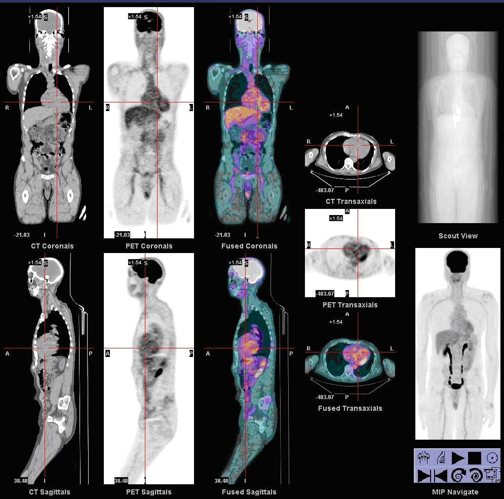
Tissues that are normally hot. The brain is an obligate glucose user, so it is the most FDG-avid normal tissue, which limits the assessment of intracranial lesions. Myocardial uptake varies with the fasting state; a prolonged fast and a low-carbohydrate, high-fat preparation can suppress it. The liver and blood pool show moderate, relatively uniform uptake, and the normal liver is the standard internal background against which lesions and response are judged.
Excretion. Unlike natural glucose, which is reabsorbed, FDG is renally excreted, so the kidneys, ureters and bladder are intensely active. That urinary activity can obscure lesions in the pelvis and bony pelvis, and hydration and bladder emptying before imaging help.
Variable and avoidable uptake. This is the source of many false positives. Skeletal muscle takes up FDG if recently exercised, hence rest before and after injection; the larynx and vocal cords do so if the patient is talking, hence "avoid talking"; bowel uptake is variable.
Brown fat. Brown adipose tissue gives symmetric supraclavicular, paraspinal and mediastinal uptake in fat density (CT-negative). Keeping the patient warm and beta-blockade or a benzodiazepine reduce it, and it should not be mistaken for nodal disease.
Bone marrow. Marrow shows mild physiological uptake that is diffusely increased after G-CSF or marrow recovery from chemotherapy. This reactive uptake can both mimic and mask marrow disease in the very sarcoma patients being imaged, so correlate before calling marrow metastases.
Controversies & Areas of Uncertainty
Prognostic or predictive? Whether baseline or interim SUV should change treatment (risk-adapted chemotherapy) is unproven. Studies show an association with histological necrosis and survival, but no randomised trial has demonstrated that altering therapy on PET findings improves outcomes, and PET-guided treatment escalation remains investigational.
Clinical Decision Scenarios
Practise clinical reasoning and management decisions out loud
“A 16-year-old with newly diagnosed osteosarcoma of the distal femur is being staged. The oncologist requests PET-CT.”
“A patient with known high-grade soft tissue sarcoma of the thigh has PET-CT showing intense uptake in the primary tumour (SUVmax 12) and a 1.5cm inguinal lymph node with SUVmax 4.”
“A patient with a cartilage tumour in the proximal humerus has PET-CT. The lesion shows SUVmax of 3.5.”
FDG-PET Principles
- FDG = Fluorodeoxyglucose (glucose analogue)
- Warburg effect: Tumours use glycolysis
- Higher grade = higher SUV generally
- SUVmax = maximum standardised uptake value
Indications
- Staging: Metastases, skip lesions
- Response: SUV decrease greater than 30-40%
- Recurrence: Symptomatic patients
- Biopsy guidance: Target active areas
FDG Avidity by Tumour
- High: Osteosarcoma, Ewing, high-grade STS
- Variable: Chondrosarcoma (grade-dependent)
- Low: Low-grade STS, enchondroma
Limitations
- False +: Infection, inflammation, surgery
- False -: Low-grade tumours, small lesions
- Glucose control essential
- Cannot replace biopsy for diagnosis
Evidence
PERCIST 1.0: standardised metabolic response criteria
- Practical guide to PET Response Criteria in Solid Tumors (PERCIST 1.0), defining quality control needed to compare FDG-PET across time points.
- Response is measured using SUL peak (lean-body-mass corrected) in a reference lesion, not raw SUVmax, to reduce variability.
- Defines complete, partial, stable and progressive metabolic disease and clarifies measurement of unequivocal progression.
FDG-PET correlates with histological necrosis in osteosarcoma
- 11 osteosarcoma patients had FDG-PET around neoadjuvant chemotherapy (9 imaged both before and after) correlated with histological necrosis.
- Post-chemotherapy SUV (SUV2) was much lower in good responders (mean 1.93) than poor responders (5.86); change in tumour size on MRI did not correlate with response.
- An SUV2 under 2.5 had 100% positive and negative predictive value for good histological response in this small series.
Baseline tumour SUV predicts response and survival in bone sarcoma
- 77 patients with localised Ewing sarcoma (45) and osteosarcoma (32) staged with FDG-PET/CT.
- Lower baseline SUVmax (under 6) predicted a higher good-response rate (72% vs 30% in Ewing; 64% vs 29% in osteosarcoma).
- Baseline SUVmax was the only independent pre-treatment prognostic factor for event-free survival on multivariate analysis.
SUVmax stratifies grade and prognosis in soft tissue sarcoma
- 50 adults with primary high-grade extremity soft tissue sarcoma and preoperative FDG-PET; mean SUVmax 12.9 (range 2.2 to 33.4).
- Lower SUVmax (under 10.3) was associated with better overall survival and lower local recurrence.
- Myxoid liposarcoma and synovial sarcoma were consistently low-uptake, limiting PET sensitivity in these subtypes.
FDG-PET differentiates benign from malignant chondroid tumours
- Systematic review of 8 studies and 166 chondroid lesions correlating SUVmax with histological grade.
- Mean SUVmax was lower for benign (1.6) than malignant (4.4) lesions and rose with grade (grade 0/1 = 2.0 vs grade 2/3 = 6.0).
- An SUVmax of 4.4 or greater was 99% specific for grade 2/3 chondrosarcoma.
Meta-analysis: PET/CT accuracy in chondrosarcoma diagnosis and grading
- Meta-analysis of 12 studies evaluating FDG-PET/CT for chondrosarcoma diagnosis and grading.
- Pooled PET/CT sensitivity 0.94 and specificity 0.89 for diagnosing chondrosarcoma.
- SUVmax separated low- from intermediate/high-grade chondrosarcoma but was limited at the benign-vs-G1 and G2-vs-G3 boundaries.
FDG-PET/CT outperforms bone scintigraphy for skeletal metastases in paediatric sarcoma
- Review of FDG-PET/CT in paediatric osteosarcoma, Ewing sarcoma and rhabdomyosarcoma.
- PET/CT has consistently better sensitivity and specificity than bone scintigraphy for detecting skeletal metastases.
- Its value for pulmonary metastases is limited (CT chest remains superior for small lung nodules) and its prognostic role outside osteosarcoma is unproven.