Winquist-Hansen Classification of Femoral Comminution
The Winquist-Hansen classification
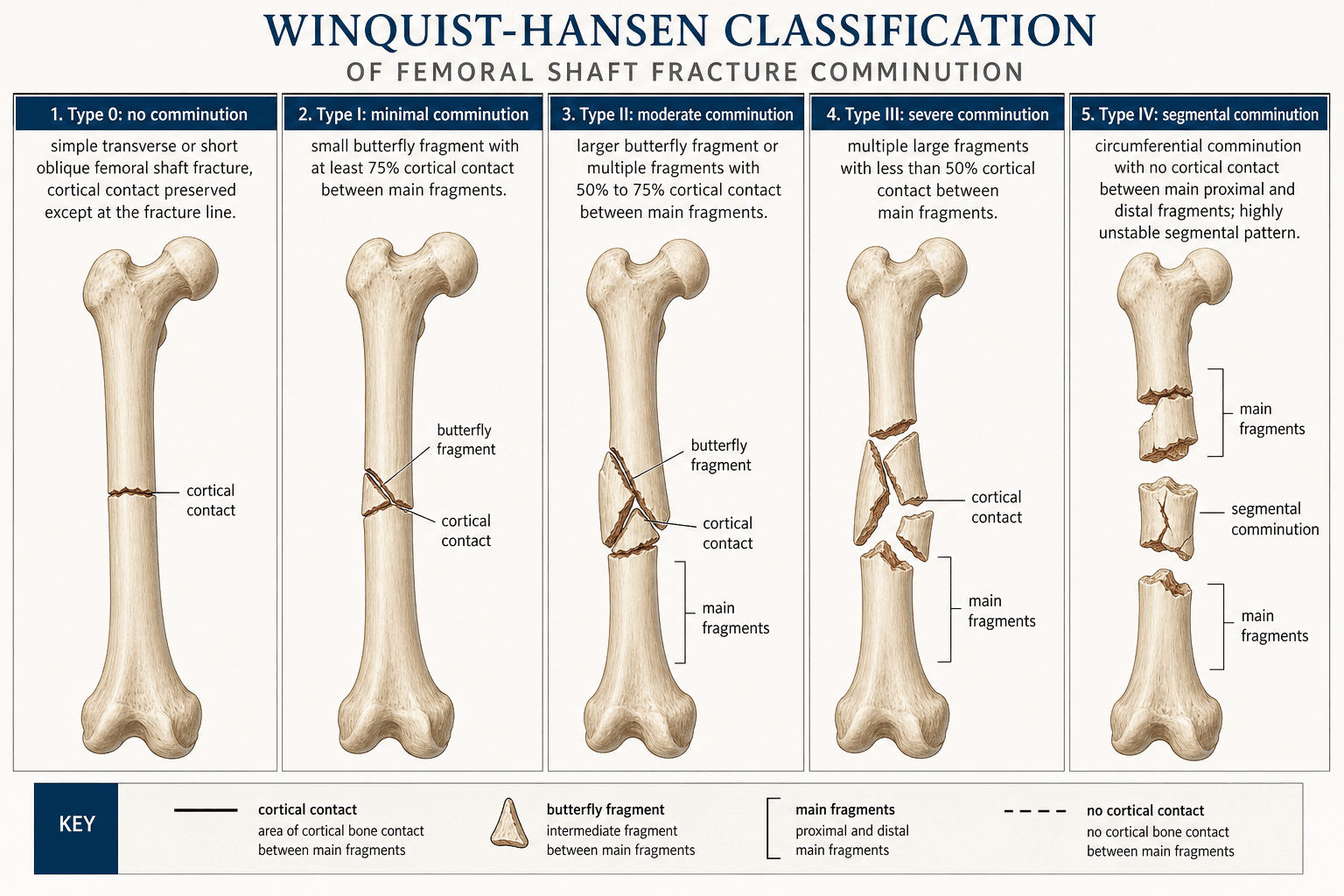
The five-type scheme grades the circumferential cortical contact between the proximal and distal main fragments after reduction, judged on AP and lateral projections (the worst view counts).
- Cortical contact
- 100% (intact cortices)
- Comminution pattern
- No comminution; simple or wedge with intact buttress
- Mechanical behaviour
- Bone bears the load; behaves like a simple fracture
- Cortical contact
- ~75% or more
- Comminution pattern
- Tiny butterfly; both cortices still in continuity
- Mechanical behaviour
- Bone still bears the load; behaviour unchanged
- Cortical contact
- At least 50% (≈50–75%)
- Comminution pattern
- Larger butterfly; at least half the circumference in contact
- Mechanical behaviour
- Bone still shares load; dynamic lock acceptable
- Cortical contact
- Less than 50%
- Comminution pattern
- Larger comminuted zone; less than half the cortex in contact
- Mechanical behaviour
- Implant bears most of the load; static lock mandatory
- Cortical contact
- None
- Comminution pattern
- Segmental or circumferential comminution; no contact
- Mechanical behaviour
- Implant is the only load-bearing structure; shortening follows if the nail fails
Contact falls as the number risesThe five types
Hook:0 → IV: cortical contact goes from full to none, and load transfers from bone to implant.
The grade is judged on the post-reduction image, not the pre-op film. Traction restores length and the image intensifier shows the comminuted zone in true AP and lateral. A butterfly that looked attached on the trauma series often hinges free once the fracture is out to length — so a type II can declare itself a type IV on the table. Document the grade with the saved image-intensifier picture (inter-observer agreement is only modest).
Surgical decision-making
The grade answers three questions at nailing: do I need a static lock, do I need an adjunct, and can the patient weight-bear through the construct?
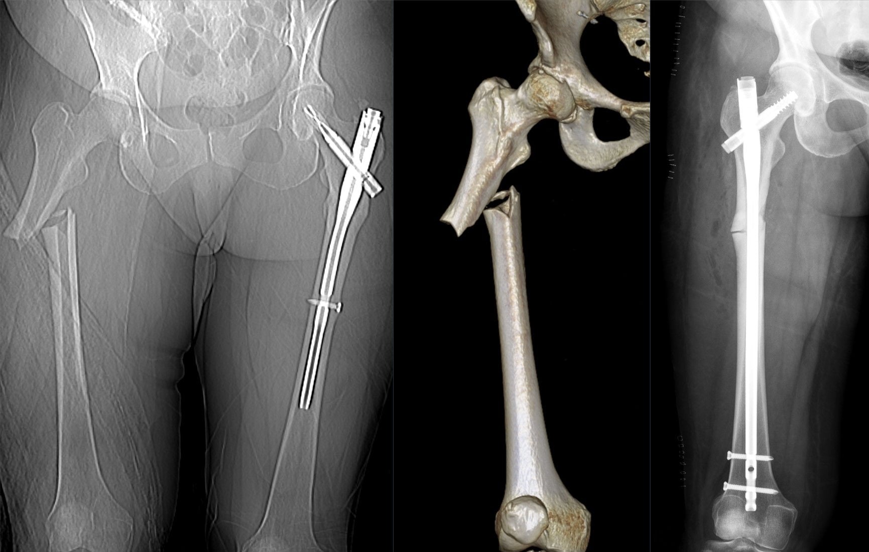
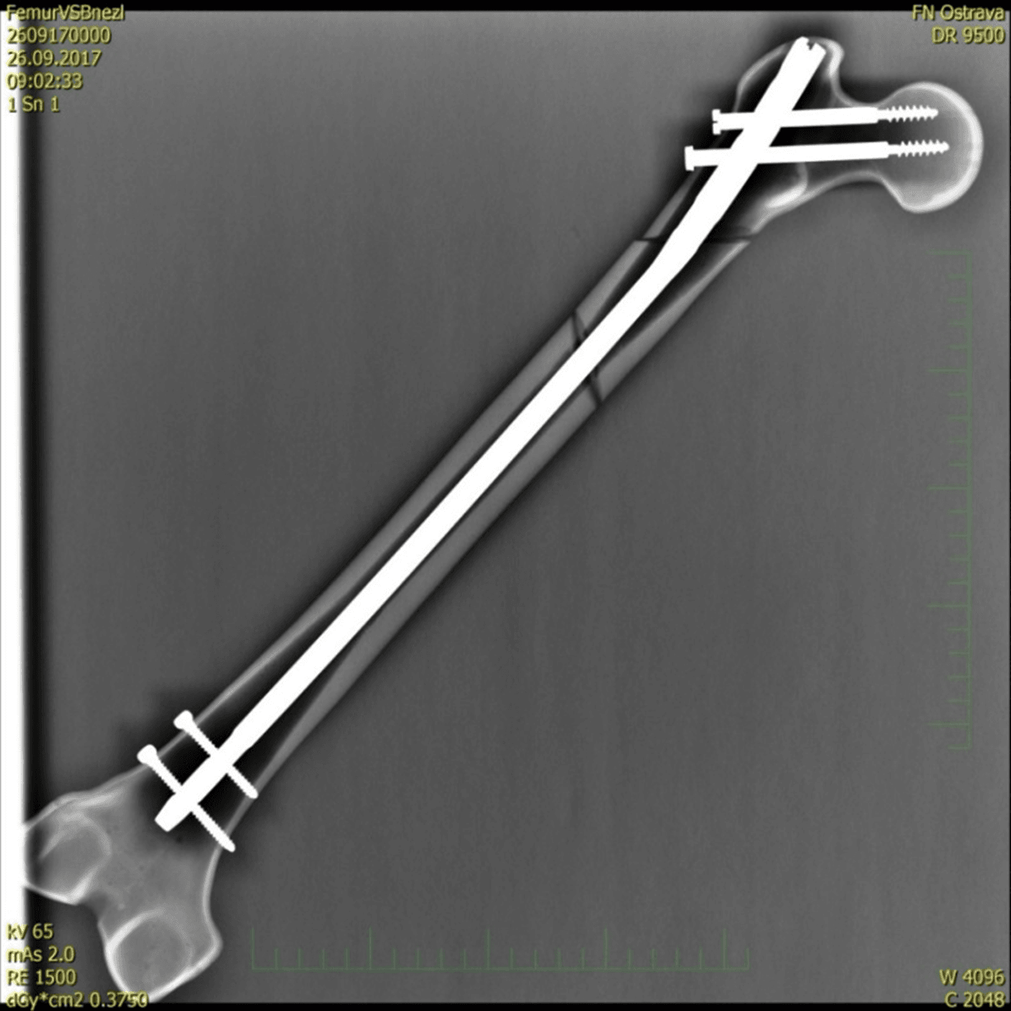
- 1Re-grade on the tableAfter reduction on the image intensifier, decide the type from cortical contact on both views — the worst view counts. A higher grade than the pre-op film is common.
- 2Lock by buttressTypes 0–II retain a cortical buttress: dynamic OR static locking both hold length (static is the modern default). Types III–IV have no buttress: static locking is mandatory, with at least one proximal and one distal screw.
- 3Add adjuncts for the comminuted/metaphyseal patternSize the nail to fill the isthmus; use blocking (Poller) screws to re-establish a buttress in metaphyseal III/IV patterns, and cerclage or a small auxiliary plate for very proximal/distal patterns.
- 4Prescribe weight-bearing by gradeTypes 0–II: weight-bear as tolerated once comfortable. Types III–IV: protected weight-bearing (≈6–8 weeks III, 8–12 weeks IV) until bridging callus on two views; dynamise only after callus.
- Locking strategy
- Dynamic or static (surgeon preference)
- Adjunct fixation
- Rarely needed
- Weight-bearing
- As tolerated
- Locking strategy
- Static preferred
- Adjunct fixation
- Occasional cerclage for a displaced butterfly
- Weight-bearing
- As tolerated once comfortable
- Locking strategy
- Static mandatory; consider blocking screws at the metaphysis
- Adjunct fixation
- Blocking (Poller) screws, percutaneous clamp, or cerclage
- Weight-bearing
- Protected (touch-down/partial) ~6–8 weeks until callus
- Locking strategy
- Static mandatory; nail sized to fill the canal
- Adjunct fixation
- Blocking screws, cerclage, or a small auxiliary plate for very proximal/distal patterns
- Weight-bearing
- Protected ~6–12 weeks; dynamise only after bridging callus on two views
In type IV fractures the nail is the only structure resisting shortening. Two proximal and two distal locking screws, a nail that fills the isthmus, and protected weight-bearing are not optional — a short or narrow nail, or premature dynamisation, will lose length and rotation before callus forms.
A blocking (Poller) screw placed in the short metaphyseal fragment narrows the effective canal and re-establishes a cortical buttress, converting a metaphyseal type III/IV into a mechanically simpler pattern. It is the single most useful adjunct for comminuted proximal-third and distal-third femoral shaft fractures treated by antegrade or retrograde nailing.
Most femoral shaft fractures are nailed antegrade, and the entry point matters: the piriformis fossa is collinear with the canal but is technically harder and risks the medial femoral circumflex blood supply (and, in children, the capital epiphysis), whereas the trochanteric-tip entry is easier but - being lateral to the canal axis - risks varus/translation in proximal-third fractures unless a trochanteric-entry nail and correct start point are used. Retrograde nailing (entry through the intercondylar notch) is preferred for the distal-third fracture, the obese patient, the polytrauma patient (fast, supine, no traction table), the floating knee (one approach for femur and tibia), bilateral femurs, an ipsilateral femoral neck or acetabular fracture, and late pregnancy; it needs an intact knee and risks knee pain/stiffness/ sepsis and distal malalignment. Malalignment is highest for proximal-third fractures regardless of route (Ricci). Whatever the route, always exclude a (commonly-missed) ipsilateral femoral neck fracture.
Two choices sit alongside the locking plan and depend on the patient, not the comminution grade:
- Reamed vs unreamed: reamed nailing allows a larger, stronger nail and gives higher union rates (the standard for isolated femoral shaft fractures), but reaming raises intramedullary pressure and embolises marrow fat to the lungs - a theoretical "second hit" in the chest-injured polytrauma patient (though it is well tolerated in the isolated injury).
- Timing - early total care vs damage control: in the physiologically stable patient, early definitive nailing (within ~24 h) reduces pulmonary complications and is preferred. In the unstable or "borderline" polytrauma patient (hypothermia, coagulopathy, high lactate/base deficit, significant chest or head injury), damage-control orthopaedics - temporary spanning external fixation, converting to a nail once the physiology recovers - avoids the second hit and ARDS.
So the Winquist grade tells you how to build the construct; the patient's physiology tells you when (and whether to ream).
Limitations & modern context
- Inter-observer agreement is only modest, particularly around the II–III boundary — document the grade with the saved image, not from memory.
- The 50% cut-off is a visual estimate, not a measured arc; CT can quantify contact but the bedside call is visual.
- Modern statically locked nails have widened immediate weight-bearing, but the Winquist principle holds: with no cortical contact, the nail bears the load and unprotected weight-bearing in poor bone risks cut-out and shortening.
- It is a diaphyseal classification — subtrochanteric and distal-third (metaphyseal) patterns sit at the edge of validity; adjuncts and construct length matter more there.
- It is silent on biology — open fracture, contamination, smoking and diabetes modify union risk independently.
- It does not transfer to paediatric flexible-nail or plate fixation.
Viva practice
Exam viva
Practise clinical reasoning and management decisions out loud
“A 25-year-old motorcyclist has an isolated closed midshaft femoral fracture. On the traction table, after reduction, the image intensifier shows a large butterfly fragment with about a third of the cortex in contact. How would you Winquist-Hansen grade this, and what does that mean for your operation and post-operative instructions?”
“A 35-year-old pedestrian has a comminuted mid-shaft femoral fracture, Winquist-Hansen grade II. One registrar says it can be dynamically locked with full weight-bearing from day one; another argues for static locking regardless. Which approach would you take, and why?”
Exam & revision
Everything below condenses the Winquist-Hansen classification for revision and viva practice.
- Five types 0–IV by cortical contact; the 50% line is the key threshold.
- Grade AFTER reduction on the image intensifier — a butterfly hinges free under traction.
- 0–II retain a buttress → dynamic or static lock, weight-bear as tolerated.
- III–IV have no buttress → static lock mandatory + protected weight-bearing until callus.
- Blocking (Poller) screws re-establish a buttress in metaphyseal III/IV patterns.
- Static interlocking does not impair healing (Brumback) — no routine dynamisation needed.
Two-and-under vs three-and-overLocking & weight-bearing rule
Hook:Two and under, the bone helps — three and over, the nail is on its own.
Exam cheat sheet
The five types (0 to IV)
- Type 0: no comminution, both cortices intact, full buttress
- Type I: tiny butterfly, both cortices in continuity (~75%+ contact)
- Type II: larger butterfly, at least 50% of the cortex in contact
- Type III: less than 50% cortical contact, nail bears the load
- Type IV: segmental/circumferential comminution, no cortical contact
Locking & adjuncts
- Types 0/I/II: dynamic or static lock both acceptable (static is the default)
- Types III/IV: static lock mandatory (≥1 proximal + ≥1 distal screw)
- Blocking (Poller) screw for metaphyseal III/IV patterns
- Cerclage or small auxiliary plate for very proximal/distal patterns
Weight-bearing
- Types 0/I/II: weight-bear as tolerated once comfortable
- Type III: protected ~6–8 weeks until callus
- Type IV: protected ~8–12 weeks; dynamise only after bridging callus on two views
- Extend the protected phase in osteoporotic bone
Caveats
- Grade AFTER reduction on the image intensifier, not the pre-op film
- Inter-observer agreement modest; document with the saved image
- 50% is a visual estimate, not a measured arc
- Diaphyseal classification; metaphyseal patterns need separate planning
Evidence Base
Closed intramedullary nailing of femoral fractures. A report of five hundred and twenty cases
- 520 femoral fractures (261 comminuted) treated by closed intramedullary nailing, with the five-type comminution grading (0–IV) by cortical contact.
- Union rate 99.1%; shortening over 2 cm in 2.0% and malrotation over 20° in 2.3% — instability (and the need for interlocking) tracked with the comminution grade.
- Established that the comminution grade predicts whether the bone or the implant bears the load.
According to PubMed: the classification and its union/instability data come from Winquist et al. 1984 (PMID 6707031); the locking rule (reserve dynamic for stable isthmal type I/II) from Brumback Part I 1988 (PMID 3198668); that static interlocking does not impair healing from Brumback Part II 1988 (PMID 3198669); the reliability of reamed statically locked nailing from Wolinsky et al. 1999 (DOI); and the malalignment-by-location data (proximal-third highest at 30%) from Ricci et al. 2001 (DOI). These are retrospective series and radiographic outcome studies (Level IV) - the working evidence base for this classic technique-driven classification. The blocking (Poller) screw principle is standard intramedullary-nailing technique.