Genu Valgum One Side, Varum the Other
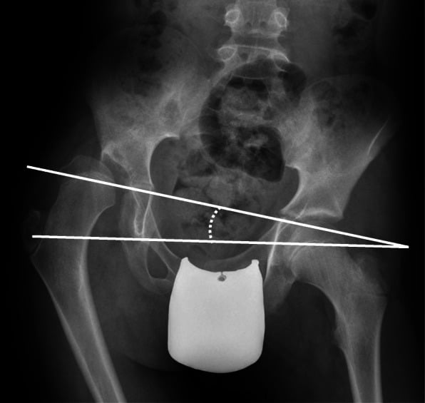
- WINDSWEPT DEFORMITY is an ASYMMETRIC coronal-plane lower-limb deformity in which ONE leg has GENU VALGUM (knock-knee) and the OTHER has GENU VARUM (bow-leg), so the legs appear 'swept' to one side; the analogous HIP windswept pattern (ABDUCTION of one hip, ADDUCTION of the other) is classically seen in cerebral palsy (and contributes to hip displacement).
- The crucial conceptual point is that windswept deformity is a SYMPTOM/SIGN with MULTIPLE possible aetiologies, NOT a single disease; the clinician's task when faced with it is to identify the UNDERLYING DISORDER, which then directs management.
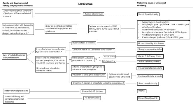
- The AETIOLOGIES fall into FOUR groups described in the systematic review: RICKETS and other METABOLIC bone disease (the LARGEST group - nutritional/hypophosphataemic rickets, renal osteodystrophy); SKELETAL DYSPLASIAS and other GENETIC disorders; TRAUMA (physeal injury/malunion causing asymmetric growth); and a 'descriptive'/idiopathic group without a specific underlying disorder - in which ALL patients were otherwise-healthy children of African descent presenting between about 2 and 3 years of age.
- BE PRECISE ABOUT 'LARGEST GROUP' - it is a count of ARTICLES, not of patients. The review pooled 45 publications, most of which its own authors classify as LOW level of evidence, and reports no patient-level denominator. So rickets generated the most literature; NO study has established what proportion of children with windswept deformity actually have it. The English-language-only search compounds this, since nutritional rickets is concentrated in settings that publish in other languages. Screen for it because it is common, treatable and dangerous to miss - not because a prevalence figure supports it.
- NEUROMUSCULAR disease, particularly CEREBRAL PALSY, is a classic cause of the windswept pattern (especially at the HIPS - one hip abducted/externally rotated and the other adducted/internally rotated), reflecting asymmetric muscle tone, and it is relevant to hip surveillance/displacement in CP.
- The WORK-UP therefore aims to FIND THE CAUSE: a careful history and examination, full-length standing ALIGNMENT RADIOGRAPHS to characterise the deformity (and localise it to bone/physis), and a METABOLIC SCREEN (calcium, phosphate, alkaline phosphatase, vitamin D, renal function/PTH) to detect rickets/metabolic disease, with genetic assessment where a dysplasia is suspected.
- MANAGEMENT is AETIOLOGY-DIRECTED: treat RICKETS/metabolic disease MEDICALLY (and much of the deformity may correct or stabilise), correct residual or structural deformity ORTHOPAEDICALLY (guided growth/hemiepiphysiodesis or osteotomy as appropriate), and manage the neuromuscular (CP) or dysplastic context accordingly - the error to avoid is correcting the deformity surgically while missing a treatable underlying metabolic cause.
- “Windswept deformity = genu VALGUM in one leg + genu VARUM in the other (legs 'swept' one way); at the HIPS = abduction of one + adduction of the other (classic in cerebral palsy).
- “It is a SYMPTOM with many causes, not a disease - FIND the underlying cause. FOUR review groups: RICKETS/metabolic (largest), skeletal dysplasias/genetic, trauma/physeal injury, idiopathic (all African-descent, healthy, ~2-3y); neuromuscular/CP is separately recognised.
- “'Largest group' counts ARTICLES, not patients - the review pooled 45 mostly LOW-level-evidence papers, English-language only, with no patient denominator. No prevalence for any aetiology has been measured. Screen for rickets because it is treatable and dangerous to miss, not because a figure supports it.
- “Work-up: alignment radiographs + METABOLIC SCREEN (Ca/PO4/ALP/vitamin D/renal-PTH) + history/exam (+/- genetics). Treat the CAUSE (medical for rickets) then correct residual deformity (guided growth/osteotomy). Don't operate while missing a treatable metabolic cause.
What It Is
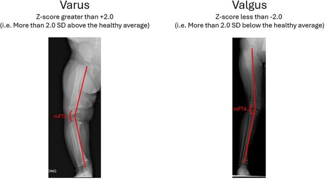
The pattern. Windswept deformity is an asymmetric coronal-plane deformity: genu valgum (knock-knee) in one leg and genu varum (bow-leg) in the other, so both knees appear swept to one side. At the hips the analogous pattern is abduction of one hip with adduction of the other, classic in cerebral palsy.
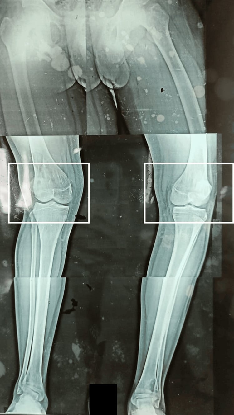
A sign, not a disease. Windswept deformity is best regarded as a symptom with multiple possible aetiologies rather than a single disease. The causes differ so much that management must be aetiology-directed, so the first task is to identify the underlying disorder, and to screen before you correct.
Causes
The review's four groups. A systematic review by Jansen and colleagues sorted the published literature into four aetiological groups. Neuromuscular disease is a separately recognised cause outside them.
- Examples
- Nutritional/hypophosphataemic rickets, renal osteodystrophy
- Note
- Largest group - treat medically (deformity may correct)
- Examples
- Various dysplasias/genetic disorders
- Note
- Genetic assessment; deformity correction in context
- Examples
- Physeal injury / malunion (asymmetric growth)
- Note
- Localise to the physis; guided growth/osteotomy
- Examples
- Otherwise-healthy children (all of African descent, ~2-3 y)
- Note
- No specific underlying disorder identified
- Examples
- Cerebral palsy (esp. hip windswept)
- Note
- Not one of the review's four groups; asymmetric tone, relevant to hip surveillance
What "largest" means. Rickets and other metabolic bone disease produced the most published articles, which is an article count, not a prevalence. The review reports no patient-level denominator, so no study has established what proportion of children with windswept deformity actually have rickets. Screen for it because it is common, treatable and dangerous to miss, not because a prevalence figure supports it.
Cerebral palsy. Neuromuscular disease, particularly cerebral palsy, is a classic cause of the windswept pattern, especially at the hips: one hip abducted and externally rotated, the other adducted and internally rotated. It reflects asymmetric muscle tone and is relevant to hip surveillance and displacement in CP.
Work-up
Finding the cause. The work-up aims to identify the underlying disorder:
- A careful history and examination
- Full-length standing alignment radiographs, to characterise the deformity and localise it to bone or physis
- A metabolic screen, which is essential: calcium, phosphate, alkaline phosphatase, vitamin D, renal function and PTH
- Genetic assessment where a dysplasia is suspected
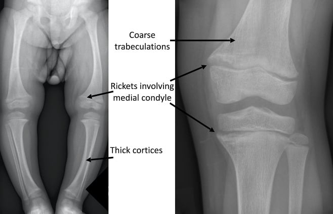
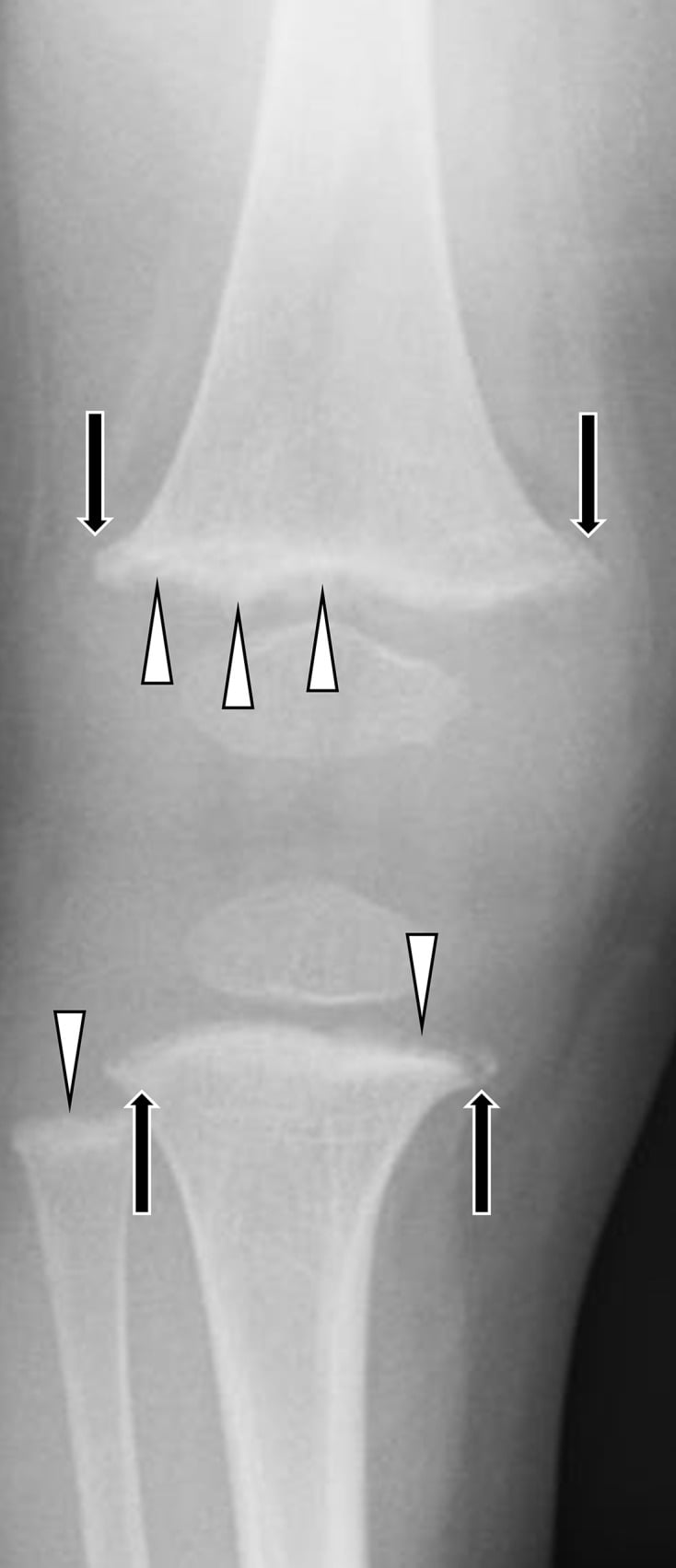
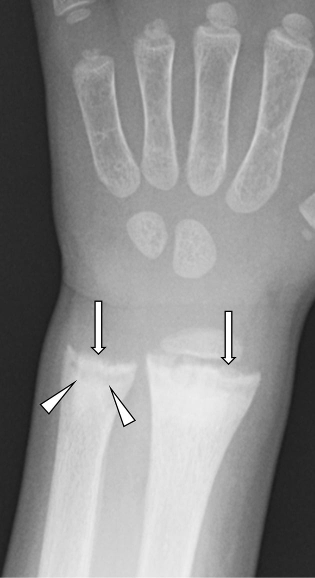
What rickets looks like. Radiographs also show whether the angular deformity sits within systemic metabolic bone disease. Wrist films are a sensitive screen, and metaphyseal cupping and fraying there support active rickets rather than an isolated mechanical knee deformity.
Management
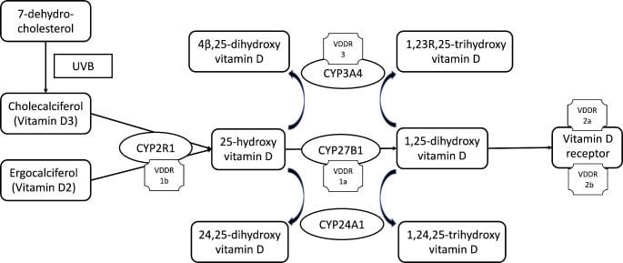
Treat the cause. Rickets and other metabolic disease is treated medically, and correcting the metabolic abnormality may resolve, stabilise or substantially improve much of the deformity. The dysplastic or neuromuscular context is managed in its own right.
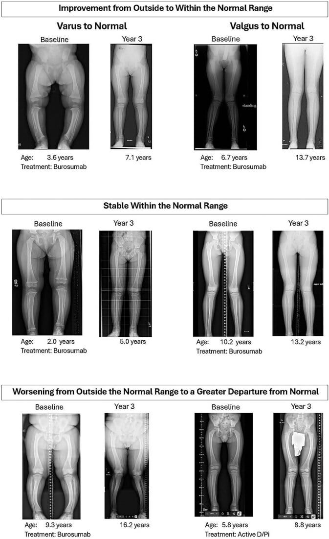
Then correct what remains. Residual or structural deformity is corrected orthopaedically, choosing a method that suits the aetiology and the physes. Guided growth (hemiepiphysiodesis) is the tool in the growing child.
Opposite plates on the two legs. Because one knee is valgus and the other varus, the guided growth is side-specific and opposite. The valgus knee is tethered on the medial physis, with a tension-band plate on the medial distal femur, with or without the medial proximal tibia, so the lateral side catches up. The varus knee is tethered on the lateral physis, so the same child receives a medial plate on one leg and a lateral plate on the other.
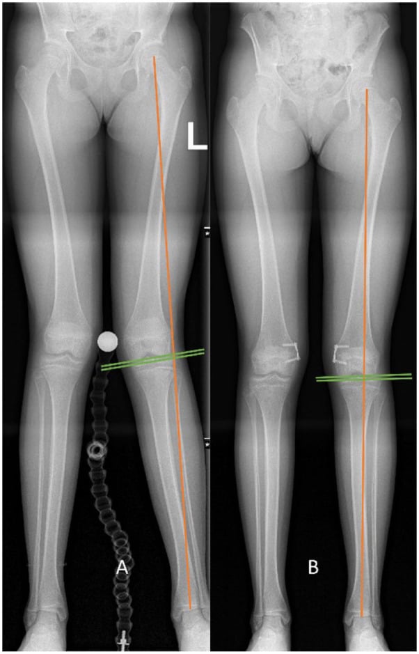
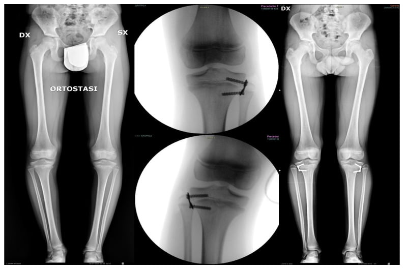
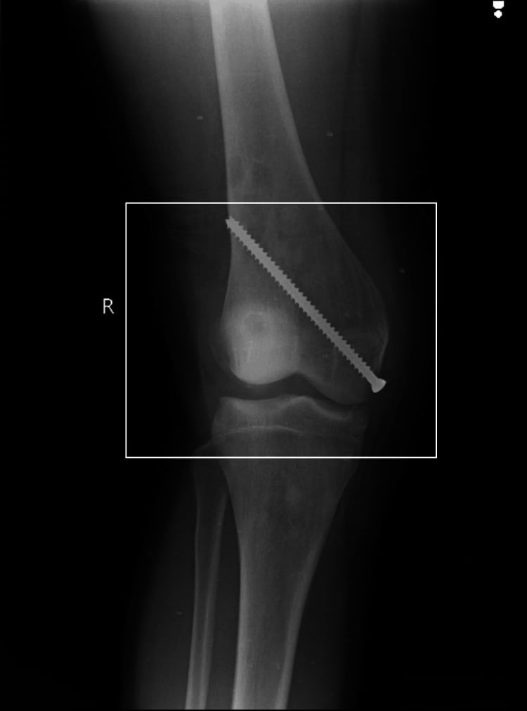
Control the metabolic disease first. Guided growth in active rickets is unreliable: the soft physes tether poorly and rebound after plate removal. The vitamin D or phosphate abnormality should therefore be optimised first or concurrently, and much of the deformity may then remodel without surgery.
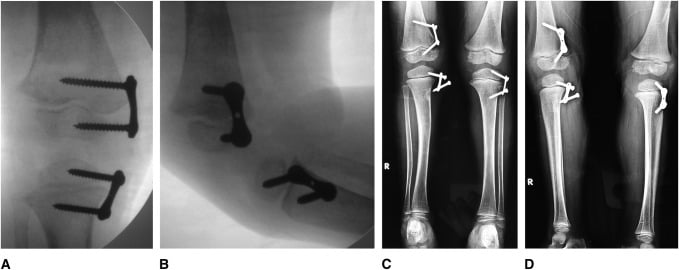
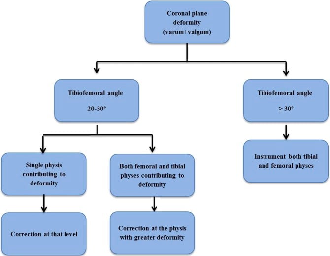
Osteotomy. Corrective osteotomy at the deformity apex is for established deformity: the skeletally mature, or severe deformity, or failed guided growth. In the young child treated with guided growth, monitor for rebound after plate removal.
The windswept hip in CP. This is addressed within cerebral palsy hip surveillance and management, through tone management and soft-tissue or bony surgery.
The error to avoid is proceeding to surgical correction of a windswept deformity while missing a treatable metabolic cause such as rickets.
Why the Deformity Is Asymmetric
The paradox. A systemic bone problem such as rickets weakens both physes equally, so it is not obvious why one knee should go valgus and the other varus rather than both the same way. The asymmetry is thought to arise where a generalised bone-quality problem is expressed asymmetrically by asymmetric mechanical loading.
The Hueter-Volkmann principle. Increased compression across a physis slows its growth, and reduced compression accelerates it. Asymmetric weight-bearing or posture can therefore drive one physis into valgus growth and the other into varus. The loading may come from habitual asymmetric sitting or lying, gait asymmetry, or a leg-length discrepancy shifting load.
A unilateral insult or dominance. A preceding unilateral problem (trauma, infection, or a Blount-type medial-physeal insult on one side) or limb dominance can tip one leg while the other follows the systemic or postural tendency. In cerebral palsy, asymmetric muscle tone (adductor/abductor imbalance) drives the hip windswept and, in turn, the knees.
Mnemonics & Memory Aids
SWEPT
Hook:SWEPT: Symptom not disease, Windswept (valgum+varum), Evaluate metabolically (rickets), Plus other causes, Treat the cause then correct.
Clinical Decision Scenarios
Practise clinical reasoning and management decisions out loud
“A child presents with one knock-knee and one bow-leg. What is this called, how do you think about it, and how do you manage it?”
What it is
- Genu valgum in one leg + genu varum in the other (legs swept one way)
- Hip windswept: one hip abducted + the other adducted (classic in cerebral palsy)
- A SIGN/symptom with many causes - not a single disease
Aetiologies
- Four review groups: rickets/metabolic (largest by ARTICLE count, not patients)
- Skeletal dysplasias/genetic disorders; trauma (physeal injury/malunion)
- Idiopathic (all African descent, otherwise healthy, ~2-3 y)
- Neuromuscular (CP) is separately recognised, not one of the four groups
- No prevalence measured for any aetiology; review pooled 45 mostly low-level, English-only papers
Work-up & management
- History/exam + full-length alignment radiographs + metabolic screen (Ca/PO4/ALP/vit D/renal-PTH)
- Treat the cause (medical for rickets - deformity may correct)
- Correct residual/structural deformity (guided growth/osteotomy); don't miss a treatable metabolic cause
Evidence & Key Studies
Windswept deformity - a disease or a symptom? A systematic review of aetiologies
- A PRISMA systematic review of 45 articles - but read its own closing caveat: the authors state that, by the Oxford criteria, MOST OF THE INCLUDED ARTICLES ARE OF LOW LEVEL OF EVIDENCE. The systematic method is sound; the material it pools is largely case reports and small series, so this is not high-level evidence about aetiology.
- Windswept deformity (simultaneous genu valgum and genu varum) is best regarded as a symptom with multiple possible aetiologies rather than a single disease.
- FOUR groups were described: 'rickets and other metabolic disorders', 'skeletal dysplasias and other genetic disorders', 'trauma', and 'descriptive articles without a specific underlying disorder' - with rickets the largest.
- CRITICAL QUALIFICATION ON 'LARGEST': the groups count ARTICLES, not patients. No patient-level denominator or prevalence is reported, so 'rickets is the largest group' means it generated the most publications - it does NOT establish that most children with windswept deformity have rickets. No study has measured the true aetiological distribution in any population.
- In the group without a specific underlying disorder, ALL patients were of African descent, otherwise healthy, presenting between two and three years of age.
- Further limitations: the search was restricted to ENGLISH-LANGUAGE articles, which matters greatly for a deformity whose major causes include nutritional rickets in low- and middle-income settings, and articles with windswept deformity as an incidental ancillary finding were included alongside those studying it directly.
The conceptualisation of windswept deformity as a symptom rather than a disease, the four aetiological groups with rickets the largest, and the observation that all patients in the group without a specific underlying disorder were otherwise-healthy children of African descent presenting at two to three years come from Jansen (DOI), a PRISMA systematic review of 45 articles restricted to English-language publications, whose authors state that most included articles are of LOW level of evidence. The definition, the hip windswept pattern in cerebral palsy, the metabolic work-up, the Hueter-Volkmann explanation for the asymmetry, and aetiology-directed management are standard, well-established teaching.
What is not established: the groups above count articles, not patients, so there is no measured prevalence for any aetiology and no published figure for what proportion of children with windswept deformity have rickets, a dysplasia, or no identifiable cause. Cerebral palsy is a separately recognised cause rather than one of the review's four groups. There is no validated severity classification for windswept deformity, no threshold defining when it warrants correction, no comparative trial of guided growth against osteotomy in this pattern, and no published rebound rate after plate removal specific to windswept limbs - the caution about operating during active rickets is a reasoned principle from physeal biology, not a measured failure rate.