Intrasutural ossicles in the lambdoid suture — a normal variant until they are too many, too big and too mosaic
- Wormian bones are accessory ossicles that develop within a cranial suture, overwhelmingly the lambdoid, then the posterior sagittal and occipitomastoid. They are NOT fracture fragments.
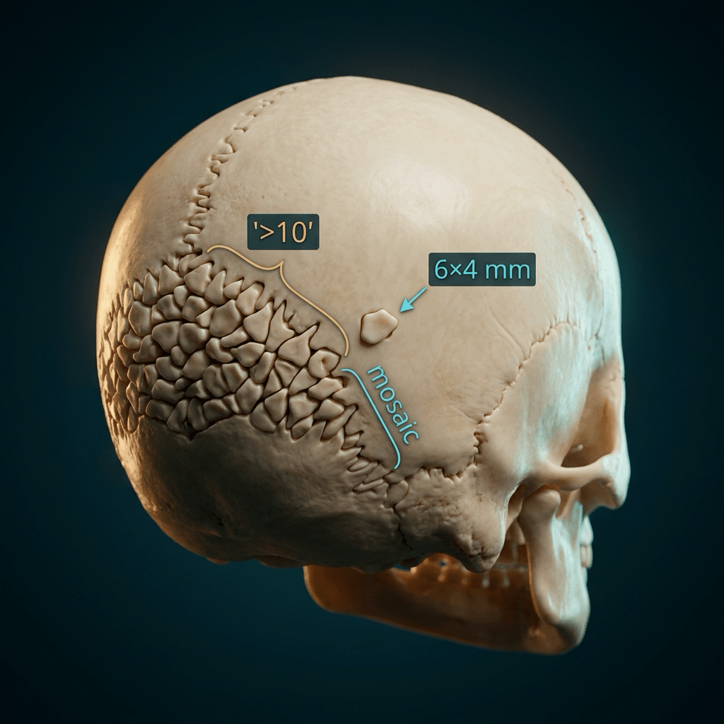
- Isolated small wormian bones are a NORMAL VARIANT AND THEY ARE COMMON - present in 53 per cent of unselected children under three on CT, with 10 per cent having four or more and no relationship to why the scan was done. The finding is only meaningful when it crosses the size, number and pattern threshold together.
- The single most useful clinical pairing in the exam is wormian bones plus multiple fractures with normal or reduced bone density equals osteogenesis imperfecta until disproven.
- Cleidocranial dysplasia is the pattern where wormian bones sit with a wide open anterior fontanelle, midline metopic defect, absent or hypoplastic clavicles and supernumerary teeth.
- Pyknodysostosis reverses the density rule: wormian bones with DENSE sclerotic bone, acro-osteolysis of the terminal phalanges and an obtuse mandibular angle.
- Hypothyroidism and rickets are the treatable, reversible causes — always check them before labelling a syndrome.
- PORKCHOPS is the established mnemonic and is expected verbatim in a viva.
- “Say the threshold out loud: 'more than ten, each larger than six by four millimetres, arranged in a mosaic pattern' — that sentence alone scores.
- “Wormian bones track OI SEVERITY, and the numbers are worth memorising: present in 96 per cent of type III, 78 per cent of type IV, but only 35 per cent of mild type I - the very form that presents as unexplained fractures in a normal-looking infant.
- “Absence of wormian bones NEVER excludes osteogenesis imperfecta.
- “A single large intrasutural bone at lambda is an interparietal or Inca bone — a normal variant, not a wormian bone pattern.
- “Menkes kinky hair disease is the trap: wormian bones, metaphyseal spurring and fractures in a boy — mimics both OI and NAI. Check hair and serum copper/caeruloplasmin.
Small solitary ossicles in the lambdoid suture are found in normal infants and in normal skulls at any age. Without the number, size and mosaic criteria you have described a variant, not a sign. Never launch into PORKCHOPS before you have justified that the pattern is genuine.
Wormian bones have sclerotic corticated margins on all sides and interlock with the adjacent suture. A fracture has sharp non-corticated margins, does not respect the suture line and usually crosses it. Get this wrong in a suspected NAI case and the whole answer collapses.
Wormian bones raise the possibility of OI but do not exclude inflicted injury, and the two can coexist. Equally, their absence does not exclude OI. Never present them as a discriminator between OI and NAI on their own — the discriminators are fracture pattern, sclerae, dentinogenesis, family history and genetics.
Hypothyroidism, rickets in the healing phase and hypophosphatasia all produce wormian bones. Thyroid function, calcium, phosphate, alkaline phosphatase and vitamin D are cheap and change management immediately. Alkaline phosphatase that is LOW rather than high is the hypophosphatasia giveaway.
Recognising the Pattern
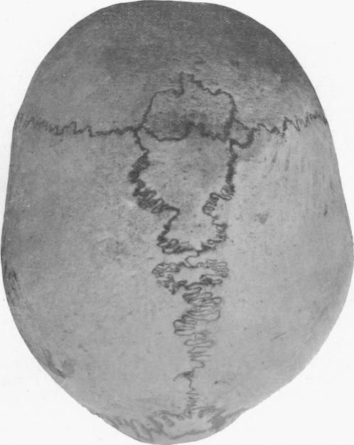
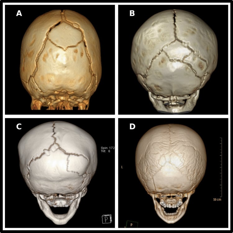
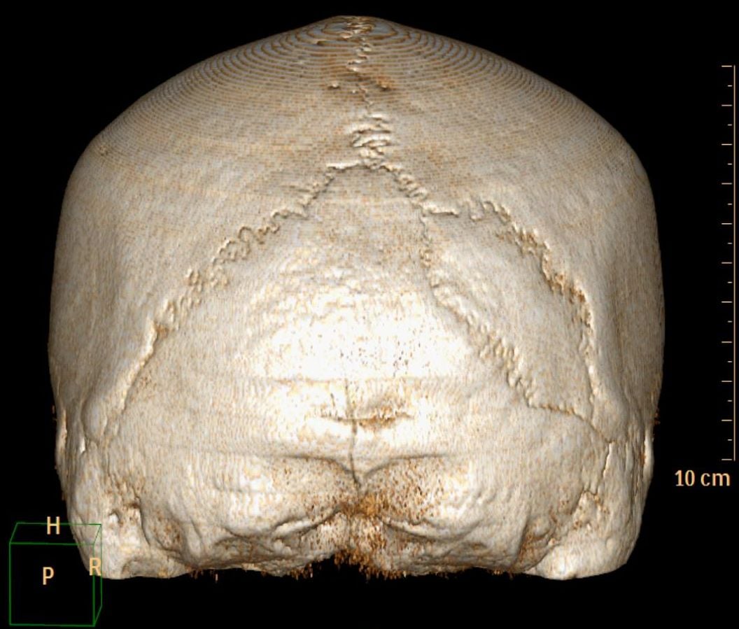
Definition. Wormian bones (sutural or intrasutural bones, named after Ole Worm) are irregular islands of independent ossification that develop within a cranial suture, separated from the adjacent skull plates by their own suture lines. They arise from accessory ossification centres in the sutural mesenchyme and are most numerous where sutural growth is most complex — the lambdoid suture above all, then the posterior third of the sagittal suture and the occipitomastoid sutures. They are essentially never seen in the coronal suture in normal children, so a coronal distribution should raise suspicion of a disorder of ossification.
How to confirm they are genuinely present and pathological. Three criteria, all required:
- Number — more than 10 discrete ossicles.
- Size — each greater than 6 x 4 mm.
- Pattern — arranged in an irregular interlocking mosaic rather than as isolated islands, and extending beyond the lambdoid suture.
Beneath that threshold, describe them as "a few small intrasutural ossicles, within normal limits".
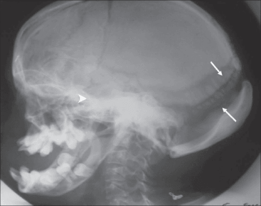
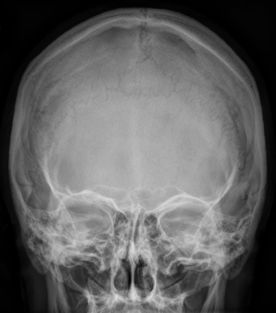
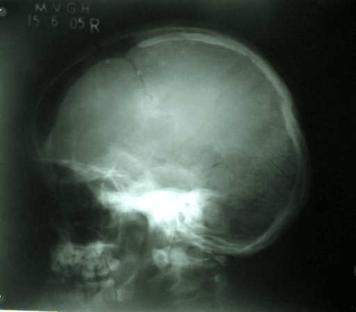
The best projection. A lateral skull radiograph is the workhorse: the lambdoid suture is thrown into profile and the ossicle mosaic is seen behind and above the posterior fossa. A Towne view or frontal view demonstrates the occipitomastoid and posterior sagittal extent. CT with 3D reformats is definitive if there is genuine doubt, but is rarely required in a well child.
Exact viva words. "This is a lateral radiograph of the skull of a young child. There are numerous small independent ossicles within the lambdoid suture, extending into the posterior sagittal suture. There are more than ten, each measures more than six by four millimetres, and they are arranged in an interlocking mosaic. These are pathological wormian bones. The calvarial thickness appears reduced and the vault is undermineralised. I would like to examine the child for blue sclerae, dentinogenesis imperfecta, joint hypermobility and a family history, and I would review any previous imaging for fractures. My leading diagnosis is osteogenesis imperfecta."
What mimics the pattern (false positives).
- Interparietal (Inca) bone — a single large triangular bone at lambda; a variant of squamous occipital segmentation, not a mosaic.
- Persistent metopic suture and accessory sutures — a linear lucency, not discrete corticated islands.
- Vascular grooves and diploic venous channels — branch, taper and lack a full sclerotic border.
- Skull fracture — sharp, non-corticated, does not follow the suture, may cross suture lines; acute clinical context.
- Lacunar skull (Lückenschädel) — scalloped lucencies in the inner table associated with Chiari II and myelomeningocele; these are membranous vault defects, not sutural ossicles, and resolve by 6 months.
- Copper-beaten skull — diffuse convolutional markings from raised intracranial pressure; a texture, not islands.
- Craniolacunia and the "hair-on-end" of chronic anaemia — perpendicular trabecular spiculation, entirely different texture.
- Widened sutures with resorption — as in leukaemia or neuroblastoma metastases, the sutures are splayed and blurred, not filled with corticated ossicles.
Next Investigation
PORKCHOPS
Hook:The established mnemonic for the causes of wormian bones — expected verbatim in a paediatric radiology viva.
DFRThe Three Sorting Questions - DFR
Hook:Ask these three questions in order before naming any cause of wormian bones — they reproduce the classification groups and stop you reaching for PORKCHOPS before the pattern is justified.
The Differential
Group your answer: undermineralised bone, dense bone, metabolic/endocrine, and syndromic/chromosomal. State the group before you list the causes.
- Typical age / setting
- Infancy to childhood; fractures from minimal trauma, positive family history in dominant types
- Discriminating feature
- Wormian bones PLUS generalised osteopenia, gracile bowed long bones, protrusio acetabuli, vertebral biconcavity, blue sclerae and dentinogenesis imperfecta; wormian bones near universal in type III
- What confirms it
- COL1A1/COL1A2 sequencing; skeletal survey; ophthalmology and dental assessment
- Typical age / setting
- Infant under 1 year with unexplained fractures; wormian bones typically ABSENT
- Discriminating feature
- Classic metaphyseal lesions, posterior rib fractures, fractures of differing ages with NORMAL bone density and NORMAL sclerae — none of which OI produces preferentially
- What confirms it
- Full skeletal survey with 11–14 day repeat, ophthalmology for retinal haemorrhage, child protection multidisciplinary process
- Typical age / setting
- Male infant, 2–3 months, developmental regression, seizures, hypothermia
- Discriminating feature
- Pale sparse steely twisted hair (pili torti) with metaphyseal spurring and fractures mimicking OI and NAI simultaneously; tortuous cerebral vessels on angiography
- What confirms it
- Low serum copper and low caeruloplasmin; ATP7A mutation; hair microscopy
- Typical age / setting
- Perinatal or infantile forms present in the first months with respiratory compromise; milder forms in childhood
- Discriminating feature
- Wormian bones with profound undermineralisation, metaphyseal 'tongues' of lucency, premature loss of deciduous teeth WITH the root intact, and a LOW alkaline phosphatase
- What confirms it
- Serum alkaline phosphatase low for age, raised urinary phosphoethanolamine, ALPL mutation
- Typical age / setting
- Diagnosed in childhood; short stature, ability to appose the shoulders anteriorly
- Discriminating feature
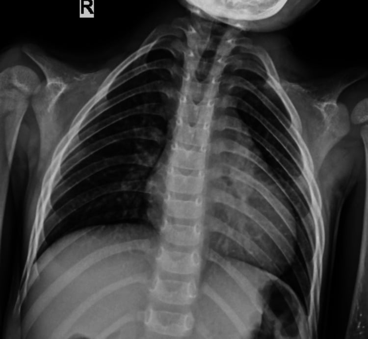
- Wormian bones with a persistently wide anterior fontanelle and midline calvarial defect, absent or hypoplastic clavicles, supernumerary unerupted teeth, wide pubic symphysis
- What confirms it
- Chest radiograph for clavicles, orthopantomogram for supernumerary teeth, RUNX2 mutation
- Typical age / setting
- Neonate to toddler; lethargy, constipation, umbilical hernia, prolonged jaundice
- Discriminating feature
- Wormian bones with DELAYED and fragmented (stippled) epiphyseal ossification and delayed bone age far behind chronological age — the delay is the discriminator
- What confirms it
- Thyroid function tests; newborn screening result; bone age radiograph of the hand and knee
- Typical age / setting
- 6 months to 3 years; nutritional or vitamin D dependent; also renal osteodystrophy
- Discriminating feature
- Wormian bones with craniotabes, frayed cupped splayed metaphyses at the wrists and knees, rachitic rosary; the wormian bones appear as sutures re-mineralise
- What confirms it
- Calcium, phosphate, high alkaline phosphatase, high PTH, low 25-OH vitamin D; wrist radiograph
- Typical age / setting
- Neonate onwards; recognisable dysmorphology
- Discriminating feature
- Wormian bones as a minor feature alongside 11 pairs of ribs, hypoplastic middle phalanx of the fifth finger, flared iliac wings with flat acetabular angles, atlantoaxial instability
- What confirms it
- Karyotype; lateral cervical spine for atlantodental interval
- Typical age / setting
- Childhood; short stature, frontal bossing, receding chin
- Discriminating feature
- Wormian bones with DENSE sclerotic bone (the density reversal), acro-osteolysis of the terminal phalanges, obtuse mandibular angle, non-union of the mandibular symphysis
- What confirms it
- Cathepsin K (CTSK) mutation; hand radiograph showing terminal tuft resorption
- Typical age / setting
- Boys, infancy; conductive deafness, cleft palate
- Discriminating feature
- Wormian bones with thick frontal bone and supraorbital ridging, absent frontal sinuses, and characteristic short broad 'tree-frog' thumbs and great toes with abnormal carpal/tarsal shapes
- What confirms it
- FLNA mutation; hand and foot radiographs; audiology
- Typical age / setting
- Childhood to adolescence, progressive
- Discriminating feature
- Wormian bones with BAND acro-osteolysis of the mid-terminal phalanges (transverse lysis leaving a distal tuft island), basilar invagination, early tooth loss, severe osteoporosis
- What confirms it
- NOTCH2 mutation; hand radiograph; craniocervical junction imaging
- Typical age / setting
- Adolescent or young adult male
- Discriminating feature
- Wormian bones with coarse thickened facial skin, digital clubbing and irregular periosteal new bone along the diaphyses — periosteal reaction is the discriminator
- What confirms it
- HPGD or SLCO2A1 mutation; long bone radiographs; exclude secondary HOA with chest imaging
- Typical age / setting
- Onset in the first 1–2 years
- Discriminating feature
- Wormian bones with alopecia, loss of subcutaneous fat, clavicular and distal phalangeal resorption, coxa valga, and premature atherosclerosis
- What confirms it
- LMNA mutation; clinical phenotype
- Typical age / setting
- Any age after ventriculoperitoneal shunting for hydrocephalus in infancy
- Discriminating feature
- Wormian bones appearing with thickened vault and small ventricles after rapid decompression — the shunt hardware on the film is the discriminator
- What confirms it
- Shunt series and prior imaging showing the ventricles before and after
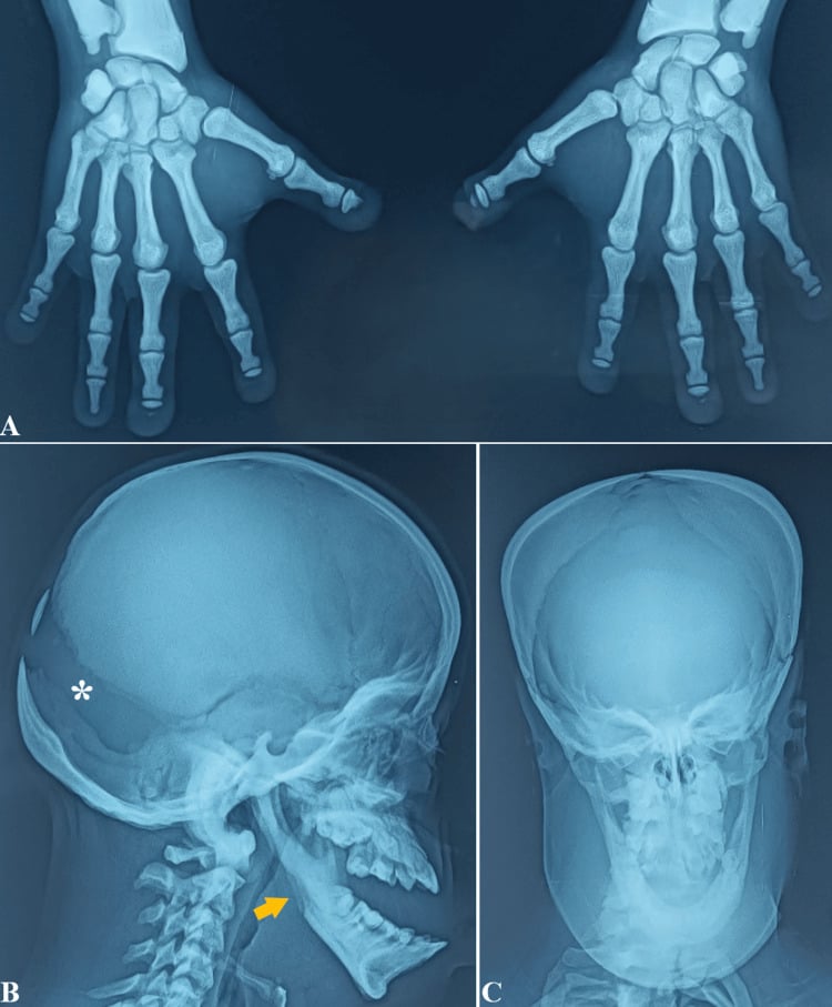
Narrowing It Down
- 1Step 1 — Are they genuinely pathological?
Apply the three Cremin criteria on a lateral skull: more than 10 ossicles, each greater than 6 x 4 mm, in a mosaic arrangement extending beyond the lambdoid suture. Confirm each ossicle has a complete sclerotic margin and lies within a suture, excluding fracture.
If any criterion fails, call it a normal variant, look for another explanation for the referral and stop. Know how weak the count alone is: in 605 CT brain scans of children under three with no suspected bone disease, wormian bones were present in 53 PER CENT, and 10 per cent had four or more. Number by itself is almost meaningless - it is the combination of number, size and mosaic pattern that carries the signal.
- 2Step 2 — What is the bone density?
Assess vault and long-bone mineralisation before considering any syndrome.
This is the single most powerful branch point. Undermineralised or osteopenic vault and long bones points to osteogenesis imperfecta, hypophosphatasia, rickets, Menkes or Hajdu-Cheney. DENSE sclerotic bone points to pyknodysostosis and is essentially diagnostic with acro-osteolysis. Normal density points to cleidocranial dysplasia, Down syndrome, otopalatodigital syndrome or a variant.
- 3Step 3 — Are there fractures, and of what type?
Characterise every fracture by pattern and site, and take a family history, before attributing causation.
Fractures with osteopenia, bowing and a positive family history equals OI. Fractures with normal density, classic metaphyseal lesions and posterior rib fractures equals suspected inflicted injury and mandates a child protection pathway. Fractures plus metaphyseal spurring plus abnormal hair in a boy equals Menkes. NEVER PRESENT OI AND INFLICTED INJURY AS MUTUALLY EXCLUSIVE - and note that the mild OI types most often confused with inflicted injury are precisely those in which wormian bones are LEAST reliable, present in only 35 per cent of type I.
- 4Step 4 — What is happening at the metaphyses and the growth plates?
Look specifically at metaphyseal contour, lucency and epiphyseal maturation on the survey.
Cupped, frayed and splayed metaphyses equals rickets. Lucent metaphyseal tongues with profound demineralisation equals hypophosphatasia. Stippled, fragmented epiphyses with markedly delayed bone age equals hypothyroidism. Normal metaphyses shifts you toward a syndromic or chromosomal cause.
- 5Step 5 — What do the clavicles, hands and teeth show?
Examine clavicles, terminal phalanges, mandibular angle and dentition, and ask about deafness.
Absent or hypoplastic clavicles plus supernumerary teeth equals cleidocranial dysplasia. Acro-osteolysis of terminal tufts plus obtuse mandibular angle equals pyknodysostosis. Band acro-osteolysis of the mid-phalanx plus basilar invagination equals Hajdu-Cheney. Short broad thumbs and great toes with conductive deafness equals otopalatodigital syndrome. Premature loss of deciduous teeth with intact roots equals hypophosphatasia; opalescent worn teeth equals dentinogenesis imperfecta of OI.
- 6Step 6 — Send the reversible bloods before you commit to a syndrome
Thyroid function, calcium, phosphate, alkaline phosphatase, PTH and 25-OH vitamin D in every case; copper and caeruloplasmin if the hair is abnormal.
A LOW alkaline phosphatase is hypophosphatasia - the one metabolic bone disease where the classic test runs downward, and the trap in every exam. A high alkaline phosphatase with low phosphate is rickets. A delayed bone age with high TSH is hypothyroidism. These are the treatable causes, and they are cheap to exclude.
- 7Step 7 — Confirm genetically and plan surveillance
Targeted or panel sequencing (COL1A1/2, ALPL, RUNX2, CTSK, NOTCH2, FLNA, ATP7A), then set the surveillance plan.
Where inflicted injury is in question, sequence COL1A1, COL1A2 AND IFITM5 simultaneously with duplication/deletion testing - Pepin and Byers recommend exactly that, because clinical features including wormian bones may be absent at the age of assessment. If no causative variant is found and there is no pathological phenotype, no further gene testing is indicated; a variant of uncertain significance needs parental segregation studies. Then move to management: bisphosphonates and rodding in OI, cervical spine surveillance in Down syndrome, craniocervical junction imaging in Hajdu-Cheney and severe OI, dental and audiology follow-up in cleidocranial dysplasia.
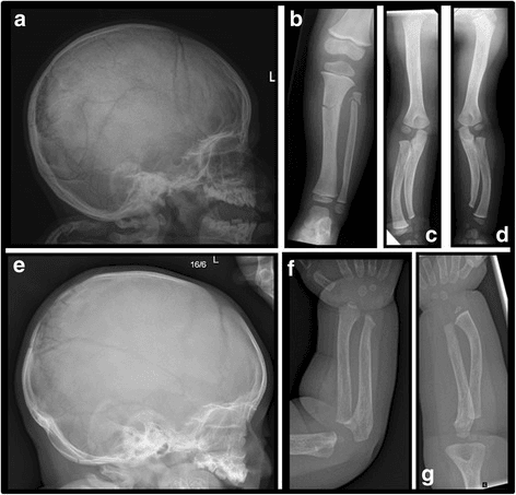
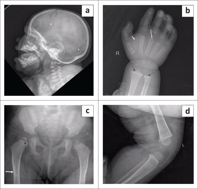
MCQ Practice Points
Q: What defines wormian bones as pathological rather than a normal variant?
A: All three of Cremin's criteria: more than 10 ossicles, each greater than 6 × 4 mm, in an irregular interlocking mosaic arrangement extending beyond the lambdoid suture. Fewer or smaller isolated ossicles confined to the lambdoid suture are a normal variant. Know why all three are needed: on CT of 605 unselected children under three, wormian bones were present in 53 per cent and 10 per cent had four or more — the count alone separates nothing.
Q: In which suture are wormian bones most commonly found?
A: The lambdoid, overwhelmingly — 343 of 476 ossicles (72 per cent) in a series of 13,519 cranial CTs, followed by the posterior fontanelle (11 per cent), then the posterior sagittal and occipitomastoid sutures. A distribution concentrated away from the lambdoid is unusual and should prompt a search for an underlying disorder of ossification.
Q: Which single blood test separates hypophosphatasia from rickets in a child with wormian bones and undermineralised bone?
A: Serum alkaline phosphatase. It is LOW for age in hypophosphatasia and HIGH in rickets — the one metabolic bone disease where the marker falls. Urinary phosphoethanolamine is raised in hypophosphatasia; the causative gene is ALPL.
Q: A boy of 3 months has fractures, metaphyseal spurring, seizures and sparse pale twisted hair. What is the diagnosis and what confirms it?
A: Menkes kinky hair disease, an X-linked disorder of copper transport due to ATP7A mutation. Confirmed by low serum copper and low caeruloplasmin, pili torti on hair microscopy and tortuous cerebral vessels on vascular imaging. It mimics both osteogenesis imperfecta and inflicted injury — always look at the hair.
Q: Does the absence of wormian bones exclude osteogenesis imperfecta?
A: No, and the numbers matter in a safeguarding setting. In 195 patients with OI they were present in 96 per cent of type III and 78 per cent of type IV but only 35 per cent of type I — and type I is the mild form that presents with unexplained fractures in an otherwise normal-looking infant. The sign tracks severity, so it is least reliable exactly where the diagnostic question is hardest. Blue sclerae, dentinogenesis imperfecta and family history may equally be absent at the age of assessment, which is why Pepin and Byers recommend sequencing COL1A1, COL1A2 and IFITM5 together with deletion/duplication testing. And their presence does not exclude coexisting inflicted injury.
Q: Which cause of wormian bones is associated with sclerotic rather than osteopenic bone?
A: Pyknodysostosis (cathepsin K deficiency, CTSK). Distinguished from osteopetrosis by a preserved medullary cavity, acro-osteolysis of the terminal phalanges and an obtuse mandibular angle. It reverses the density rule that governs the rest of the differential, which is why it is an exam favourite.
Exam Viva Scenarios
Practise clinical reasoning and management decisions out loud
“You are shown this lateral skull radiograph of a 7-month-old infant, referred after a spiral femoral fracture sustained during a nappy change. There are numerous ossicles in the lambdoid suture. What do you see and how will you proceed?”
“You are shown this skull radiograph of a 6-year-old with short stature. There are wormian bones and a wide midline calvarial defect with a persistently open anterior fontanelle. The mother demonstrates that the child can appose both shoulders in front of the chest. What is the diagnosis?”
“You are shown a lateral skull radiograph of a 9-year-old with short stature and a receding chin. There are wormian bones, the vault is diffusely sclerotic, the frontal sinuses are absent and the mandibular angle is obtuse. The hand radiograph shows resorption of the terminal phalangeal tufts. Diagnosis and pitfalls?”
The threshold (say it verbatim)
- More than 10 ossicles
- Each greater than 6 x 4 mm
- Irregular interlocking mosaic pattern
- Extending beyond the lambdoid suture
- Below threshold equals normal variant — stop
PORKCHOPS
- Pyknodysostosis
- Osteogenesis imperfecta
- Rickets (healing) and renal osteodystrophy
- Kinky hair (Menkes)
- Cleidocranial dysplasia
- Hypothyroidism / Hypophosphatasia
- Otopalatodigital syndrome
- Primary acro-osteolysis (Hajdu–Cheney), Pachydermoperiostosis, Progeria
- Syndrome of Down
Density branch point
- Osteopenic: OI, hypophosphatasia, rickets, Menkes, Hajdu–Cheney
- Sclerotic: pyknodysostosis
- Normal: cleidocranial dysplasia, Down syndrome, otopalatodigital syndrome
One-feature discriminators
- Blue sclerae plus dentinogenesis imperfecta — OI
- Absent clavicles plus supernumerary teeth — cleidocranial dysplasia
- Acro-osteolysis of tufts plus obtuse mandible — pyknodysostosis
- Band acro-osteolysis plus basilar invagination — Hajdu–Cheney
- Low alkaline phosphatase plus early tooth loss with intact root — hypophosphatasia
- Delayed bone age plus stippled epiphyses — hypothyroidism
- Cupped frayed metaphyses — rickets
- Pili torti plus low caeruloplasmin — Menkes
- Tree-frog thumbs plus conductive deafness — otopalatodigital syndrome
Mimics to exclude
- Skull fracture — non-corticated, crosses sutures
- Interparietal (Inca) bone — single large bone at lambda
- Lacunar skull — inner table scalloping with Chiari II
- Copper-beaten skull — raised intracranial pressure
- Vascular grooves and diploic channels
- Splayed sutures from marrow infiltration
Investigation ladder
- Lateral skull plus Towne view
- Skeletal survey if under 2 with fractures, with 11–14 day repeat
- Hand and wrist film — acro-osteolysis, metaphyses, bone age
- Bloods: TFT, Ca, PO4, ALP, PTH, vitamin D, copper/caeruloplasmin
- Chest film and OPG if cleidocranial dysplasia suspected
- Targeted genetics: COL1A1/2, ALPL, RUNX2, CTSK, NOTCH2, FLNA, ATP7A
- MRI craniocervical junction if basilar invagination risk
- No further imaging if well child, no fractures, normal bloods, sub-threshold ossicles
Evidence Base
Wormian Bones in Osteogenesis Imperfecta and Other Disorders
- THE SOURCE OF THE THRESHOLD EVERY EXAM ASKS FOR: skull radiographs of 81 patients with osteogenesis imperfecta compared against 500 skull radiographs of normal children
- Significant wormian bones, as opposed to normal developmental variants, were defined as MORE THAN 10 IN NUMBER, each measuring GREATER THAN 6 mm BY 4 mm, and arranged in a GENERAL MOSAIC PATTERN
- On that definition they were found in ALL of the osteogenesis imperfecta cases and in NONE of the normal skulls
- The authors open by conceding the difficulty: recognition varies with the age of the patient, radiographic objectivity and personal subjectivity
- The occurrence of significant wormian bones in other bone dysplasias was catalogued from their own material and the literature
Wormian Bones in Osteogenesis Imperfecta: Correlation to Clinical Findings and Genotype
- 195 patients with osteogenesis imperfecta, median age 11.8 years, skull radiographs analysed for a significant number of wormian bones (defined here as 10 or more)
- THE NUMBERS THAT KILL 'PATHOGNOMONIC': present in 35 PER CENT of OI type I, 96 PER CENT of type III and 78 PER CENT of type IV
- By genotype: 28 per cent with COL1A1 haploinsufficiency (nonsense and frameshift) mutations, 96 per cent with helical glycine substitutions in the alpha-1 chain, 72 per cent in the alpha-2 chain
- Height z-score, an indicator of disease severity, was INVERSELY related to the prevalence of wormian bones on multivariate analysis - the sicker the child, the more likely the sign
- They were visible in 19 of the 26 patients radiographed in the first year of life, INCLUDING A TWO-WEEK-OLD NEWBORN, so they form largely in utero
Wormian Bones in a General Paediatric Population
- 605 CT brain scans in children aged 0 to 3 years, performed for a range of indications, with suspected constitutional bone disease excluded - a denominator for what NORMAL looks like
- Wormian bones were present in 53 PER CENT of children: 43 per cent had one to three, 10 PER CENT HAD FOUR OR MORE, and 6 per cent had five or more
- The lambdoid suture carried by far the greatest numbers
- There was NO significant relationship between the number of wormian bones and the indication for the scan
- The authors' conclusion: wormian bones are common and can be numerous without pointing to osteogenesis imperfecta
Wormian Bones: Expanded Differential Diagnosis and Implications for Abnormal Head Shape in Infancy
- 13,519 children aged 0 to 18 undergoing cranial CT for any indication; 77 (0.57 per cent) had radiologically confirmed wormian bones, totalling 476 ossicles - rising to 2.1 per cent in the craniofacial clinic cohort
- Distribution: LAMBDOID suture 343 of 476 (72.1 per cent), then the posterior fontanelle (11.1 per cent)
- Multiple ossicles (two or more) were the commonest phenotype, in 52 of 77 cases (67.5 per cent)
- 13 patients (16.8 per cent) had an associated skeletal or craniofacial condition, and CRANIOSYNOSTOSIS accounted for 10 of those 13
- Cephalic index showed a predominance of BRACHYCEPHALY (70.1 per cent)
- Because wormian bones are commoner than craniosynostosis (0.57 to 2.1 per cent against about 0.05 per cent), the authors argue they belong in the differential of abnormal head shape in infancy
What Every Clinical Geneticist Should Know About Testing for Osteogenesis Imperfecta in Suspected Child Abuse Cases
- A review addressing the exact clinical dilemma: an infant with unexplained fractures, where mild osteogenesis imperfecta is the genetic differential to non-accidental injury
- STATES PLAINLY THAT THE CLINICAL SIGNS MAY ALL BE ABSENT: blue sclerae, dentinogenesis imperfecta, WORMIAN BONES ON X-RAY and a positive family history 'may not be present or apparent at the age of evaluation'
- Infants with OI type I or type IV - the mild forms - may be MISSED if clinical evaluation alone is used
- Recommended testing: simultaneous DNA sequencing of COL1A1, COL1A2 and IFITM5, plus duplication/deletion testing
- If no causative variant is found and there is no pathological clinical phenotype, NO further gene testing is indicated; a variant of uncertain significance requires parental segregation studies
- Draws on the authors' own Collagen Diagnostic Laboratory data from 2008 to 2014 for cases where non-accidental injury was noted on the test request
Wormian Bones: Thinking Beyond Osteogenesis Imperfecta
- A short Archives of Disease in Childhood piece from a paediatric metabolic medicine and radiology group, whose title is its argument: wormian bones should prompt a differential wider than osteogenesis imperfecta
- PubMed indexes no abstract for this article, so nothing beyond the title, authors and journal is verified here
- It is cited on this page as an example of the specialist paediatric position, not as a source of data