Resorption of the distal phalanx — tuft or band — and how to name the cause from the film
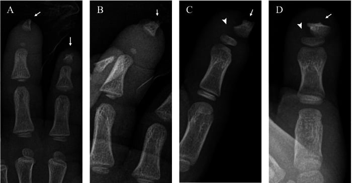
- Acro-osteolysis means resorption of bone in the distal phalanx. Terminal (tuft) resorption sharpens or amputates the tip; band-like (transverse) resorption dissolves a mid-phalangeal segment leaving the tuft as a floating island.
- Band-like resorption is a short, hard list: polyvinyl chloride exposure, Hajdu–Cheney syndrome, hyperparathyroidism and a handful of dysplasias. Tuft resorption is the long list.
- Scleroderma is tuft resorption plus soft-tissue pulp atrophy plus subcutaneous calcinosis — the calcinosis is the giveaway, not the lysis.
- Hyperparathyroidism resorbs the radial margins of the middle phalanges of the index and middle fingers before it takes the tufts. Always inspect the radial cortex on a magnified hand film.
- Psoriatic arthropathy erodes AND proliferates — fluffy new bone, whiskering and ankylosis alongside the lysis. Nothing else on the list makes bone while it destroys it.
- A neuropathic pattern (leprosy, diabetes, syringomyelia, congenital insensitivity to pain) gives a smooth, tapered 'licked candy stick' phalanx with ulceration and soft-tissue loss, often asymmetric.
- Solitary acro-osteolysis in one digit is a different problem — think infection, glomus tumour, epidermoid inclusion cyst or trauma, not systemic disease.
- “Say the pattern before the diagnosis: 'There is bilateral, symmetric resorption of the terminal tufts with soft-tissue tapering and punctate subcutaneous calcification.'
- “Thumb involvement argues for occupational vinyl chloride exposure and against frostbite, which characteristically spares the thumb.
- “Wormian bones plus band-like acro-osteolysis plus basilar invagination equals Hajdu–Cheney until proven otherwise.
- “Thyroid acropachy is a mimic: it adds fluffy periosteal new bone to the diaphyses, it does not remove bone.
Recognising the Pattern
Definition. Acro-osteolysis is resorption of bone within the distal (terminal) phalanx of the hand or foot. It is a descriptive pattern, not a diagnosis, and it has two structurally distinct forms that carry different differentials. A mixed form, with features of both in the same hand, is also recognised; it does not exclude either list, so both are worked up.
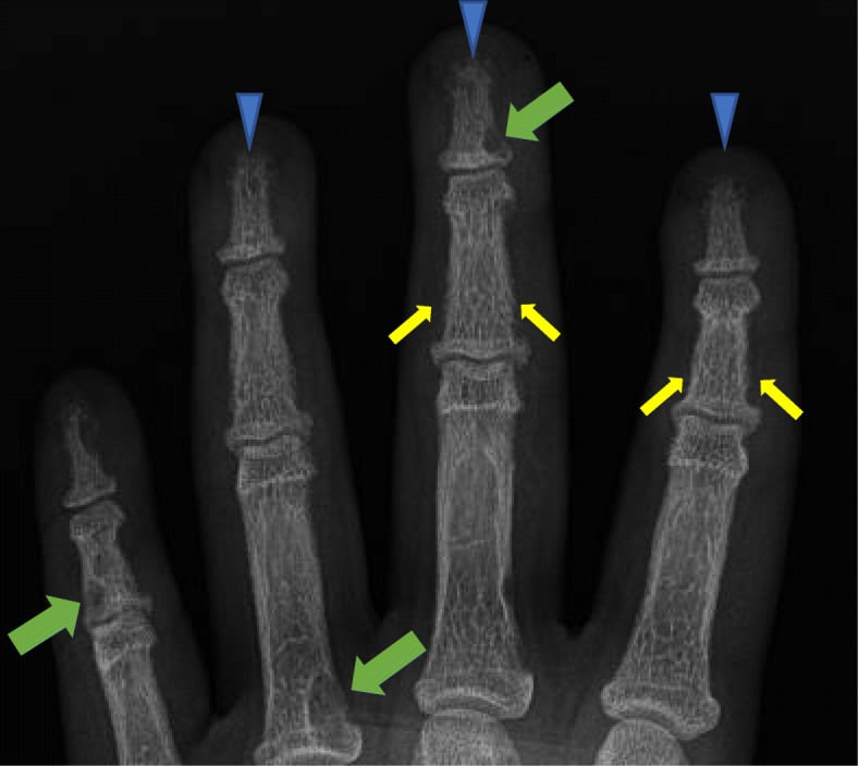
Terminal (tuft) resorption. Loss begins at the ungual tuft and progresses proximally. The tip becomes pencilled, pointed or frankly amputated, the phalanx shortens, and the overlying pulp tapers with it. This is the long differential.
Band-like (transverse) resorption. A horizontal lucent band appears through the mid-portion or base of the distal phalanx while the tuft survives distally as an isolated ossific fragment. This is the pattern that should stop you, because the list is short and hard: polyvinyl chloride exposure, Hajdu–Cheney syndrome, hyperparathyroidism and a handful of rare dysplasias.
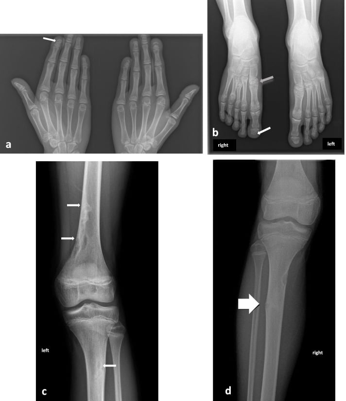
Solitary or symmetric. Count the involved digits on both hands and check for symmetry. One digit, whatever the pattern, means a local process: infection or felon, glomus tumour, epidermoid inclusion cyst or prior trauma. Multiple, bilateral and roughly symmetric digits mean systemic disease, whether connective tissue, metabolic, neuropathic or occupational.
Confirming it is genuinely present. Tuft morphology varies widely, small tuft nutrient foramina and ungual spurs are normal, and an overpenetrated or digitally over-processed image amputates tufts artefactually. Demand a dedicated, coned, well-exposed PA hand film (a wrist or chest film is not adequate), compare with the adjacent digits and the opposite hand, and accept the finding only when three things are confirmed:
- The cortical margin of the tuft is absent or irregular rather than simply thin
- The phalanx is shortened relative to the adjacent digits and the middle phalanx of the same ray
- The soft-tissue pulp contour follows the bone loss
If the soft tissues are normal and full, suspect a technical artefact.
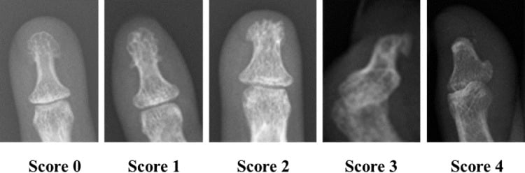
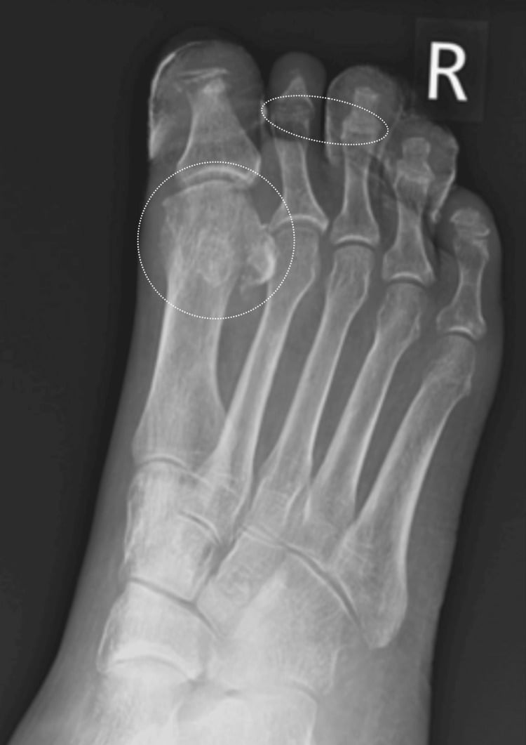
Mimics. Several processes destroy, or appear to destroy, the fingertip without being acro-osteolysis:
- Normal variant tuft shape, ungual spurs and nutrient foramina
- Over-exposure or aggressive digital post-processing burning off the tuft
- Erosive arthropathies (gout, rheumatoid, erosive osteoarthritis), which destroy juxta-articular bone rather than the tuft; destruction that respects the joint and has overhanging edges is an erosive arthropathy, and this differential is abandoned entirely
- Thyroid acropachy, which lays fluffy periosteal new bone along the phalangeal and metacarpal diaphyses, an additive process
- Amputation or old crush injury, with a smooth remodelled stump and an obvious surgical or traumatic history
- Sarcoidosis, whose lace-like intramedullary trabecular destruction can secondarily collapse the tuft
How to say it out loud. "This is a coned PA radiograph of the hand. There is resorption of the distal phalanges. The pattern is terminal, affecting the tufts of the index, middle and ring fingers bilaterally and symmetrically, with tapering of the overlying soft-tissue pulp and punctate subcutaneous calcification. The joint spaces are preserved and there is no periosteal reaction or new bone formation. The appearances are those of acro-osteolysis, and the distribution with calcinosis favours systemic sclerosis. I would like to review the chest radiograph and check calcium, phosphate and parathyroid hormone."
Next Investigation
The radiographs. Coned PA films of both hands, and of the feet if symptomatic, are the first and often the only imaging required. They establish tuft versus band pattern, symmetry, distribution and the presence of calcinosis, subperiosteal resorption or proliferative new bone, and they change management by directing the blood tests rather than further imaging.
The blood tests. Corrected calcium, phosphate, alkaline phosphatase, PTH, urea and creatinine are mandatory in any band-like or bilateral symmetric case. They detect primary or renal hyperparathyroidism, the one fully reversible and systemically dangerous cause: a raised PTH triggers parathyroid localisation and referral, and normal values effectively remove the metabolic branch.
The connective-tissue branch. Anti-centromere and anti-Scl-70 antibodies, with capillaroscopic dropout and giant loops on nailfold capillaroscopy, confirm systemic sclerosis. The diagnosis prompts HRCT of the chest for interstitial disease, echocardiography for pulmonary hypertension and vasodilator therapy for digital ischaemia.
The chest radiograph. Cheap and high yield: bilateral hilar lymphadenopathy for sarcoidosis, basal interstitial fibrosis for scleroderma. It alters the whole diagnostic direction for the cost of one film.
MRI. Reserved for solitary acro-osteolysis. It differentiates osteomyelitis (confluent marrow oedema, cortical breach, rim-enhancing collection) from a glomus tumour (a small, avidly enhancing subungual nodule) or an epidermoid inclusion cyst, and it changes management directly: antibiotics and debridement versus excision.
Genetic testing and skeletal survey. For band-like resorption in a child or young adult with dysmorphism: NOTCH2 for Hajdu–Cheney, CTSK for pyknodysostosis. A lateral skull and cervical spine film for wormian bones and basilar invagination is the practical first step and may pre-empt neurosurgical catastrophe.
When nothing more is needed. Retrieve and compare any previous hand films, however old. A stable, sharply corticated deformity in a patient with an established diagnosis (documented scleroderma, treated leprosy, old frostbite or prior amputation), unchanged on comparison films, is old injury or treated disease and requires nothing more; repeating cross-sectional imaging in burnt-out disease adds cost and no information. Progressive loss over months demands metabolic screening and rheumatological referral.
The Differential
Reading the table. The pattern decides which list you are in and the distribution decides whether the problem is local or systemic. Within the long tuft list, the discriminating feature is what separates one cause from the next, and the last column is the test that settles it.
- Typical age / setting
- Adult; dialysis unit, renal clinic, hypercalcaemia
- Discriminating feature
- Subperiosteal resorption of the RADIAL cortex of the middle phalanges of index and middle fingers precedes tuft loss; salt-and-pepper skull; brown tumours
- What confirms it
- Corrected calcium, phosphate, alkaline phosphatase and PTH; renal function
- Typical age / setting
- Any age; diabetic, penetrating injury, paronychia
- Discriminating feature
- SOLITARY digit, hot swollen pulp, permeative destruction with soft-tissue swelling and sometimes gas
- What confirms it
- MRI marrow oedema with rim-enhancing collection; bone biopsy and culture
- Typical age / setting
- Adult reactor-cleaner, plastics industry, years of exposure
- Discriminating feature
- BAND-like transverse resorption with a preserved distal tuft island; THUMB involved; associated Raynaud, sclerodactyly, portal fibrosis and angiosarcoma risk
- What confirms it
- Occupational history; regression after removal from exposure; liver imaging
- Typical age / setting
- Female 30-60; Raynaud phenomenon for years
- Discriminating feature
- Tuft resorption with tapered pulp atrophy PLUS punctate subcutaneous calcinosis; tight shiny skin; flexion contractures
- What confirms it
- Anti-centromere or anti-Scl-70 antibody; nailfold capillaroscopy; HRCT chest
- Typical age / setting
- Adult 30-50; nail pitting, skin plaques
- Discriminating feature
- Destruction WITH bone production — fluffy periostitis, whiskering, pencil-in-cup at the DIP and ankylosis; ray distribution
- What confirms it
- Skin and nail examination; DIP-predominant distribution; seronegative serology
- Typical age / setting
- Any age; cold exposure, homelessness, mountaineering
- Discriminating feature
- Sharply demarcated resorption in exposed digits with the THUMB SPARED (protected within the fist); may show epiphyseal destruction in children
- What confirms it
- Exposure history; sharply defined non-progressive appearance on serial films
- Typical age / setting
- Adult; long-standing diabetes, usually feet more than hands
- Discriminating feature
- Tapered 'licked candy stick' phalanx with plantar ulceration, vascular calcification and Charcot changes proximally
- What confirms it
- Glycaemic history, monofilament testing, ulcer inspection; MRI to exclude coexisting osteomyelitis
- Typical age / setting
- Endemic region or migrant; young to middle-aged adult
- Discriminating feature
- Concentric tapering plus enlarged palpable ulnar and common peroneal nerves, hypopigmented anaesthetic skin patches; nasal spine erosion
- What confirms it
- Slit-skin smear; nerve thickening on examination or ultrasound
- Typical age / setting
- Female young adult; episodic triphasic colour change
- Discriminating feature
- Tuft resorption WITHOUT calcinosis or skin thickening; digital tip ulcers
- What confirms it
- Autoantibody screen and nailfold capillaroscopy; digital pressures or arteriography
- Typical age / setting
- Child to young adult; family history, distinctive facies
- Discriminating feature
- BAND-like acro-osteolysis with wormian bones, osteoporosis, basilar invagination and premature tooth loss
- What confirms it
- NOTCH2 mutation; skull and lateral cervical radiographs
- Typical age / setting
- Child; short stature, frontal bossing
- Discriminating feature
- Acro-osteolysis in a DENSE, sclerotic skeleton with obtuse mandibular angle and persistent wide sutures — lysis in a bone-forming disorder
- What confirms it
- CTSK mutation; generalised osteosclerosis on skeletal survey
- Typical age / setting
- Adult 25-50; African-Caribbean predilection
- Discriminating feature
- Lace-like reticulated trabecular destruction and tunnelling of the middle and distal phalanges rather than pure tuft loss; associated dactylitis
- What confirms it
- Chest radiograph with bilateral hilar lymphadenopathy; serum ACE; biopsy
- Typical age / setting
- Child; lifelong blistering skin disease
- Discriminating feature
- Progressive tuft resorption with soft-tissue fusion of digits into a mitten (pseudosyndactyly) deformity
- What confirms it
- Dermatological diagnosis; skin biopsy with immunofluorescence mapping
- Typical age / setting
- Adult; single digit, subungual pain (glomus) or prior penetrating injury (cyst)
- Discriminating feature
- SOLITARY well-defined scalloped cortical defect with sclerotic margin, not true resorptive dissolution; exquisite cold sensitivity in glomus
- What confirms it
- MRI showing a small avidly enhancing subungual nodule; excision biopsy
PINCH FOCauses of Acro-osteolysis
Hook:PINCH FO generates the list; the tuft-versus-band split and the soft tissues then narrow it.
Narrowing It Down
Once the pattern and the distribution are settled, the tuft list is sorted by the questions below, asked of the film first and then of the patient.
Is bone being made as well as destroyed? Inspect the entheses and the DIP joints for periosteal new bone alongside the lysis. Fluffy periostitis, whiskering, pencil-in-cup deformity and ankylosis indicate psoriatic arthropathy, the only member of the list that proliferates while it erodes. Diffuse periosteal apposition without lysis is thyroid acropachy, a mimic rather than a member.
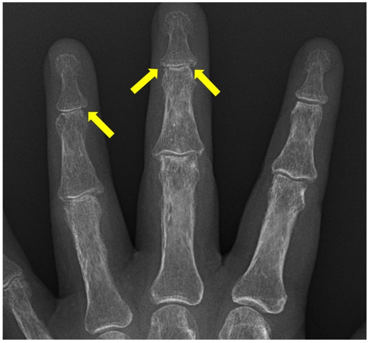
What do the middle phalanges and the rest of the skeleton show? Magnify the radial cortex of the middle phalanges of the index and middle fingers. Subperiosteal resorption there precedes tuft loss in hyperparathyroidism, and tuft resorption may be the only finding you notice until you look for it. With a salt-and-pepper skull, brown tumours or vascular calcification on any available skull, pelvis and chest films, it moves you decisively to the parathyroid axis, primary or secondary to renal failure. Generalised sclerosis with an obtuse mandibular angle suggests pyknodysostosis; wormian bones and basilar invagination suggest Hajdu–Cheney.
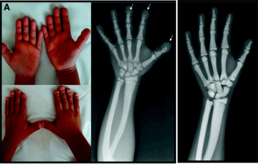
What do the soft tissues and the history say? The soft-tissue window carries the diagnosis: calcinosis for scleroderma, sausage digit for psoriasis, ulceration for neuropathy, gas for infection and pulp atrophy for a vascular cause. Examine the pulp, the skin and the nail folds on the film and at the bedside, and take an occupational, exposure and neurological history.
- Punctate subcutaneous calcinosis plus Raynaud equals systemic sclerosis; tuft resorption without calcinosis or skin thickening, with digital tip ulcers, is Raynaud phenomenon or a connective-tissue overlap short of full scleroderma
- Ulceration with anaesthesia and a smooth, tapered "licked candy stick" phalanx, often asymmetric, equals a neuropathic cause: ask about diabetes, leprosy endemicity and syringomyelia, and remember congenital insensitivity to pain
- Thumb involvement plus a plastics-industry history points to vinyl chloride; thumb sparing plus cold exposure points to frostbite, the thumb protected within the fist
- Blistering skin and mitten hands equal epidermolysis bullosa
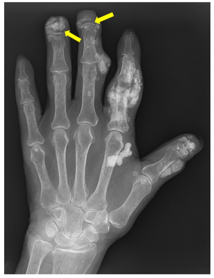
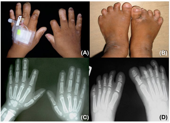
The solitary digit. When one digit alone is involved, the film sorts the local causes. Permeative destruction with a hot, swollen pulp, soft-tissue swelling and sometimes gas is osteomyelitis or a felon. A well-defined scalloped cortical defect with a sclerotic margin, not true resorptive dissolution, is a glomus tumour or an epidermoid inclusion cyst, the glomus betrayed by subungual pain and exquisite cold sensitivity and the cyst by a prior penetrating injury.
MCQ Practice Points
Q: Which single radiographic feature most reliably separates hyperparathyroidism from scleroderma as a cause of acro-osteolysis?
A: Subperiosteal resorption along the radial cortex of the middle phalanges of the index and middle fingers. It precedes tuft resorption in hyperparathyroidism and does not occur in scleroderma, where the discriminating additional feature is punctate subcutaneous calcinosis with pulp atrophy.
Q: Acro-osteolysis with thumb sparing following an environmental exposure — what is the cause and why is the thumb spared?
A: Frostbite. The thumb is habitually held within the clenched fist during cold exposure and is thereby thermally protected, producing sharply demarcated resorption of the exposed digits with a normal thumb. In children the injury may additionally destroy the distal phalangeal epiphyses and produce growth arrest.
Q: Which cause of acro-osteolysis produces bone formation alongside bone destruction?
A: Psoriatic arthropathy. Fluffy periostitis, whiskering of entheses, pencil-in-cup deformity at the distal interphalangeal joints and eventual ankylosis coexist with the resorption. This proliferative element separates it from every other entry on the differential.
Q: A patient with band-like acro-osteolysis, wormian bones and premature loss of teeth. What is the diagnosis and which complication threatens life?
A: Hajdu–Cheney syndrome, caused by a gain-of-function NOTCH2 mutation in exon 34. Basilar invagination with craniocervical instability and brainstem or cord compression is the serious complication; generalised osteoporosis with fragility fractures is also characteristic, and no antiresorptive or anabolic agent has established benefit for it.
Q: What is thyroid acropachy and why does it not belong in this differential?
A: It is a rare manifestation of Graves disease with clubbing, soft-tissue swelling and fluffy, spiculated periosteal new bone along the diaphyses of the metacarpals and phalanges. It is an ADDITIVE process — bone is laid down, not resorbed — so it mimics the clinical appearance of a deformed hand but is radiographically the opposite of acro-osteolysis.
Exam Viva Scenarios
Practise clinical reasoning and management decisions out loud
“You are shown this radiograph of both hands in a 44-year-old woman referred to the hand clinic with painful fingertip ulcers. The distal phalangeal tufts of several digits are resorbed and there are punctate dense opacities in the soft tissues of the pulps.”
“You are shown this hand radiograph of a 38-year-old man. There is a transverse lucent band through the mid-portion of several distal phalanges including the thumb, with the tufts preserved as separate distal fragments.”
“You are shown this radiograph of the index finger of a 61-year-old man with type 2 diabetes. The distal phalanx shows permeative destruction with adjacent soft-tissue swelling. The other digits are normal.”
Define the pattern first
- Terminal (tuft): tip pencilled or amputated, pulp tapers with it — long differential
- Band-like (transverse): lucent band mid-phalanx, tuft survives as a distal island — short differential
- Solitary versus multiple: solitary equals local disease, multiple symmetric equals systemic disease
PINCH FO
- Psoriasis — erosion plus proliferation, pencil-in-cup, DIP predominance
- Injury — frostbite (thumb spared), thermal, electrical, crush
- Neuropathy — diabetes, leprosy (thickened nerves), syringomyelia, congenital insensitivity to pain
- Collagen vascular — systemic sclerosis with calcinosis and Raynaud
- Hyperparathyroidism — radial subperiosteal resorption of index and middle middle-phalanges
- Familial — Hajdu–Cheney (wormian bones, basilar invagination), pyknodysostosis (dense bones, obtuse mandible)
- Occupational — polyvinyl chloride, band-like, thumb involved, hepatic angiosarcoma risk
The one-line discriminators
- Calcinosis in the pulp equals scleroderma
- Fluffy new bone equals psoriasis
- Radial middle-phalanx resorption equals hyperparathyroidism
- Thumb spared equals frostbite; thumb involved and band-like equals vinyl chloride
- Anaesthetic ulcerated licked-candy-stick digit equals neuropathy
- Dense sclerotic skeleton with lysis equals pyknodysostosis
- Lace-like tunnelling trabeculae equals sarcoidosis
Investigation
- Coned PA films of both hands — first and often only imaging
- Corrected calcium, phosphate, ALP, PTH, renal function in every bilateral or band-like case
- Autoantibodies and nailfold capillaroscopy for the connective tissue branch
- Chest radiograph — hilar nodes (sarcoid) or basal fibrosis (scleroderma)
- MRI only for solitary digit disease — infection versus glomus versus inclusion cyst
- No further imaging for a stable, corticated deformity with an established diagnosis
Mimics to exclude
- Normal tuft variants, nutrient foramina and over-exposed films
- Erosive arthropathies — gout, rheumatoid, erosive osteoarthritis
- Thyroid acropachy — periosteal apposition, an additive process
- Old amputation or remodelled crush injury
Evidence Base
Acro-osteolysis: imaging, differential diagnosis and disposition review
- Acro-osteolysis is osseous destruction of the distal phalanges of the hand or foot, categorised as terminal tuft, midshaft (band-like) or mixed patterns
- Recognition on radiographs is straightforward; the value lies in narrowing the differential and deciding when advanced imaging or tissue diagnosis is warranted
- The pattern (tuft versus band), distribution (solitary versus symmetric) and associated soft-tissue and skeletal clues drive the differential
Vinyl chloride and polyvinyl chloride (occupational disease review)
- Vinyl chloride monomer exposure during PVC production produced a sentinel occupational-health event: an excess of hepatic angiosarcoma at facilities worldwide
- The review lists acro-osteolysis among the syndromes associated with PVC - but qualifies it, stating the association is LESS CLEAR for vinyl chloride than the established angiosarcoma link
- Recognition of the occupational hazard rested on large-scale epidemiological studies of exposed workers
Hajdu-Cheney syndrome: a review
- Hajdu-Cheney syndrome combines band-like acro-osteolysis with severe osteoporosis, wormian bones, craniofacial features and polycystic kidneys
- It is caused by gain-of-function mutations in exon 34 of NOTCH2, upstream of the PEST domain, producing a truncated, stable protein with enhanced signalling
- Basilar invagination is a recognised serious complication; antiresorptive therapy has been tried for the osteoporosis but benefit is unproven