Spinal Cord Cavity | Dissociated Cape Sensory Loss | Charcot Joint | Scoliosis
- Cape-like suspended dissociated sensory loss is the classic presentation (C2-T1)
- Spinothalamic fibres cross in anterior white commissure (where central syrinx strikes first)
- Dorsal columns spared early - proprioception, vibration, light touch preserved
- MRI is the diagnostic gold standard (sagittal T2, axial T2, post-gad T1)
- Treat the cause: Chiari decompression, tumour resection, arachnoidolysis
- Charcot shoulder is painless destruction of the glenohumeral joint - classic exam sign
- “Syringomyelia + painless swollen shoulder = Charcot joint until proven otherwise
- “Every new left-thoracic scoliosis in a child needs MRI to exclude syrinx
- “Cape distribution reflects the longest crossing spinothalamic fibres (C2-T1)
- “Syringomyelia is a cause of scoliosis, NOT a complication - correct workup matters
- “Syrinx growth over 2-3 mm per year on serial MRI suggests active disease
- “Never correct scoliosis in syringomyelia without addressing the syrinx first
Overview and Epidemiology
Syringomyelia is a fluid cavity, a syrinx, within the spinal cord, and the classic exam cause of dissociated sensory loss over a cape distribution. It is also a silent driver of two orthopaedic problems, Charcot shoulder and progressive scoliosis, and a known cause of catastrophic neurological injury during scoliosis correction if it is not recognised before the operation. The orthopaedic surgeon may be the first to suspect it, faced with a painless destroyed shoulder or an atypical curve.
Who. The prevalence is estimated at 8-9 per 100,000 in the general population. Chiari-associated disease peaks at 20-50 years, and post-traumatic disease has a bimodal age distribution. There is a slight male predominance overall, and a female predominance where the cause is Chiari I.
Onset. Chiari-associated syringomyelia comes on insidiously. A post-traumatic syrinx classically presents months to years after the index injury, often in a patient whose neurology had appeared stable.
Pathophysiology
The anatomical bottleneck. Crossing spinothalamic fibres ascend 1-2 segments before they decussate through the anterior white commissure, which lies immediately anterior to the central canal. A central syrinx expands from the canal outward and strikes these crossing fibres first, so pain and temperature are lost while the dorsal columns (proprioception, vibration, fine touch) are spared until late disease. Hence the term dissociated sensory loss.
Why a cape. The C2-T1 crossing fibres are the longest, travel through the longest vertical segment of the cord and have the most extensive central representation, so C2-T1 is the classic zone of loss. The result is a bilateral, symmetrical, suspended segmental loss over the shoulders, upper arms and upper chest. It reflects damage to the crossing fibres at the level of the syrinx, and it follows no peripheral nerve territory.
How the syrinx forms. Four theories of formation and propagation are quoted:
- Proposer
- Gardner (1965)
- Mechanism
- CSF pushed from 4th ventricle into central canal via obex
- Clinical Implication
- Explains Chiari II / Dandy-Walker; less applicable to Chiari I
- Proposer
- Oldfield (1994)
- Mechanism
- Tonsils act as piston, forcing CSF into cord via Virchow-Robin spaces
- Clinical Implication
- Best supported by cine-MRI; basis for foramen magnum decompression
- Proposer
- Ball & Dayan (1972)
- Mechanism
- Spinal CSF pressure transiently exceeds cranial; CSF tracks along dorsal roots into cord
- Clinical Implication
- Explains non-Chiari syrinx; supports restoration of normal CSF dynamics
- Proposer
- Levine (1995)
- Mechanism
- Chronic epidural venous hypertension drives fluid transudation into cord
- Clinical Implication
- Particularly relevant to post-traumatic and arachnoiditic syrinx
Classification and Types
A syrinx is classified by its cause, by its anatomy (Milhorat) and by the cord level it occupies.
Chiari I is by far the most common cause in exam questions, so always look for tonsillar descent on the sagittal brain and cervical MRI.
- Frequency
- 60-80 percent
- Imaging Clue
- Tonsils greater than 5 mm below foramen magnum
- Surgical Implication
- Posterior fossa decompression, duraplasty
- Frequency
- 5-15 percent of SCI
- Imaging Clue
- Cord injury site, focal syrinx, often rostral
- Surgical Implication
- Arachnoidolysis, syringopleural shunt
- Frequency
- Syrinx in about 45 percent of intramedullary tumours (Samii 1994)
- Imaging Clue
- Enhancing nodule, expansile cord
- Surgical Implication
- Tumour resection; syrinx usually resolves
- Frequency
- Variable
- Imaging Clue
- Clumped nerve roots, CSF flow void loss
- Surgical Implication
- Subarachnoid space decompression, shunt
- Frequency
- Variable
- Imaging Clue
- Low-lying conus, lipoma, diastematomyelia
- Surgical Implication
- Detethering, dysraphism repair
Clinical Assessment
History. The sensory story is of painless burns or injuries to the hands and an inability to feel hot water. Motor symptoms are progressive upper-limb weakness, gait disturbance and falls, and the pain is deep, poorly localised and often in the cape distribution. Ask about autonomic change (hyperhidrosis or anhidrosis, bladder symptoms, Horner syndrome) and about the trigger: antecedent trauma, often years earlier, or new shoulder swelling.
Examination. The pattern is lower motor neurone signs at the level of the syrinx and upper motor neurone signs below it. At the level, the small muscles of the hand waste and the biceps and triceps reflexes are lost; below it, the knee and ankle reflexes are brisk and the Babinski sign is present. Look at the shoulder for painless swelling, instability and crepitus, the Charcot sign, and at the spine for scoliosis, abdominal reflex asymmetry and a stiff gait.
- What It Tests
- Pin-prick vs vibration
- Finding
- Lost pain, preserved vibration over cape
- Significance
- Anterior commissure involvement
- What It Tests
- Small muscles (T1)
- Finding
- Thenar/hypothenar atrophy
- Significance
- Lower motor neuron at syrinx level
- What It Tests
- Lower limb corticospinal
- Finding
- Brisk knee/ankle, Babinski
- Significance
- Upper motor neuron below syrinx
- What It Tests
- Thoracic segments
- Finding
- Asymmetric or absent
- Significance
- Suggests intraspinal pathology (Chiari, syrinx)
- What It Tests
- Charcot arthropathy
- Finding
- Massive effusion, no tenderness
- Significance
- Urgently image the cervical spine
Testing for dissociation. Test pin-prick (pain) and cold (temperature) in the upper limbs, trunk and lower limbs, then vibration with a 128 Hz tuning fork and joint position sense (toe up or down). In early syringomyelia pin-prick is lost over a cape while vibration is preserved, and a sensory level on the trunk implies thoracic extension. Always compare both sides, and document the upper limit of the sensory loss: it corresponds to the rostral end of the syrinx.
The syringomyelic hand. Wasted small muscles (LMN at T1) sit beside preserved forearm muscle bulk, because the syrinx is sparing the anterior horn cells at C5-C7. The result is the main en griffe, or intrinsic-minus, pattern, with trophic skin changes, recurrent burns and Charcot joints alongside. Recognising this combination on inspection is a viva favourite.
Syringomyelia is the classic cause of dissociated sensory loss, but the examiner who asks "what else does this?" expects a structured differential, because the lesion can be central-cord, vascular or peripheral:
- Other central-cord lesions: an intramedullary tumour (ependymoma, astrocytoma, haemangioblastoma) can give the identical suspended dissociated pattern with or without a syrinx; acute central cord syndrome (hyperextension injury in a spondylotic neck) gives a cape-like, upper-limb-predominant deficit.
- Vascular: anterior spinal artery (ventral two-thirds cord) infarction spares the dorsal columns and so produces dissociated loss (lost pain and temperature, preserved vibration and proprioception), but with a sudden onset and a clear sensory level rather than a suspended cape.
- Peripheral / infective: leprosy is the commonest cause of dissociated sensory loss worldwide (it preferentially destroys the small pain and temperature fibres in cool acral skin), and hereditary sensory and autonomic neuropathies (HSAN) and amyloid or diabetic small-fibre neuropathy give a distal, length-dependent dissociated loss, distinguished from a syrinx by the peripheral (glove) rather than suspended (cape) distribution.
- Brainstem: a lateral medullary (Wallenberg) syndrome gives crossed dissociated loss (ipsilateral face, contralateral body).
The discriminator is the shape. A suspended, bilateral cape pattern with LMN signs at the level and UMN signs below points to a central cord process (syrinx or tumour) and mandates MRI of the whole neuraxis; a glove, length-dependent pattern points peripheral; a sudden level with preserved dorsal columns points to anterior spinal artery infarction.
When to suspect it. The presentations that should send a patient for MRI:
- Key Finding
- Lost pain/temp, preserved light touch
- Imaging
- MRI whole spine + brain
- Action
- Refer to spinal neurosurgery urgently
- Key Finding
- Charcot arthropathy, no pain
- Imaging
- X-ray shoulder + MRI cervical spine
- Action
- Charcot = spinal MRI to look for syrinx
- Key Finding
- Asymmetric reflexes, abdominal reflex loss
- Imaging
- MRI whole neuraxis before any fusion
- Action
- Correct syrinx first; brace/observe scoliosis
- Key Finding
- Worsening pain, sensory level rising
- Imaging
- MRI confirms post-traumatic syrinx
- Action
- Arachnoidolysis, shunt, or detethering
Investigations
MRI, of the whole neuraxis. MRI is the only definitive investigation. Plain films are normal, and CT is unhelpful except to define associated bone abnormalities. Request the brain and the whole spine, with a CSF flow study, because an associated Chiari I, hydrocephalus or dysraphism will change management.
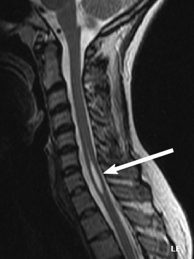
The sequences:
- Sagittal T2 of the entire spine
- Axial T2 at the level of the syrinx
- T1 before and after gadolinium
- Cine phase-contrast MRI, which confirms obstruction at the foramen magnum in Chiari-associated cases
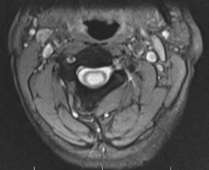
What to look for. The position and length of the cavity, nodularity of its wall (which suggests tumour), a Chiari I and loss of the craniocervical CSF flow void. Chiari I is taken as tonsillar descent greater than 5 mm, although in Milhorat's series of symptomatic patients (see Evidence) descent of less than 5 mm did not exclude the diagnosis.
When a tumour is suspected. Focal cord expansion, a mural nodule or a syrinx centred on an enhancing mass calls for MRI with contrast and delayed imaging. Each tumour has its signature:
- Ependymoma: classically central, with a cap sign (haemosiderin)
- Astrocytoma: eccentric, long segment
- Haemangioblastoma: cyst with a mural nodule; spinal angiography is reserved for its pre-operative planning
Surveillance. A confirmed syrinx under surveillance, or after treatment, is followed with serial MRI at 6 months and then annually if stable, sooner if the neurology changes. The threshold for intervention is an increase in length of more than one level, or in diameter of more than 2-3 mm per year, with symptoms.
Management Algorithm
Treat the cause: Chiari decompression, tumour resection or arachnoidolysis. Direct drainage of the syrinx is the rescue option once the cause has been addressed.
Aim. Restore normal CSF dynamics at the foramen magnum, decompress the syrinx and halt neurological decline.
Before the operation. Confirm tonsillar descent greater than 5 mm and flow obstruction on cine-MRI. Counsel that symptom stabilisation is the goal, and take consent for CSF leak, pseudomeningocele, wound infection and failure to improve.
The decompression.
- Prone, head neutral, with neuromonitoring (SSEP, MEP)
- Suboccipital craniectomy of 3 x 3 cm and C1 laminectomy, sometimes C2
- Dural opening for duraplasty is the standard; bone-only decompression is debated for paediatric cases
- Pericranial or synthetic patch graft, with a water-tight closure
When it is not enough. Tonsillar coagulation, which shrinks obstructing tonsils, and syrinx shunting are reserved for the unusual case that does not respond; shunting is for a syrinx that persists or expands after decompression. Fourth-ventricle stenting is used for persistent obstruction.
Afterwards. MRI at 3 months and again at 12 months. Expect a 50-80% decrease in syrinx size; a persistent or growing syrinx suggests failed decompression. Pain typically improves first, and sensory and motor recovery is less predictable.
The single most important predictor of success is adequate bone removal and a water-tight duraplasty. Intraoperative ultrasound demonstrating restoration of CSF pulsation across the foramen magnum is a useful end-point.
Orthopaedic Manifestations
- Mechanism
- Loss of proprioception and pain sensation
- Clinical Picture
- Painless swelling, instability, joint destruction
- Management
- Brace, arthrodesis; arthroplasty controversial
- Mechanism
- Same - upper limb neuropathic joint
- Clinical Picture
- Painless instability, crepitus, effusion
- Management
- Brace, arthrodesis for end-stage
- Mechanism
- Cord dysfunction, asymmetric paraspinal input
- Clinical Picture
- Atypical curve (left thoracic), rapid progression
- Management
- MRI first, brace, treat syrinx, then fuse
- Mechanism
- T1 anterior horn cell loss plus sympathetic loss
- Clinical Picture
- Wasted intrinsics, trophic skin, recurrent burns
- Management
- Skin care, hand therapy, protect from injury
- Mechanism
- Vertebral body denervation
- Clinical Picture
- Painless vertebral collapse, kyphosis
- Management
- Brace, address syrinx, instrumented fusion
The Charcot shoulder. Neuropathic arthropathy of the shoulder is the classic orthopaedic presentation, and it is uncommon: the entire world literature pooled in Wawrzyniak's 2023 systematic review amounted to 65 patients and 71 shoulders drawn from 39 case reports and small series, which is not the profile of a complication affecting half of all syrinx patients. The high percentage runs the other way. Syringomyelia accounts for the great majority of upper-limb Charcot joints, so an unexplained destructive shoulder should prompt MRI of the cord. The elbow is the second most common upper-limb neuropathic joint.
Why the joint fails. Three losses combine:
- Loss of proprioception, so microtrauma goes unperceived
- Loss of pain, so injury is repeated without protective guarding
- Loss of sympathetic tone, with vasodilation, hyperaemia and osteoclastic activation
The result is joint destruction, often with a massive effusion. The shoulder is the most common upper-limb site because the cord level matches.
The resorptive pattern. The 6 D's describe the hypertrophic (productive) neuropathic joint, with the exuberant sclerosis, osteophyte and debris typical of the diabetic foot and tabetic knee. The upper-limb Charcot of syringomyelia is classically the atrophic (resorptive) form, and recognising it is a distinct radiographic exam point.
- Atrophic, resorptive: gross osteolysis and resorption of the bone ends with little or no new bone. The humeral head can simply melt away, giving the tapered "licked candy stick" or "sucked candy" appearance, a surgical-looking "resorptive amputation" of the proximal humerus, scant debris and gross instability or pseudarthrosis. More common in non-weight-bearing, proximal upper-limb joints.
- Hypertrophic, productive: the 6 D's with abundant osteophyte and fragmentation. More common in weight-bearing lower-limb joints.
Why the pattern matters. The massive osteolysis of the resorptive shoulder is easily mistaken for an aggressive tumour or infection; the painless presentation, the dissociated sensory loss and a cervical syrinx on MRI make the diagnosis. It also explains why reconstruction is so hard, with little bone stock left to fuse or implant into.
6DsThe 6 D's of Charcot (Neuropathic) Joint
Hook:Destruction, Density, Dislocation, Disorganisation, Distension, Debris: the hypertrophic neuropathic joint. The syringomyelic shoulder is classically the resorptive form.
Treating the Charcot shoulder. Bracing and activity modification are first line. Treating the syrinx does not reverse established Charcot change, though it may halt progression, and the syrinx is always addressed before reconstructive surgery. For the end-stage shoulder, arthrodesis is the most reliable procedure despite the destruction; reverse total shoulder arthroplasty is controversial, with high failure from persistent denervation, and is used selectively in stable neurology.
Scoliosis: get the direction right. The two conditionals differ by an order of magnitude. Scoliosis is common among syrinx patients: it was present in 42% of 364 symptomatic Chiari I patients in Milhorat's series, and that is the direction the commonly repeated "25-50% of idiopathic scoliosis" belongs to. The yield of screening a presumed idiopathic scoliosis is small. A systematic review of 51 studies and 8,622 patients found a syrinx in 3.7%, a Chiari in 3.0% and all neural axis anomalies together in 11.4%, rising only to 14.2% in patients preselected for risk factors, not to anything like 90%. In infantile curves the figure for all anomalies is higher, around 13-22%, but still nowhere near a majority.
Who to image. The low yield sharpens the imaging argument. The recognised risk factors are early-onset curves, male sex, atypical or left-thoracic curves (right thoracic is the usual idiopathic pattern), increased thoracic kyphosis and any abnormal neurological finding, including reflex or sensory change. The other red flags:
- Painful scoliosis in a child or adolescent
- Rapid progression of the curve despite bracing
- Asymmetric reflexes, abdominal reflex loss or a change in gait
- Skin stigmata of dysraphism: a dimple, hair patch, lipoma or sinus
- Foot deformities: cavus, claw toes, leg length discrepancy
Posterior scoliosis correction in a patient with an unrecognised or untreated syrinx has a substantially increased risk of intra-operative neurological deterioration: paraplegia, hemiplegia or a new sensory level. MRI of the whole neuraxis is mandatory before any scoliosis fusion, because of that consequence rather than because the prior probability is high. If a syrinx is found, treat it first (cause-directed surgery), then proceed to deformity correction with full neuromonitoring. Sublaminar wires (Luque-Galveston) are particularly hazardous; prefer pedicle screws and incremental correction.
The sequence of care.
- MRI of the whole neuraxis to confirm the syrinx and identify its cause
- Referral to spinal neurosurgery to address the cause
- A brace for curves under 25-30 degrees while the syrinx is treated
- Fusion only after the syrinx has stabilised or resolved
- Follow-up for neurological change and curve progression
Complications
- Incidence
- Untreated or progressive syrinx
- Risk Factors
- Syrinx growth greater than 2-3 mm per year
- Management
- Cause-directed surgery, shunting as salvage
- Incidence
- Uncommon (see Orthopaedic Manifestations)
- Risk Factors
- Long-standing syrinx, delayed diagnosis
- Management
- Brace, arthrodesis, address syrinx
- Incidence
- Up to 50 percent in paediatric series
- Risk Factors
- Atypical curve, untreated syrinx
- Management
- Brace, fuse after syrinx treated
- Incidence
- 5-10 percent
- Risk Factors
- Inadequate duraplasty, poor wound healing
- Management
- Re-explore, resuture, lumbar drain
- Incidence
- Up to 10 percent
- Risk Factors
- Inadequate dural closure, raised ICP
- Management
- Compression, re-suture, lumbar drain
- Incidence
- 20-50 percent at 5 years
- Risk Factors
- Proteinaceous fluid, scarring
- Management
- Revision, alternative shunt, cause retreatment
- Incidence
- Rare but catastrophic
- Risk Factors
- Unrecognised syrinx, sublaminar wires
- Management
- Wake-up test, neuro-monitoring, abort and stage
The most common medicolegal claims involving syringomyelia centre on (1) failure to image the cervical spine in a patient with a painless Charcot shoulder, (2) failure to perform a pre-operative MRI in atypical scoliosis, and (3) failure to recognise an ascending sensory level years after spinal cord injury. Document the dissociated sensory exam, image the neuraxis when the cape pattern is present, and ensure the scoliosis workup includes MRI of the entire spine and brain.
Outcomes and Prognosis
- Treatment
- Foramen magnum decompression + duraplasty
- Pain Relief
- 70-80 percent improved
- Sensory / Motor Recovery
- 30-50 percent improved; 80 percent stabilised
- Long-term
- Good, durable if syrinx collapses on MRI
- Treatment
- Arachnoidolysis, shunt, detethering
- Pain Relief
- 70 percent improved
- Sensory / Motor Recovery
- Stabilised rather than recovered in most series
- Long-term
- Shunt failure common; revision often required
- Treatment
- Tumour resection
- Pain Relief
- Syrinx usually resolves
- Sensory / Motor Recovery
- Depends on tumour biology and resection extent
- Long-term
- Syrinx recurrence implies tumour recurrence
- Treatment
- Subarachnoid decompression, shunt
- Pain Relief
- Variable
- Sensory / Motor Recovery
- Stabilised in most
- Long-term
- Shunting often needed, prone to obstruction
Who does well. The best prognosis belongs to a Chiari I-associated syrinx with a short duration of symptoms, no fixed neurological deficit, an intact gait and normal hand function.
Who does not. A long-standing dense motor deficit, established Charcot joints, a post-traumatic aetiology, post-arachnoiditic scarring and a syrinx recurring after multiple shunts all carry a poor prognosis.
What improves. Pain is the most responsive symptom. Fixed sensory and motor deficits are stabilised more often than reversed.
Guidelines, Registries & Global Practice
- Prevalence: estimated 8-9 per 100,000 population, with higher rates in tertiary referral centres
- Chiari I: 0.5-1% of MRI series, with smaller numbers progressing to symptomatic syringomyelia
- Post-traumatic: 5-15% of spinal cord injury patients, often delayed years after index injury
- Scoliosis link: scoliosis affects roughly 42% of symptomatic Chiari I patients, while screening a presumed idiopathic scoliosis yields a syrinx in about 3.7% and any neural axis anomaly in 11.4% (14.2% where risk factors are present)
- High-resource: cine phase-contrast MRI, intraoperative ultrasound, full neuro-monitoring, and dedicated CSF flow clinics
- Limited-resource: structural MRI plus clinical follow-up; bone-only decompression may be used where duraplasty grafts are limited
- Universal principle: treat the cause first; never fuse a curve through an unrecognised syrinx
- Surgery: foramen magnum decompression is concentrated in tertiary spinal units globally
- Diagnostic emphasis
- Whole-spine MRI plus cine flow study; tonsillar descent greater than 5 mm
- Treatment emphasis
- Foramen magnum decompression plus duraplasty for Chiari I; syrinx shunting reserved for salvage
- Key caution
- Bone-only decompression debated; lower response in adults
- Diagnostic emphasis
- MRI whole neuraxis; brain included to identify hydrocephalus or Chiari
- Treatment emphasis
- Multidisciplinary spina bifida and Chiari clinics; syringo-pleural shunt for non-Chiari syrinx
- Key caution
- Shunt failure rates 20-50% at 5 years - counsel patients carefully
- Diagnostic emphasis
- Standardised MRI protocols; cine flow study where available
- Treatment emphasis
- Cause-directed surgery with selective shunting
- Key caution
- Scoliosis correction requires pre-operative MRI; pedicle screws over sublaminar wires
- Diagnostic emphasis
- MRI mandatory for atypical, painful, or left-thoracic curves
- Treatment emphasis
- Brace while syrinx is treated; staged fusion only after syrinx resolved
- Key caution
- Intra-operative neuro-monitoring; abort and stage if signals change
There is no dedicated international registry for syringomyelia comparable to NJR or AJRR for arthroplasty. The evidence base is dominated by single-centre surgical series and a few landmark imaging-pathogenesis studies (Oldfield, Heiss, Milhorat, Samii). Registry data is therefore drawn from condition-specific databases (Chiari and Syringomyelia Foundation, the Park-Reeves Syringomyelia Research Consortium) rather than from arthroplasty platforms. For orthopaedic outcomes, the Scoliosis Research Society database remains the most useful source for complication rates after scoliosis surgery in syrinx-positive patients.
Record in every patient with suspected or confirmed syringomyelia:
- Dissociated sensory exam with sensory level clearly mapped on a body diagram
- Reflex chart including abdominal reflexes
- MRI whole neuraxis (brain and full spine) with and without contrast
- Cine phase-contrast CSF flow study if Chiari I is suspected
- Multidisciplinary referral to spinal neurosurgery
- Pre-operative MRI and neurology status before any scoliosis correction
A missed syrinx in a patient undergoing scoliosis correction is a recurring source of complaints and claims worldwide. Theatres that perform deformity correction should have a standing rule that any atypical, painful, or left-thoracic curve has a documented whole-neuraxis MRI.
Controversies & Areas of Uncertainty
Some paediatric centres advocate bone-only suboccipital decompression without duraplasty to reduce CSF-related complications, but adults and patients with established syrinx tend to have higher response rates with duraplasty. There is no large randomised trial; choice is surgeon-led and tailored to age, severity, and syrinx behaviour.
Direct syrinx shunting (syringo-subarachnoid, syringo-pleural, syringo-peritoneal) is a salvage option for persistent or expanding syrinx after adequate cause-directed surgery. Shunt failure rates of 20-50% at 5 years limit enthusiasm, and shunting should not be the primary procedure for Chiari I or tumour-associated cases.
Reverse total shoulder arthroplasty is increasingly used for end-stage Charcot shoulder in selected patients with stable neurology, but the failure rate remains higher than in primary osteoarthritis. Glenohumeral arthrodesis remains the most reliable option for the heavily destroyed joint in a young, high-demand patient.
There is no high-quality evidence on how long to wait after syrinx surgery before correcting scoliosis. Most centres perform serial MRI and clinical review, with fusion once the syrinx has stabilised or resolved (often 6-12 months) and the curve is no longer responding to bracing. Decisions remain patient-specific.
MCQ Practice Points
Q: Why does syringomyelia classically cause a cape-distribution sensory loss? A: The crossing spinothalamic fibers in the anterior white commissure are damaged by a central cord cavitation. The C2-T1 fibers travel through the longest vertical segment of the central cord and have the most extensive central representation, so the loss is suspended, bilateral, and cape-shaped. Dorsal column modalities (proprioception, vibration) are spared early.
Q: What is the gold-standard investigation for suspected syringomyelia, and what additional sequences would you request? A: MRI of the whole spine and brain with sagittal T2 of the whole spine, axial T2 at the level of the cavity, T1 pre- and post-gadolinium (to exclude tumour), and cine phase-contrast CSF flow imaging (to assess for Chiari I and foramen magnum obstruction).
Q: A 35-year-old presents with a painless, swollen, warm shoulder. Examination shows preserved range of motion despite gross instability. What is the most likely underlying diagnosis? A: Charcot (neuropathic) arthropathy of the shoulder secondary to syringomyelia. The combination of painless swelling, gross destruction, and preserved movement despite instability is the hallmark. Urgent cervical spine MRI is mandatory to confirm the syrinx.
Q: A 12-year-old presents with a rapidly progressive left-thoracic scoliosis. What imaging must you arrange before considering any surgical correction, and why? A: MRI of the whole neuraxis (brain and entire spine) with contrast. Atypical, painful, or rapidly progressive curves - and left-thoracic curves in particular - have a high prevalence of intraspinal pathology, most commonly syringomyelia with or without Chiari I malformation. Unrecognised syrinx during posterior fusion is a recognised cause of catastrophic intra-operative spinal cord injury.
Q: What is the surgical treatment of Chiari I malformation with associated syringomyelia? A: Foramen magnum decompression (suboccipital craniectomy approximately 3 x 3 cm, C1 laminectomy, sometimes C2) with water-tight duraplasty, often augmented by tonsillar coagulation. The goal is restoration of normal CSF dynamics at the foramen magnum, which leads to syrinx collapse in the majority of cases. Direct syrinx shunting is reserved for the persistent or expanding syrinx despite adequate decompression.
Q: Which symptom of syringomyelia is most likely to improve after surgical treatment? A: Pain is the most responsive symptom (70-80% improvement after foramen magnum decompression). Fixed motor and sensory deficits are stabilised rather than recovered. Established Charcot joint destruction is permanent and does not reverse with syrinx treatment.
Exam Viva Scenarios
Practise clinical reasoning and management decisions out loud
“A 32-year-old teacher presents with 6 months of progressive difficulty using her right hand - she has dropped mugs of hot coffee twice without noticing the temperature. On examination, there is dissociated sensory loss over both shoulders and upper arms, with loss of pin-prick and temperature but preserved vibration and joint position sense. Reflexes are absent in the upper limbs and brisk in the lower limbs with bilateral Babinski signs. The right hand shows small muscle wasting. What is the most likely diagnosis, how do you confirm it, and what is the definitive treatment?”
“A 14-year-old boy is referred with a left-convex thoracic scoliosis. His mother says the curve has progressed rapidly over 6 months despite bracing. He denies pain but his school teacher has noticed that he occasionally drags his left foot. On examination there is a left thoracic rib hump, asymmetric abdominal reflexes (absent on the right), and a subtle broad-based gait. There are no skin stigmata of dysraphism. What is the most important next step, and why does it change the surgical plan?”
Definition and Pathology
- Syringomyelia = fluid cavity within the spinal cord (syrinx)
- Hydromyelia = true dilation of the central canal lined by ependyma
- Most cases are mixed; both are managed identically
- Causes: Chiari I (60-80%), post-traumatic, tumour, arachnoiditis, dysraphic, idiopathic
Clinical Pattern - The Cape
- Dissociated suspended sensory loss over C2-T1 (cape distribution)
- Pain and temperature lost; vibration and proprioception preserved
- Crossing spinothalamic fibers in anterior white commissure damaged first
- LMN signs at syrinx level (wasted hand, absent upper limb reflexes)
- UMN signs below (brisk lower limb reflexes, Babinski)
Imaging
- MRI whole neuraxis (brain plus entire spine) is the gold standard
- T2 sagittal and axial; T1 pre- and post-gadolinium to exclude tumour
- Cine phase-contrast flow study for Chiari I and foramen magnum obstruction
- Serial MRI to track growth (greater than 2-3 mm per year suggests progression)
Management Principles
- Treat the cause: Chiari decompression, tumour resection, arachnoidolysis
- Foramen magnum decompression plus water-tight duraplasty for Chiari I
- Syrinx shunting reserved for persistent or progressive syrinx despite cause treatment
- Surgery stabilises deficits and relieves pain; fixed deficits often do not recover
Orthopaedic Manifestations
- Charcot shoulder: painless swelling, instability, joint destruction - image cervical spine
- Charcot elbow: second most common upper limb neuropathic joint
- Syrinx and scoliosis - know the direction: scoliosis in about 42% of symptomatic Chiari I patients, but a syrinx in only about 3.7% of presumed idiopathic scoliosis (11.4% for any neural axis anomaly, 14.2% with risk factors)
- MRI whole neuraxis is mandatory before scoliosis surgery in atypical cases
- Sublaminar wires are hazardous in unrecognised syrinx; use pedicle screws
Complications and Red Flags
- Progressive myelopathy, ascending sensory level, neurogenic bladder
- Charcot joint destruction and recurrent burns from loss of protective sensation
- Syrinx scoliosis progression if underlying cause not addressed
- CSF leak and pseudomeningocele after posterior fossa decompression
- Shunt obstruction in 20-50% of syrinx shunts at 5 years
Exam Pearls
- Cape dissociated sensory loss = syringomyelia until proven otherwise
- Painless destroyed shoulder in a young adult = MRI cervical spine
- Left-thoracic curve in a child = MRI whole neuraxis before any fusion
- Pain is the most treatment-responsive symptom; fixed deficits stabilise
- Never correct scoliosis in a patient with an untreated syrinx
Evidence Base and Key Trials
Pathophysiology of syringomyelia associated with Chiari I malformation of the cerebellar tonsils: implications for diagnosis and treatment
- Cine phase-contrast MRI demonstrated obstructed CSF flow at foramen magnum in Chiari I with syrinx
- Cadaveric and clinical evidence supports a piston mechanism - cerebellar tonsils compress cord on each cardiac systole
- Restoration of normal flow after foramen magnum decompression correlates with syrinx collapse
Elucidating the pathophysiology of syringomyelia
- Intraoperative ultrasound and pre/post-operative cine MRI in Chiari I patients
- Successful foramen magnum decompression restored normal CSF dynamics and led to syrinx shrinkage
- Demonstrated that abnormal CSF flow, not congenital central canal dilation, drives syrinx propagation
Chiari I malformation redefined: clinical and radiographic findings for 364 symptomatic patients
- Largest single-centre series redefining Chiari I: 364 symptomatic patients (275 female, 89 male), mean onset 24.9 years
- Associated problems: syringomyelia 65%, scoliosis 42%, basilar invagination 12% - the source of the 'scoliosis in 42% of symptomatic Chiari I' figure quoted on this page
- Chiari I defined as tonsillar herniation of at least 3-5 mm; 332 of 364 had 5 mm or more, and less than 5 mm did NOT exclude the diagnosis
- Posterior fossa volume reduced (mean 13.4 ml) with 40% less CSF volume - overcrowding of a normally developed hindbrain
Surgical results of 100 intramedullary tumors in relation to accompanying syringomyelia
- One hundred intramedullary tumours (94 patients) resected; syrinx present in 45% - more often rostral to the tumour (49% above vs 11% below; 40% both)
- Syrinx resolution correlated with extent of tumour resection
- Persistent or recurrent syrinx was a marker of residual or recurrent tumour