Ligament | alignment | reducibility | cartilage | demand
- Pain is not instability. Link symptoms to ligament injury, carpal alignment, reducibility and cartilage status.
- Scapholunate injury tends to DISI. The scaphoid flexes; the lunate follows the triquetrum into extension.
- Lunotriquetral injury may produce VISI, but isolated LT instability is less common than combined ulnar-sided or midcarpal pathology.
- Neutral radiographs can be normal. Stress views, dynamic fluoroscopy, MRI arthrogram and arthroscopy may be needed.
- A chronic arthritic wrist is not a repair problem. It is a salvage procedure discussion.
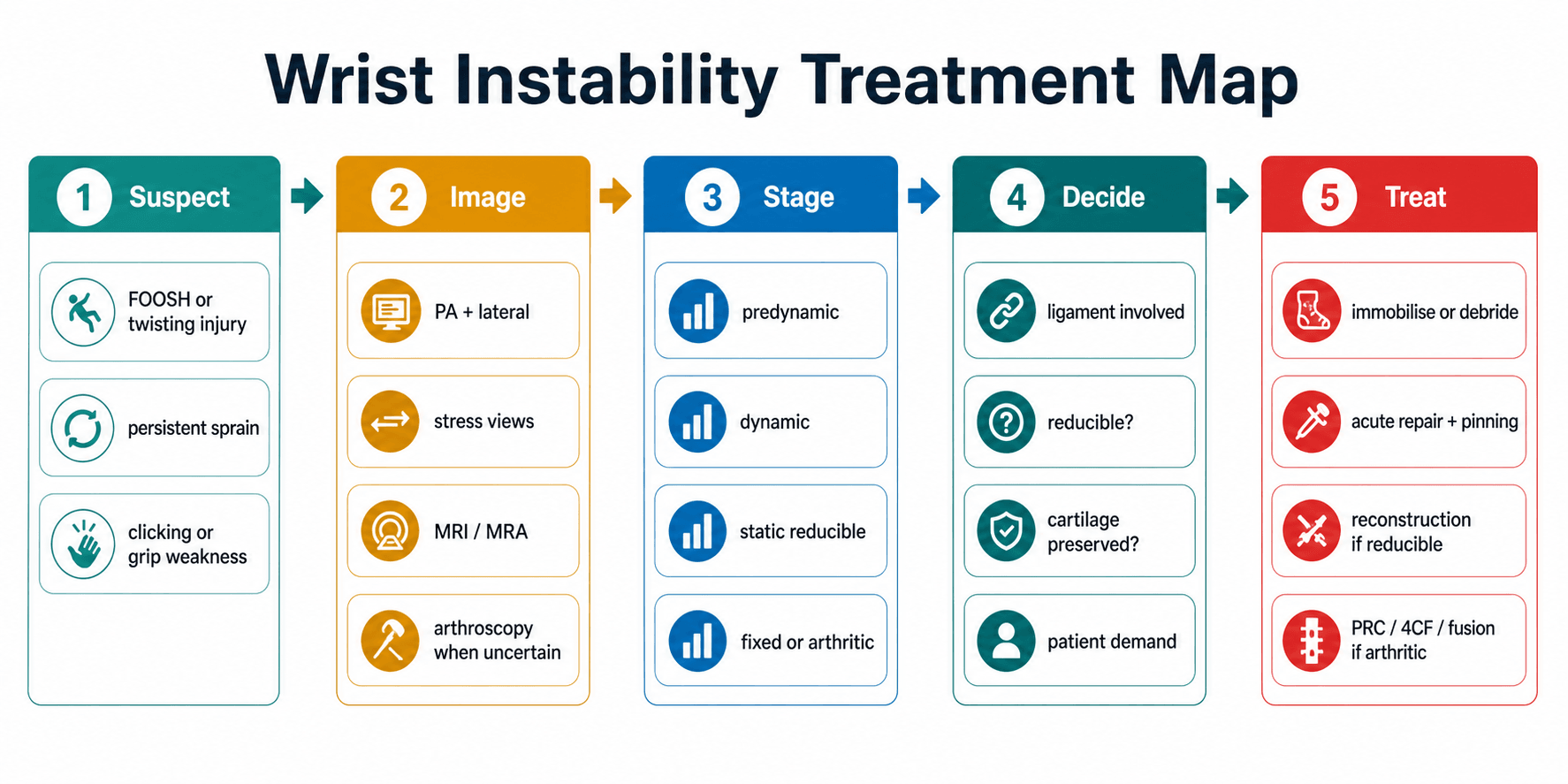
- “The safe answer starts with stage: predynamic, dynamic, static reducible, static irreducible or arthritic.
- “Every management plan must state timing, reducibility, cartilage and patient demand.
- “Watson and LT ballottement tests are screening tests; they do not replace imaging and arthroscopy.
- “SLAC progression is the late consequence of untreated chronic scapholunate dissociation.
A widened scapholunate interval, painful Watson test or VISI posture is not enough to choose an operation. Treatment depends on the ligament injured, timing, reducibility, cartilage status, associated fracture, ulnar-sided pathology and patient demand.
| Clinical Question | What You Need To Know | Why It Changes Treatment |
|---|---|---|
| Which ligament failed? | SL, LT, extrinsic ligament, TFCC, midcarpal or combined injury. | Determines whether the main pattern is DISI, VISI, ulnar-sided instability or mixed instability. |
| Is it dynamic or static? | Normal neutral films versus stress gap or fixed malalignment. | Dynamic injury can be missed; static injury needs reducibility and cartilage assessment. |
| Is it acute or chronic? | Repairable ligament tissue versus attenuated scar. | Acute repair differs from chronic reconstruction or salvage. |
| Is the carpus reducible? | Alignment corrects under fluoroscopy, traction or arthroscopy. | Reducible deformity can be reconstructed; irreducible deformity often needs salvage. |
| Is cartilage preserved? | Radioscaphoid, capitolunate, radiolunate and midcarpal cartilage. | Cartilage loss makes ligament reconstruction inappropriate. |
GATEAssessment | FLEXScapholunate | RACETreatment Choice |
|---|---|---|
G Gap Measure SL or LT interval on neutral and stress views. | F Flexed scaphoid Scaphoid flexion gives ring sign and SL gap. | R Reducible Can carpal alignment be restored? |
A Alignment Assess DISI, VISI, carpal height and capitolunate axis. | L Lunate extends DISI posture develops with dorsal lunate tilt. | A Arthritis Is there SLAC, SNAC or radiocarpal cartilage loss? |
T Timing Acute, subacute, chronic or arthritic. | E Early repair Best before chronic attenuation and arthritis. | C Chronicity Acute repair differs from chronic reconstruction. |
E End-stage Irreducible or arthritic wrists need salvage logic. | X X-ray stress Dynamic injury may need clenched-fist or stress views. | E Expectation Age, demand and occupation change the operation. |
GATE decides whether the wrist is reconstructable. | Scaphoid flexion plus lunate extension creates DISI. | Race through stage before choosing treatment. |
Overview and Epidemiology
Wrist ligament instability is failure of the soft-tissue stabilisers that keep the carpal bones aligned during load and motion. The most important intrinsic ligament injuries are scapholunate and lunotriquetral injuries, but a competent assessment must also consider the dorsal intercarpal ligament, dorsal radiocarpal ligament, volar radiocarpal ligaments, TFCC, distal radius alignment, scaphoid fracture or nonunion, distal radioulnar joint pathology and midcarpal instability.
The typical patient presents after a fall on the outstretched hand, a twisting injury, high-energy trauma, sports injury, or persistent symptoms after a wrist sprain. Symptoms may be dorsal wrist pain, ulnar-sided pain, clicking, clunking, grip weakness, push-up pain, reduced load tolerance, or late degenerative collapse. The injury is commonly missed because early radiographs may be normal and because symptoms can be attributed to a nonspecific sprain.
The core management question is not "is there a tear?" The surgeon must decide whether the wrist is stable, dynamically unstable, statically malaligned, reducible, irreducible or arthritic. That staging determines whether the answer is observation, immobilisation, arthroscopic debridement, direct repair, pinning, capsulodesis, ligament reconstruction, limited fusion, proximal row carpectomy or wrist fusion.
Pathophysiology
The proximal carpal row has no direct tendon insertions. It behaves as an intercalated segment between the radius and distal carpal row. Stability depends on carpal bone geometry, intrinsic ligaments, extrinsic ligaments, capsule and coordinated load transfer.
Causes and injury mechanisms
| Mechanism | Typical Injury Pattern | Clinical Consequence |
|---|---|---|
| Fall on the outstretched hand | Hyperextension, ulnar deviation and intercarpal supination load can tear the SL ligament or progress into a perilunate spectrum injury. | Dorsal radial wrist pain, SL tenderness, dynamic widening or acute static dissociation. |
| High-energy carpal trauma | Perilunate fracture-dislocation, trans-scaphoid pattern, capitate fracture, distal radius fracture or combined intrinsic ligament injury. | Do not stop at the ligament label; search for fracture, median nerve symptoms and carpal malalignment. |
| Twisting or axial load during sport/work | Partial SL, LT, TFCC or midcarpal injury, often with normal initial radiographs. | Persistent pain, clunking and load intolerance need stress views or arthroscopic staging. |
| Malunion or altered bony alignment | Distal radius malunion, scaphoid nonunion or carpal collapse changes load transfer. | Instability may be secondary; treatment may require correction or salvage rather than isolated ligament repair. |
| Degenerative or inflammatory wrist disease | Capsuloligamentous attenuation and cartilage loss. | Pain may come from arthritis rather than repairable instability. |
Surgically relevant ligament anatomy
| Structure | Main Role | Why It Matters |
|---|---|---|
| Dorsal scapholunate ligament | Strongest SL component; resists abnormal scaphoid-lunate separation and rotation. | Repair target in acute SL injuries; poor tissue quality pushes treatment toward reconstruction. |
| Volar and proximal SL components | Contribute to restraint but are weaker than the dorsal band. | MRI signal in these regions must be correlated with instability, not treated in isolation. |
| Dorsal intercarpal ligament | Secondary stabiliser that helps restrain scaphoid flexion and links the dorsal carpus. | Used or tensioned in capsulodesis and tenodesis concepts for chronic reducible SL instability. |
| Dorsal radiocarpal ligament | Dorsal extrinsic restraint to proximal-row malrotation and carpal translation. | Secondary stabiliser; injury or attenuation worsens chronic instability. |
| Radioscaphocapitate ligament | Volar restraint and sling supporting the scaphoid waist region. | Preserve volar ligaments during salvage such as PRC; disruption can destabilise the remaining wrist. |
| Long and short radiolunate ligaments | Volar restraints stabilising the lunate against translation and excessive rotation. | Radiolunate cartilage and stability decide whether motion-preserving salvage is possible. |
| Lunotriquetral ligament | Volar band is usually the strongest LT component; controls lunate-triquetrum motion. | Complete LT failure may produce VISI or painful ulnar-sided instability, but isolated LT disease is uncommon. |
| TFCC and ulnocarpal ligaments | Stabilise the ulnar wrist and distal radioulnar joint. | LT symptoms overlap with TFCC, ECU and ulnar impaction; missing these leads to wrong surgery. |
Scapholunate instability
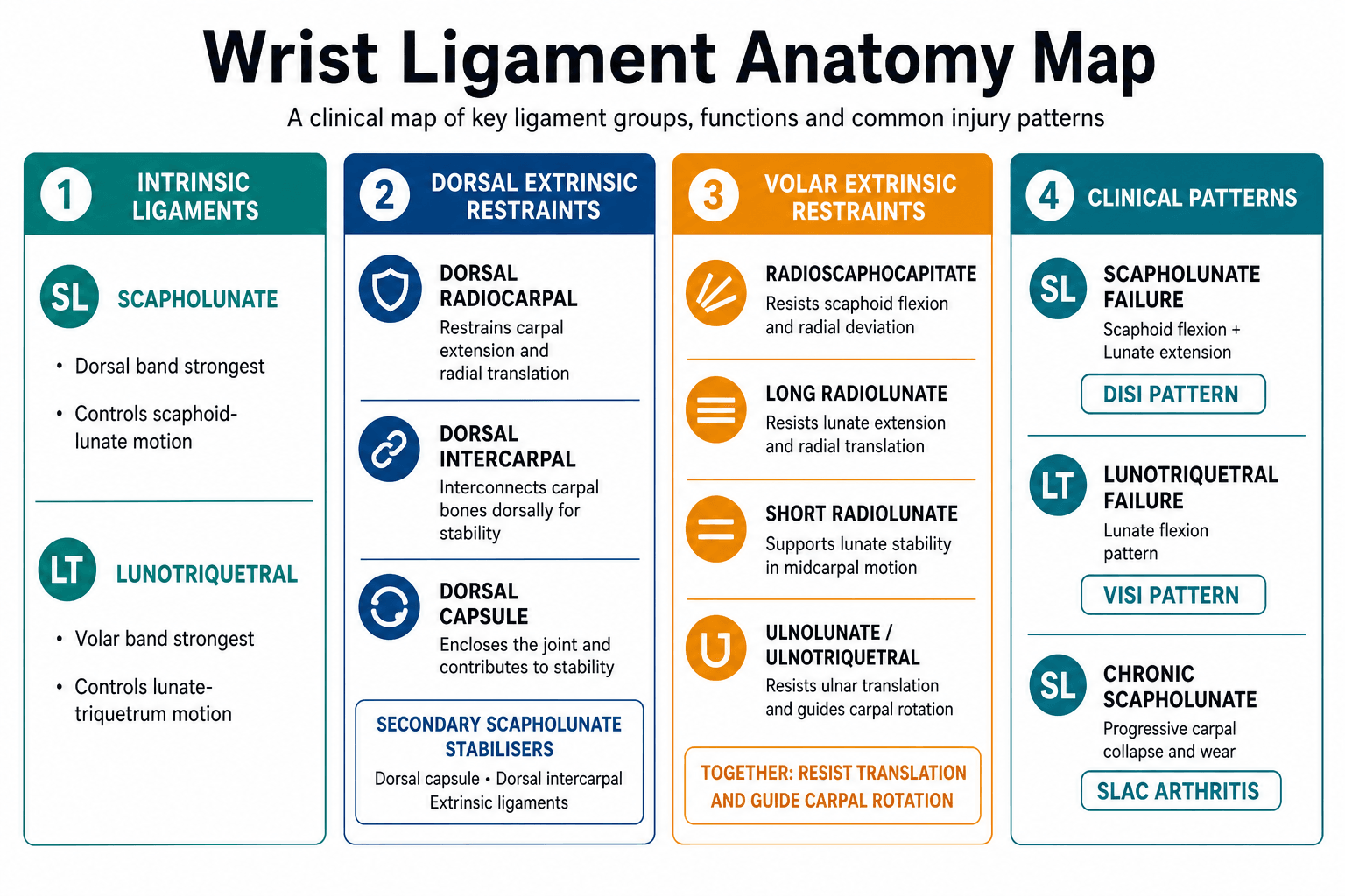
The scapholunate interosseous ligament has dorsal, proximal membranous and volar portions. The dorsal portion is the strongest and most important restraint to abnormal scaphoid-lunate motion. When the scapholunate ligament fails, the scaphoid tends to flex and pronate, while the lunate remains linked to the triquetrum and extends. The result may be a dorsal intercalated segment instability pattern.
Important associated stabilisers include the dorsal intercarpal ligament, dorsal radiocarpal ligament, radioscaphocapitate ligament and scaphotrapeziotrapezoid stabilisers. Chronic instability is therefore not simply a torn SL ligament; it is progressive failure of a stabilising complex.
Lunotriquetral instability
The lunotriquetral interosseous ligament has dorsal, proximal and volar components. The volar portion is usually the strongest. LT instability can produce ulnar-sided pain, painful clicking and sometimes volar intercalated segment instability. Isolated LT instability is less common than scapholunate instability and must be separated from TFCC injury, ulnar impaction, ECU pathology and midcarpal instability.
Degenerative collapse
Chronic scapholunate dissociation changes carpal load. The usual late pattern is scapholunate advanced collapse: radioscaphoid arthritis begins first, then scaphocapitate and capitolunate degeneration may follow while the radiolunate joint is often preserved until late. Once cartilage is lost, ligament reconstruction cannot restore a durable painless wrist.
SL and LT ligaments connect proximal-row bones. They control the immediate relationship between scaphoid, lunate and triquetrum.
Dorsal and volar extrinsic ligaments become increasingly important in chronic instability and reconstruction planning.
Chronic malalignment overloads cartilage. Once arthritis develops, treatment shifts from ligament reconstruction to salvage.
Classification
| Stage | Definition | Typical Findings | Treatment Direction |
|---|---|---|---|
| Predynamic | Partial or complete ligament injury without radiographic instability. | Pain, tenderness, normal PA/lateral and stress views; arthroscopy may show tear. | Immobilisation, therapy, arthroscopic debridement or pinning in selected symptomatic tears. |
| Dynamic | Instability appears only under load or stress. | Clenched-fist SL widening, dynamic fluoroscopy abnormality, normal resting alignment. | Repair or reconstruction if symptomatic, high-demand and repairable. |
| Static reducible | Gap or malalignment visible on resting films but correctable. | SL gap, DISI or VISI on neutral films; alignment reduces with fluoroscopy or traction. | Acute repair if early; chronic reconstruction or capsulodesis if cartilage preserved. |
| Static irreducible | Fixed malalignment without reliable reduction. | Fixed DISI/VISI, adaptive contracture, chronic deformity. | Limited fusion or salvage depending pain and cartilage. |
| Arthritic | Degenerative collapse. | SLAC, SNAC or radiocarpal/midcarpal cartilage loss. | PRC, four-corner fusion, limited fusion, total wrist fusion or selected arthroplasty. |
Clinical Presentation
History
Ask for the mechanism and the functional problem, not just the pain site.
| Question | Why It Matters | Decision Consequence |
|---|---|---|
| Was there a fall on the outstretched hand or high-energy wrist trauma? | SL injury and perilunate spectrum injuries may follow extension and ulnar deviation loading. | Triggers careful PA, lateral, stress and fracture assessment. |
| How long since injury? | Tissue repairability decreases with chronic attenuation and capsular contracture. | Acute repair, chronic reconstruction and salvage are different decisions. |
| Where is the pain? | Dorsal central pain suggests SL; ulnar-sided pain suggests LT, TFCC, ECU or ulnar impaction. | Directs examination and imaging. |
| Is there clicking, clunking or giving way? | Mechanical symptoms suggest instability rather than isolated inflammation. | Raises threshold for stress imaging or arthroscopy. |
| What does the hand need to do? | Manual work, sport, cane use and transfers increase load demands. | Influences operative threshold and salvage choice. |
Examination technique
Examine both wrists because laxity, contralateral widening and old malunion can mislead interpretation. The sequence should be practical: look, feel, move, load, then stress specific ligament intervals.
| Step | How To Do It | What You Are Looking For |
|---|---|---|
| Position | Seat the patient facing you with forearms supported on a table. Expose both wrists and hands. Compare side-to-side throughout. | Guarding, swelling, asymmetry, old scars, deformity, generalised laxity and functional confidence. |
| Look | Inspect dorsally, radially and ulnarly. Ask the patient to make a fist, extend the wrist, load through the palm if tolerated and demonstrate the painful activity. | Dorsal swelling around the SL interval, ulnar-sided fullness, ECU subluxation, apprehension with loading, clunking or inability to push up. |
| Feel | Palpate Lister's tubercle, SL interval just distal to it, scaphoid tubercle, anatomic snuffbox, LT interval, fovea, ECU sheath and DRUJ. | Localised SL tenderness, LT tenderness, foveal pain, ECU pain or DRUJ tenderness. Pain location guides the next stress tests. |
| Move | Assess active and passive flexion, extension, radial deviation, ulnar deviation, pronation, supination and grip. Compare strength and painful arcs. | Loss of extension after dorsal capsular injury, painful midcarpal clunk, grip weakness, load-related pain or motion loss from arthritis. |
| Stress | Only after localisation, perform Watson shift, LT ballottement, midcarpal shift, TFCC foveal/compression tests, ECU synergy and DRUJ stability. | The aim is to reproduce the patient's pain and demonstrate abnormal motion compared with the other side. |
| Test | Exact Technique | Positive Finding | Interpretation Limits |
|---|---|---|---|
| Watson scaphoid shift | Stabilise the distal radius with one hand. Put the thumb of the other hand firmly on the palmar scaphoid tubercle and fingers around the distal carpus. Start in ulnar deviation and slight extension, then move the wrist into radial deviation and flexion while keeping dorsal pressure through the scaphoid tubercle. Release pressure at the end to feel reduction. | Dorsal radial pain, apprehension, a clunk, or a subluxation-reduction sensation compared with the other wrist. | Pain alone is non-specific. False positives occur with laxity and painful sprains. It is a screening test; imaging and arthroscopy stage the injury. |
| SL interval palpation | Find Lister's tubercle, then palpate just distal and slightly ulnar over the dorsal SL interval with the wrist flexed slightly. | Point tenderness matching the patient's dorsal pain. | Useful localisation, but it does not prove mechanical instability. |
| LT ballottement | Stabilise the lunate between thumb and index finger of one hand. Grasp the triquetrum and pisiform with the other hand and translate the triquetrum dorsally and volarly relative to the lunate. | Pain, excessive translation, crepitus or clunk compared with the other side. | Overlap with TFCC injury, ECU pathology, ulnar impaction and midcarpal instability is common. |
| Ulnar fovea and TFCC compression | Palpate between the ulnar styloid and FCU tendon for foveal tenderness. Add ulnar deviation and axial compression with rotation if tolerated. | Deep ulnar pain suggests TFCC or ulnocarpal pathology. | A positive ulnar-sided test should make isolated LT surgery less likely until TFCC and ulnar variance are assessed. |
| ECU synergy and subluxation | Resist thumb abduction or wrist extension/ulnar deviation while palpating ECU. Supinate/pronate and look for tendon snapping. | ECU pain, bowstringing or subluxation. | ECU pathology can mimic LT or TFCC pain and changes the operative plan. |
| Midcarpal shift | Apply axial load to the hand with the wrist slightly flexed and ulnarly deviated, then move from radial to ulnar deviation while feeling for sudden reduction or clunk. | Painful clunk, catch or apprehension at the midcarpal joint. | Can reflect constitutional laxity. Interpret with symptoms and imaging rather than as an isolated diagnosis. |
Watson shift and LT ballottement are useful only when interpreted with symptoms, comparison examination, radiographs, stress imaging and cartilage status. A painful manoeuvre is not the same as surgically important instability.
Differential diagnosis
The single most examined error in this topic is anchoring on a ligament label before excluding the mimics. Dorsal-sided and ulnar-sided wrist pain each have a short, high-yield differential that changes the operation.
| Condition | Discriminating Features | Key Test or Investigation | Why It Changes Management |
|---|---|---|---|
| Scapholunate dissociation | Dorsal-radial pain, positive Watson shift, SL gap and DISI on imaging. | PA, lateral and stress PA; arthroscopy when uncertain. | True repairable or reconstructable instability if reducible and cartilage preserved. |
| Lunotriquetral instability | Ulnar-sided pain, positive LT ballottement, possible VISI. | LT ballottement, MRA, arthroscopy. | Often overlaps mimics; isolated surgery rarely indicated without confirmation. |
| TFCC tear / ulnocarpal pathology | Foveal tenderness, painful forearm rotation, pain with ulnar deviation and loading. | Fovea sign, ulnar variance, MRA, arthroscopy. | Treated by TFCC repair or ulnar shortening, not carpal ligament surgery. |
| Ulnar impaction syndrome | Ulnar-positive variance, chronic load-related ulnar pain, lunate/triquetral chondral change. | Neutral-rotation PA grip view for variance; MRI marrow oedema. | Ulnar shortening osteotomy or wafer addresses the driver; ignoring it dooms LT surgery. |
| ECU tendinopathy / subluxation | Ulnar pain, snapping with supination, positive ECU synergy test. | ECU synergy test, dynamic ultrasound. | Tendon or subsheath problem; managed conservatively or by subsheath reconstruction. |
| Midcarpal instability (CIND) | Painful clunk on ulnar deviation, often constitutional laxity, frequently no discrete tear. | Midcarpal shift test, dynamic fluoroscopy. | Reconstruction is unreliable; therapy and proprioceptive retraining come first. |
| Perilunate dislocation spectrum | High-energy injury, broken Gilula arcs, lunate or perilunate malalignment, median nerve signs. | True lateral radiograph, CT, urgent assessment. | Surgical emergency-level reduction and stabilisation, not elective ligament work. |
| Scaphoid fracture / nonunion (SNAC) | Snuffbox tenderness, scaphoid waist pain, humpback deformity. | Scaphoid views, CT. | Fixation or salvage of the scaphoid drives treatment, with its own collapse pattern (SNAC). |
| Kienbock disease | Central dorsal pain, lunate sclerosis or collapse, possible ulnar-negative variance. | Radiographs (Lichtman stage), MRI. | Lunate osteonecrosis needs unloading or salvage, not interosseous ligament repair. |
Investigations
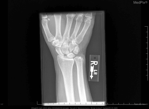
Plain radiographs
Write a specific imaging request. A useful request is: PA wrist, true lateral wrist, oblique wrist, clenched-fist or pencil-grip stress PA view, and contralateral comparison if subtle instability is suspected. Add scaphoid views if scaphoid fracture is possible and traction or dynamic fluoroscopy if a perilunate spectrum injury or reducibility question exists.
| View | Why You Request It | What To Check |
|---|---|---|
| PA wrist | Baseline carpal alignment. | SL interval, LT interval, Gilula arcs, cortical ring sign, fractures, ulnar variance and arthritis. |
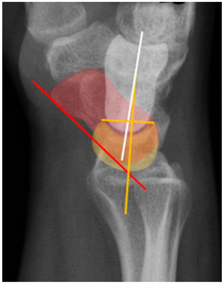
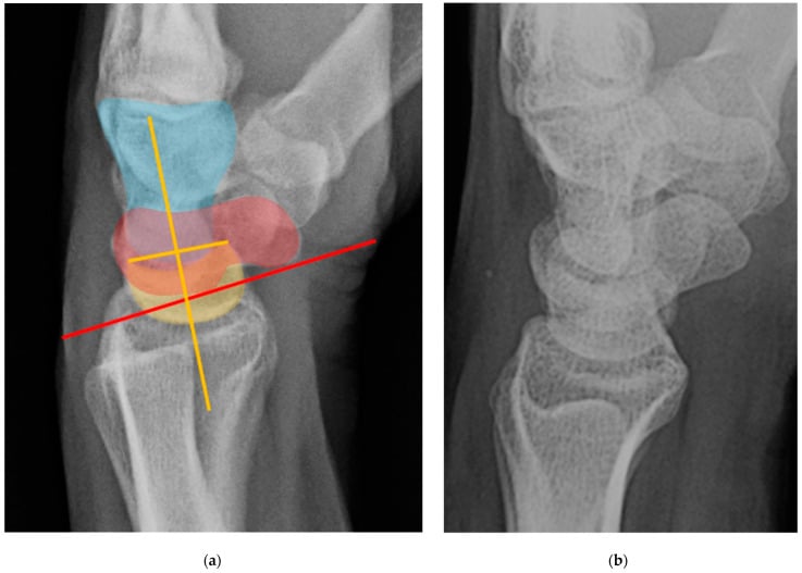
| True lateral wrist | Intercalated segment alignment. | SL angle, capitolunate angle, radiolunate angle, DISI, VISI and perilunate alignment. |
| Oblique views | Occult carpal fracture and joint overlap clarification. | Scaphoid, triquetrum, hamate, capitate and distal radius articular injury. |
| Clenched-fist or pencil-grip stress PA | Loads the carpus to reveal dynamic SL widening. | Side-to-side SL gap difference and dynamic dissociation. |
| Contralateral comparison | Separates abnormal widening from constitutional laxity. | Compare gap, angles and symptoms rather than treating an isolated number. |
| Dynamic fluoroscopy | Shows gap opening, clunk and reducibility in real time. | Helpful when examination and static radiographs disagree. |
| Finding | How To Assess | Meaning |
|---|---|---|
| Scapholunate gap | Measure the interval between the scaphoid and lunate on PA and stress PA views. Greater than 3 mm is suspicious; around 5 mm or more is strongly abnormal when symptomatic. | SL dissociation or dynamic SL instability. |
| Terry Thomas sign | Visible widening between scaphoid and lunate on neutral PA view. | Static SL dissociation if present at rest. |
| Cortical ring sign | Look for the distal scaphoid seen end-on because the scaphoid is flexed. | Scaphoid flexion caused by loss of SL control. |
| Scapholunate angle | On the true lateral, draw the scaphoid long axis and the lunate axis. Normal is roughly 30 to 60 degrees; greater than 70 degrees supports DISI. | Dorsal lunate extension pattern after SL failure. |
| Radiolunate angle | Draw the radial shaft axis and lunate axis on the lateral. The lunate should sit close to neutral relative to the radius. | Dorsal or volar lunate tilt helps define DISI or VISI. |
| Capitolunate angle | Draw the capitate axis and lunate axis. A small angle is expected; widening indicates carpal malalignment. | Perilunate spectrum, DISI, VISI or midcarpal instability. |
| Gilula arcs | Smooth arcs across proximal and distal carpal rows on PA view. | Broken arcs suggest carpal malalignment or fracture-dislocation. |
| SLAC arthritis | Radioscaphoid narrowing, styloid arthritis, scaphocapitate/capitolunate involvement. | Ligament reconstruction becomes inappropriate when cartilage is lost. |
Advanced imaging
| Investigation | Best Use | Limitations |
|---|---|---|
| Stress radiographs | Dynamic SL gap and comparison with the contralateral wrist. | Can be normal in predynamic injury and can overcall laxity. |
| Dynamic fluoroscopy | Real-time clunk, gap opening and reducibility. | Operator-dependent and requires clinical correlation. |
| MRI | Ligament signal, occult fracture, cartilage, marrow oedema and associated pathology. | May miss partial tears or overcall incidental signal change. |
| MR arthrogram / CT arthrogram | Contrast passage through SL or LT interval and cartilage assessment. | Invasive; still does not replace arthroscopy when treatment depends on exact grade. |
| CT | Fracture, scaphoid nonunion, carpal alignment, arthritis and surgical planning. | Poor for ligament integrity unless arthrographic. |
| Arthroscopy | Direct ligament, interval, reducibility and cartilage assessment. | Invasive, but often the reference standard for uncertain symptomatic instability. |
Management
Management is chosen by stage, timing, reducibility, cartilage, associated pathology and patient demand. A repairable acute SL tear, a chronic reducible SL dissociation and an arthritic SLAC wrist are different problems.
| Clinical Situation | Best Treatment Logic | Do Not Do |
|---|---|---|
| Stable sprain or partial tear | Immobilisation, analgesia, oedema control, hand therapy and reassessment. Arthroscopic debridement only for persistent symptomatic synovitis or partial tear. | Do not reconstruct a stable wrist because MRI shows signal change. |
| Acute complete SL tear, reducible, no arthritis | Direct dorsal SL repair, temporary pinning and possible dorsal capsulodesis augmentation. | Do not repair without correcting the SL gap and carpal angles first. |
| Dynamic SL instability | Stress-view or arthroscopy-confirmed instability: acute repair if early and repairable; reconstruction if chronic and symptomatic. | Do not reassure solely because neutral radiographs look normal. |
| Chronic reducible SL instability, preserved cartilage | Capsulodesis, tenodesis or ligament reconstruction depending tissue, demand and surgeon technique. | Do not promise normal wrist kinematics; counsel stiffness and recurrence risk. |
| Irreducible DISI or established SLAC | Salvage based on cartilage: PRC, four-corner fusion, limited fusion or total wrist fusion. | Do not offer ligament reconstruction when cartilage is already damaged. |
| LT-type ulnar-sided symptoms | Exclude TFCC, ECU, ulnar impaction and midcarpal instability before repair, pinning or fusion. | Do not diagnose isolated LT instability from tenderness alone. |
Non-operative management is appropriate only when the wrist is stable or when the patient's symptoms, demand, comorbidity or preference do not justify surgery.
| Situation | Treatment | Review Trigger |
|---|---|---|
| Acute sprain without instability | Short immobilisation, analgesia, oedema control, hand therapy and reassessment. | Persistent dorsal or ulnar-sided pain, clicking or inability to load. |
| Partial SL or LT tear | Immobilisation, protected loading and therapy; arthroscopic debridement if persistent mechanical synovitis. | Positive stress imaging or persistent mechanical symptoms. |
| Low-demand chronic symptoms | Splinting, activity modification, injections only as diagnostic or temporary tools, therapy. | Progressive deformity, loss of function or degenerative change. |
| Arthritic wrist not ready for surgery | Splint, analgesia, activity modification, corticosteroid injection and shared decision-making. | Pain, function and radiographic progression. |
Do not call non-operative care complete unless follow-up is planned. Dynamic instability can declare itself later.
Delayed diagnosis can convert a repairable ligament injury into a reconstruction or salvage problem. Persistent wrist pain after trauma needs reassessment, not reassurance alone.
Surgical Technique Details
Purpose: confirm ligament grade, dynamic instability, cartilage condition and associated TFCC or chondral injury.
Steps
- Supine position, arm table, traction tower or wrist traction setup.
- Mark Lister's tubercle, EPL, 3-4 portal, 4-5 portal, 6R/6U portals and midcarpal portals.
- Establish 3-4 portal for radiocarpal viewing.
- Inspect radius, scaphoid fossa, lunate fossa, proximal scaphoid, lunate, TFCC and SL interval.
- Probe SL and LT ligaments; assess step-off and whether a probe or scope passes through the interval.
- Use midcarpal portals to assess SL/LT interval from the opposite side, capitolunate joint and cartilage.
- Treat according to grade: debridement for stable partial tears, pinning/repair for unstable acute tears, reconstruction or salvage if chronic.
Hazards: superficial radial nerve, dorsal sensory branches, ECU subsheath, extensor tendons and iatrogenic chondral injury.
| Question | Proximal Row Carpectomy | Four-Corner Fusion |
|---|---|---|
| Cartilage required | Capitate head and lunate fossa must be usable. | Radiolunate joint must be preserved. |
| Main advantage | No fusion site; simpler rehabilitation when cartilage is suitable. | Maintains carpal height better and may suit heavier demand in selected patients. |
| Main risk | Radiocapitate arthritis and reduced grip strength if cartilage selection is poor. | Nonunion, hardware irritation and stiffness. |
| Do not choose if | Capitate head cartilage or lunate fossa is poor. | Radiolunate arthritis is present or the patient cannot tolerate fusion protection. |
Complications
| Problem | Cause | Prevention or Response |
|---|---|---|
| Missed dynamic instability | Normal neutral films interpreted as normal wrist. | Use stress views, reassessment and arthroscopy when symptoms persist. |
| Stiffness | Immobilisation, capsular surgery, tendon tethering or pain. | Counsel preoperatively, protect tendons and use staged hand therapy. |
| Recurrent SL gap | Chronic tissue attenuation, failed reconstruction or inadequate reduction. | Stage properly, reduce before fixation and counsel limitations. |
| Progression to SLAC | Persistent malalignment and cartilage overload. | Recognise when reconstruction is too late; shift to salvage. |
| Median or sensory nerve symptoms | Trauma, swelling, portals or surgical exposure. | Document pre/post-op nerve status; decompress if acute carpal tunnel syndrome is present. |
| Pin or hardware complications | K-wire migration, infection, irritation, broken implants. | Appropriate pin care, removal timing and follow-up. |
| Nonunion after partial fusion | Poor preparation, fixation, smoking, biology or early loading. | Meticulous joint preparation, grafting, stable fixation and protected rehabilitation. |
| Persistent pain after salvage | Adjacent joint disease, nonunion, expectations or residual impingement. | Match procedure to cartilage pattern and patient goals. |
Controversies and Areas of Uncertainty
This is a field with strong opinions and weak comparative evidence. Naming the honest controversies is a fast way to demonstrate maturity in a viva.
| Question | The Debate | Pragmatic Position |
|---|---|---|
| Best operation for chronic reducible scapholunate instability | Capsulodesis, three-ligament tenodesis, bone-ligament-bone graft and screw-based constructs (RASL/SLAM) all have advocates; comparative high-level data are lacking and recurrent radiographic gap is common with every technique. | Match technique to tissue, demand and surgeon experience; counsel that no method reliably restores normal kinematics and that radiographic recurrence does not always mean clinical failure. |
| Does fixing the ligament prevent SLAC? | The assumption that early stabilisation prevents degenerative collapse is biologically plausible but not proven by long-term controlled data. | Treat symptomatic, repairable, reducible injury early, but avoid over-promising arthritis prevention. |
| Predynamic and incidental MRI tears | High-resolution MRI and arthroscopy detect partial SL/LT tears whose natural history in asymptomatic wrists is unknown; overtreatment is a real risk. | Treat the patient, not the scan; reserve intervention for mechanical symptoms with corroborating examination and stress imaging. |
| PRC versus four-corner fusion | Meta-analysis now favours PRC for outcomes and complications, yet many surgeons still prefer fusion for younger, heavy-demand patients citing carpal-height preservation. | Let cartilage pattern decide first (capitate head and lunate fossa for PRC; radiolunate for fusion); use demand and age as tie-breakers. |
| Role of proprioceptive rehabilitation | Cadaver and clinical work suggests selective muscle retraining can dynamically stabilise ligament-deficient wrists, but robust trial evidence is limited. | A reasonable adjunct for dynamic instability and low-demand patients; not a substitute for surgery in established static instability. |
| LT arthrodesis | Reported non-union rates up to 57% and persistent pain make LT fusion contentious. | Use only after exhaustively excluding TFCC, ulnar impaction and midcarpal drivers, and counsel the high failure rate. |
Guidelines, Registries & Global Practice
Carpal instability is a low-volume, high-complexity problem with no single randomised gold-standard operation, so practice is driven by expert society consensus, surgeon experience and resource setting rather than by registry-mandated implants.
Global epidemiology
- Scapholunate injury is the most frequent carpal instability and a common occult injury after fall-on-the-outstretched-hand trauma (Kitay & Wolfe, J Hand Surg Am 2012).
- Up to two-thirds of displaced intra-articular distal radius fractures carry an associated intracarpal ligament or TFCC injury (Geissler, JBJS Am 1996), so instability is frequently a missed companion of a common fracture.
- SLAC is the most common pattern of wrist osteoarthritis, accounting for around 57% of degenerative wrists in the original radiographic series (Watson & Ballet 1984).
Side-by-side society positions
| Body / Source | Emphasis | Practical Take-Home |
|---|---|---|
| AAOS (US) | Evidence-based management of distal radius fractures highlights screening for associated carpal/TFCC injury; instability itself has no dedicated high-level guideline. | Have a low threshold to look for SL/LT injury when treating wrist trauma. |
| BSSH / BOA (UK) | Specialist hand-surgery referral pathways for suspected carpal instability and ulnar-sided wrist pain; emphasis on staged assessment and arthroscopy. | Refer persistent post-traumatic wrist pain to a hand unit rather than repeatedly reassuring. |
| IWAS / EWAS (wrist arthroscopy societies) | Standardised arthroscopic staging of SL and LT tears and arthroscopic-assisted treatment algorithms. | Arthroscopy is the reference standard when clinical and radiographic findings disagree. |
| AO Foundation | Reduction-first principles for the perilunate spectrum and articular distal radius; carpal alignment must be restored before soft-tissue work. | Reduce and align the carpus before judging or repairing ligaments. |
| EFORT / European consensus | Recognition that no single reconstruction is superior and that staged, cartilage-led decision-making prevails across Europe. | Tailor the operation to stage, reducibility and cartilage, not to a fixed protocol. |
Registry note
Unlike hip and knee arthroplasty, wrist ligament reconstruction is not tracked by national joint registries because it uses no standardised implant. Total wrist arthroplasty — a salvage option in selected low-demand wrists — is captured in some national registries (for example the Norwegian and Australian arthroplasty registries report wrist implant survival), and these data consistently show inferior longevity compared with hip and knee implants, reinforcing caution in high-demand patients.
High- versus limited-resource practice variation
| Setting | Typical Pathway | Reason |
|---|---|---|
| Well-resourced unit | Stress imaging, MR arthrography, diagnostic and therapeutic wrist arthroscopy, full salvage menu including arthroplasty. | Access to arthroscopy and advanced imaging allows precise staging and motion-preserving options. |
| Limited-resource setting | Plain radiographs with contralateral comparison, clinical staging, immobilisation, open repair and fusion-based salvage. | Reliance on robust low-cost techniques; arthrodesis is durable and forgiving where arthroscopy and implants are scarce. |
| Both settings | Reduction of perilunate injuries, recognition of acute carpal tunnel syndrome and avoidance of reconstructing an arthritic wrist. | These principles are universal and do not depend on resources. |
Evidence Base
The papers below are the ones most worth being able to cite by name in a viva. Each statistic has been checked against the original PubMed record. According to PubMed, the verified sources are as follows.
Watson & Ballet — the SLAC pattern (landmark natural history)
- Review of 4,000 wrist radiographs identified 210 cases of degenerative arthritis after excluding other arthritides; the scapholunate advanced collapse (SLAC) sequence was the single most common pattern at 57%, beginning at the radioscaphoid joint.
- A further 27% involved the scaphotrapezium-trapezoid joint and 15% combined both patterns; 18 of 19 operated patients had less pain postoperatively.
Geissler — arthroscopic classification of carpal ligament tears
- Prospective arthroscopic study of 60 displaced intra-articular distal radius fractures: 41 patients (68%) had associated intracarpal soft-tissue injury, including 26 TFCC tears, 19 scapholunate and 9 lunotriquetral interosseous ligament tears.
- Intracarpal injuries clustered with fractures involving the lunate facet; the paper introduced the arthroscopic grading of interosseous ligament tears now known as the Geissler classification.
Kitay & Wolfe — scapholunate instability current concepts
- Comprehensive review establishing scapholunate injury as the most frequent cause of carpal instability and proposing a widely used treatment algorithm based on stage of injury, secondary ligament damage and arthritic change.
- An isolated SLIL tear may show normal static radiographs yet progress to abnormal kinematics, cartilage wear and degenerative collapse.
Common Pitfalls
Persistent wrist pain after trauma needs reassessment. Dynamic SL instability can have normal neutral radiographs.
MRI signal does not equal clinically important instability. Correlate with stress imaging, arthroscopy and symptoms.
SLAC or fixed arthritic collapse is a salvage problem, not a ligament repair problem.
LT pain overlaps with TFCC injury, ECU pathology, ulnar impaction and midcarpal instability.
Viva Scenarios
Practise clinical reasoning and management decisions out loud
“A young manual worker has dorsal wrist pain three months after a fall. Neutral radiographs look normal.”
“A high-demand patient has an acute complete scapholunate ligament tear with reducible SL widening and no arthritis.”
“A patient has chronic scapholunate dissociation, DISI deformity and radioscaphoid arthritis.”
“A 35-year-old presents with chronic ulnar-sided wrist pain, a painful click and a positive lunotriquetral ballottement test. They want to know if they need a lunotriquetral fusion.”
MCQ Practice Points
Q: What does a normal PA wrist radiograph exclude? A: It excludes obvious static dissociation, but it does not exclude predynamic or dynamic scapholunate instability.
Q: What radiographic pattern is classically associated with scapholunate dissociation? A: DISI, with scaphoid flexion and lunate extension.
Q: What is the key contraindication to ligament reconstruction? A: Fixed irreducible deformity or established arthritis where salvage is required.
Core diagnosis
- SL injury: dorsal pain, Watson shift, SL gap, DISI.
- LT injury: ulnar-sided pain, LT ballottement, possible VISI.
- Dynamic injury may need stress views or arthroscopy.
Measure
- SL gap on PA and stress views.
- Scapholunate angle and capitolunate angle on lateral.
- Gilula arcs, ring sign, DISI/VISI and arthritis pattern.
Stage
- Predynamic: symptoms, no radiographic instability.
- Dynamic: stress instability.
- Static reducible: visible malalignment that corrects.
- Irreducible or arthritic: salvage logic.
Treat
- Stable partial tear: immobilisation, therapy, selected arthroscopic debridement.
- Acute complete reducible SL: repair, pinning, possible capsulodesis.
- Chronic reducible SL: reconstruction or capsulodesis.
- Arthritic collapse: PRC, four-corner fusion, limited fusion or total wrist fusion.
Must not miss
- Perilunate spectrum injury in high-energy trauma.
- TFCC, ECU and ulnar impaction in LT-type symptoms.
- Cartilage loss before offering reconstruction.
- Median nerve symptoms after carpal trauma.