How to Examine the Hand: Nerves, Tendons and a Look-Feel-Move Script That Scores
A hand examination script for the clinical exam: look, feel and move, isolating each tendon, a 60-second nerve screen and localising nerve lesions by level.
By OrthoVellum Editorial TeamPublished 14 min read
Educational content for clinicians, not medical advice. Editorial policy
Key points
- A hand examination is a localisation exercise. Compare active with passive movement, and the patient's other hand, before you call anything a rupture or a palsy.
- Isolate every tendon. A composite fist can look normal over a divided FDP or FDS, and the little-finger FDS looked absent on standard testing in 35% of 200 hands examined but in 4% with a modified test.
- Screen each nerve with one muscle and one patch of skin: APB and the index pulp (median), first dorsal interosseous and the little-finger pulp (ulnar), EPL retropulsion and the dorsal first web (radial).
- Three manoeuvres localise most nerve lesions: dorso-ulnar sensation, FDP to the ring and little fingers, and palmar sensation. A striking ulnar claw points to the wrist, not the elbow.
- No single carpal tunnel provocation test rules the diagnosis in or out. In the JAMA review, median-territory hypalgesia, the hand diagram and thumb abduction strength carried more weight than Phalen or Tinel.
On this page11 sections
The hand examination station separates candidates who know the tests from candidates who can say which structure has failed and at what level. Marks go on the same errors: one hand examined instead of two, a composite fist accepted as proof of tendon continuity, "weak grip" offered as a finding, and a Phalen test presented as the diagnosis. The answer is a fixed script in which every step localises, always against the other hand. This post sets out that script, what each test can and cannot tell you, and a model answer for the clawed hand. The full reference is the hand examination and clinical localisation topic page.
What is the hand examination station testing?
Three things: which structure (tendon, nerve, joint, skin, vessel or pain), which level, and how urgent. A tendon problem loses active motion through one tendon while passive motion is preserved. A nerve problem follows a motor and sensory territory. A joint problem restricts active and passive motion alike. Pain can mimic all three, so do not call pain-limited movement a rupture.
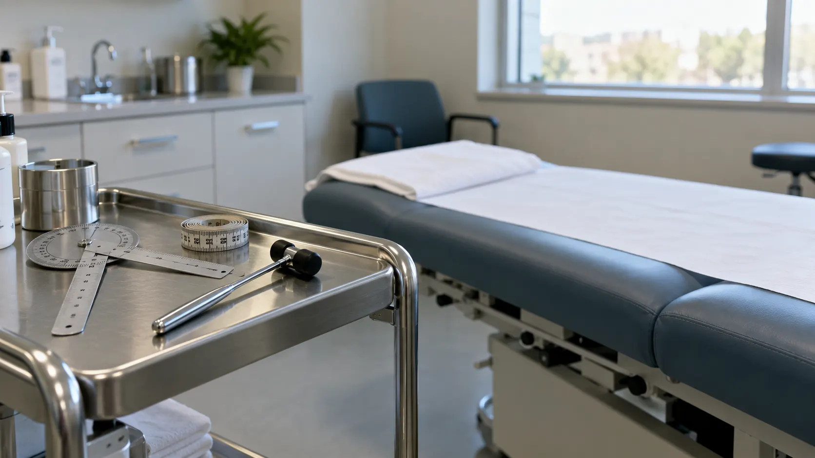
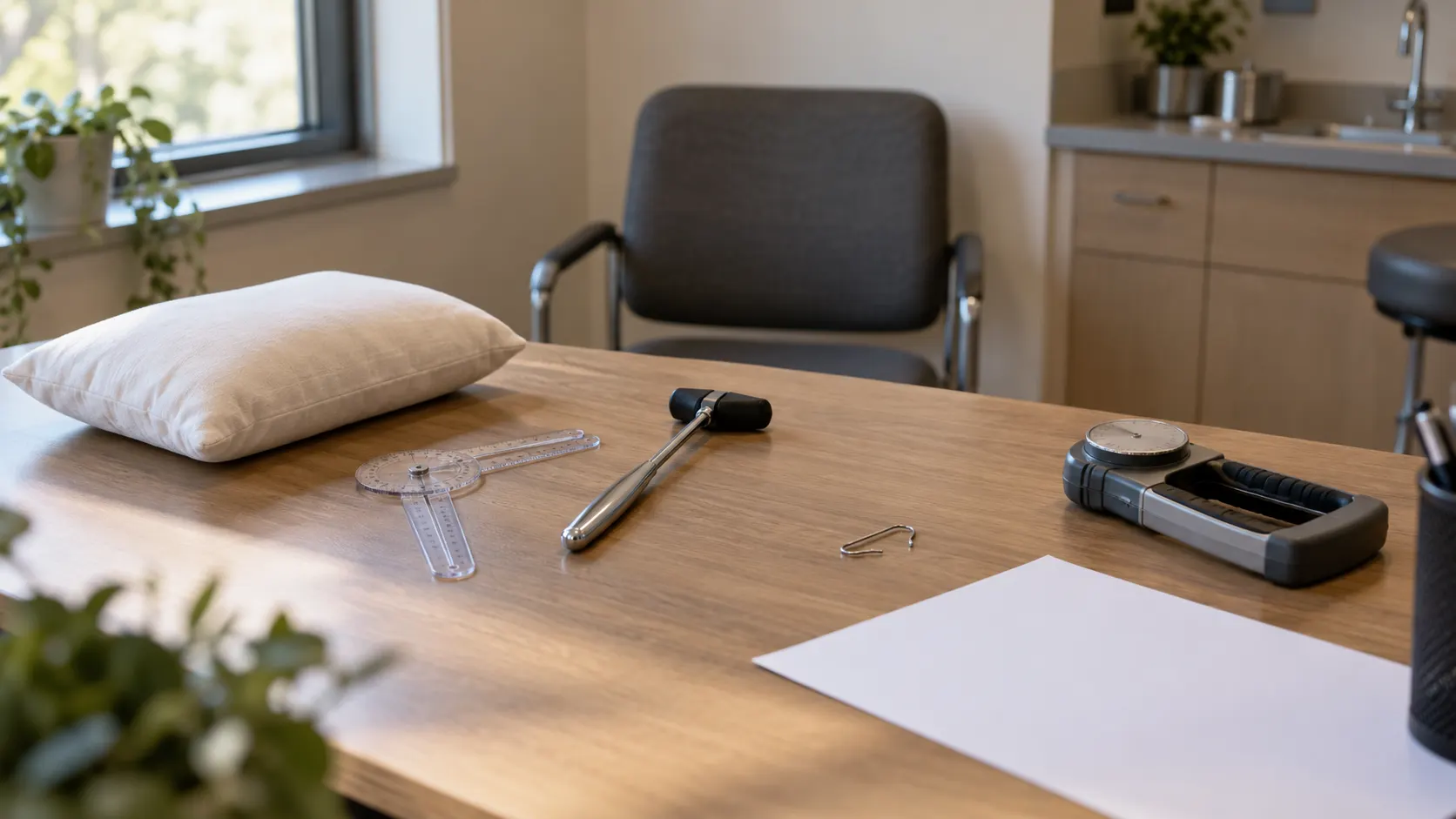
Set-up. Expose both hands and forearms to above the elbow, with rings and watch off if they can be removed safely, and rest the hands on a pillow so they lie relaxed in their true posture. A sleeve left on hides the forearm wasting and the cubital tunnel. You need a small goniometer, a bent paperclip or two-point discriminator, a sheet of paper, a tendon hammer, and a button, coin and pen for function.
The other hand is your control. Cascade, range, bulk, sweating, strength and sensibility are all comparisons. Hold the hands side by side and say that you are comparing.
Time budget for a five-minute station. A workable split is about 45 seconds to look, 45 to feel, 60 to move, 60 for tendons and nerves, 60 for special tests and 30 for grip, function and the closing sentence. The station is lost by the candidate still describing nails at three minutes.
Look: palms down, then palms up
Palms down. Look at the resting cascade first. Relaxed fingers sit in a spiral of increasing flexion from index to little finger. A finger that sits proud of the cascade has a flexor tendon injury, a malunion or a joint disruption until proven otherwise. Sight down the fingers end-on for rotation. Then read the dorsum:
- Swelling by joint row. Boggy MCP swelling suggests synovitis; firm PIP swellings are Bouchard nodes; DIP swellings are Heberden nodes or mucous cysts.
- Wasting, and its distribution. A hollow first web (first dorsal interosseous) and guttering between the metacarpals are ulnar. Wasting takes weeks, so its presence dates the lesion.
- Deformity, named by joint. Mallet is DIP flexion from a terminal extensor injury. Boutonnière is PIP flexion with DIP hyperextension from central slip failure. Swan neck is the mirror image. Measure each in degrees.
- Scars as an operation record: a palmar carpal tunnel scar, Bruner zigzags from a flexor repair or fasciectomy, straight dorsal scars over the MCP joints after joint replacement.
Palms up. Compare thenar and hypothenar bulk side to side. Look for Dupuytren nodules, cords and skin pits, a dry shiny territory (sympathetic fibres travel with the nerve), and wounds in relation to tendon and nerve lines. A fusiform, semi-flexed finger, tender along the sheath and painful on passive extension, is a flexor sheath infection: name the Kanavel signs and stop examining.
Feel: temperature, joints, sheaths and Tinel sites
Run the back of your fingers over both hands; a hot finger is inflamed and changes what you do next. Then stroke your pulps across both palms to compare sweating. Palpate each MCP joint with your thumbs either side of the extensor tendon, then the PIPs, DIPs and the thumb CMC joint, watching the patient's face rather than your hands.
Run a finger pulp along each flexor sheath from the distal palm. A nodule that clicks at the distal palmar crease is a trigger finger catching at the A1 pulley. Percuss the median nerve across the carpal tunnel, the ulnar nerve in Guyon's canal and behind the medial epicondyle, working from healthy nerve towards the suspected level. A positive Tinel sends tingling into the nerve's territory. Local pain under your finger does not count.
Move: active against passive
Ask for a fist and full extension, then take each joint passively and feel the end point. Measure MCP, PIP and DIP with a goniometer and separate an extension lag from a fixed contracture. The comparison of active with passive motion is the most useful step in the examination.
| Pattern | Most likely | Next step |
|---|---|---|
| Active lost, passive full | Tendon rupture, nerve palsy, pain inhibition or poor effort | Isolate the tendon; test the nerve pattern |
| Active and passive both restricted | Joint stiffness, contracture, swelling, adhesions or a mechanical block | End feel, radiographs, look for inflammation or infection |
| Passive full but severe pain | Infection, fracture, instability or CRPS | Do not force; investigate the urgent cause |
| Motor deficit with sensory loss | Peripheral nerve lesion above its sensory branch | Map motor and sensory territories |
| Motor deficit without sensory loss | AIN or PIN lesion, tendon rupture, central cause or muscle injury | Nerve-specific motor tests and tendon isolation |
Composite grips localise the short structure. A full fist flexes every joint. A straight fist (MCP and PIP flexed, DIP straight) is the FDS arc, so a full straight fist with an incomplete full fist points to the DIP or FDP. A hook fist (MCPs straight, IPs flexed) shows adhesions between the two flexors. The tabletop test, with the palm and fingers flat on the table, exposes a Dupuytren cord or a joint contracture. Score thumb opposition with the Kapandji score from 0 to 10, by how far along the fingers and palm the thumb tip reaches, and score both hands.
How do you test the flexor and extensor tendons?
One at a time. A finger may still flex weakly through its neighbours, the lumbricals or tenodesis, so a fist can look intact over a divided tendon. If you can, examine before any local anaesthetic block removes the motor testing.
| Structure | How to test | Positive finding |
|---|---|---|
| FDP | Hold the PIP extended; ask for DIP flexion | No active DIP flexion |
| FDS | Hold the other three fingers fully extended; ask for PIP flexion | No isolated PIP flexion |
| FPL | Stabilise the thumb MCP; ask for IP flexion | No active thumb IP flexion |
| EPL | Hands flat; lift both thumbs off the table (retropulsion) | The thumb cannot be lifted clear on one side |
| Central slip | Elson test: PIP at 90° over the table edge, resisted PIP extension | Weak PIP extension with a rigid, extended DIP |
| Terminal extensor | Active DIP extension | DIP extensor lag (mallet) |
The FDS trap is the little finger. Holding the three neighbouring fingers straight blocks their FDP, which shares one muscle belly, so the free finger can flex its PIP only through FDS. In Austin's 50 normal subjects, little-finger FDS was independent in 58%, shared with the ring finger in 21% and functionally absent in 21%, and the two hands differed in 26%, yet all 40 dissected cadaver hands had the tendon. In Stein's 200 hands, standard testing suggested absent FDS in 35% and modified testing in 4%. Without a wound, a failed little-finger test is usually anatomy: test the other hand.
EPL is tested by retropulsion, not IP extension. The intrinsics can extend the thumb IP joint with EPL ruptured, but only EPL lifts the thumb off the table. Test it after every distal radius fracture. In a survey of 596 hand surgeons, 33% reported at least one flexor tendon injury after volar plating in the previous year, FPL accounted for 75% of those, and 36% had seen extensor ruptures.
The Elson test catches the boutonnière before it forms. A fresh central slip injury can look almost straight because the lateral bands still extend the PIP. With the PIP flexed to 90° the lateral bands are slack, so a ruptured central slip leaves weak PIP extension and a DIP pulled rigidly straight. In a cadaver study of 20 fingers it was the only one of four non-invasive tests that detected both simulated injuries; the Boyes test was unreliable. There are no clinical accuracy figures, an acutely painful PIP will guard, and one technique review recommends testing under a digital block. See boutonnière deformity and, for lacerations, flexor tendon lacerations.
The 60-second three-nerve screen
Test each nerve where it is purest: one muscle and one patch of autonomous skin.
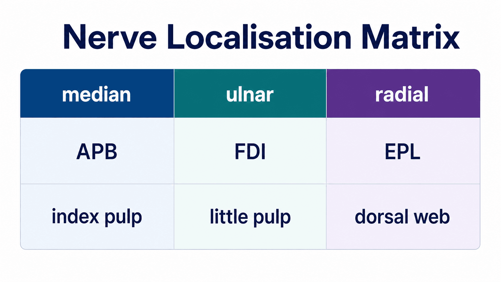
- Median: abductor pollicis brevis and the index pulp. Palm up, the patient points the thumb at the ceiling while you resist and palpate APB, so flexor pollicis brevis cannot trick the movement. Then the OK sign for the anterior interosseous nerve: FPL and FDP to the index, with normal sensation.
- Ulnar: first dorsal interosseous and the little-finger pulp. Palm down, the patient spreads the fingers against your resistance on the radial side of the index while you feel the first web.
- Radial: EPL retropulsion, wrist extension and the dorsal first web. The posterior interosseous nerve carries no skin sensation, so the dorsal web separates a sensory from a motor problem at the same level.
Grade power on the MRC scale with the muscle named ("APB 4 on the right, 5 on the left"). For sensation, light touch compared side to side is the screen. Static two-point discrimination and Semmes-Weinstein monofilaments quantify it, but in digital nerve repairs both had poor reproducibility and the monofilament was the more reliable of the two, so record the method and read the number alongside the motor findings.
How do you localise an ulnar nerve lesion?
Froment's sign tests adductor pollicis. The patient holds a sheet of paper between the thumb and the side of the index while you pull it away. Watch the thumb, not the paper.
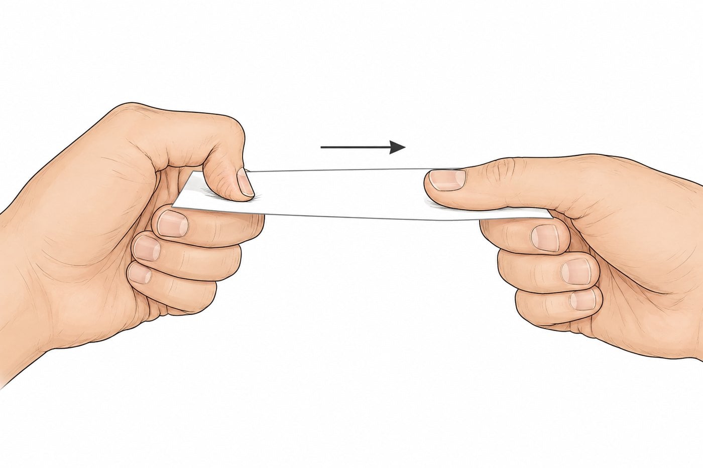
Froment and Wartenberg (the little finger resting abducted) confirm an ulnar nerve palsy; neither has formal accuracy data, so do not quote any. The level comes from three steps:
- The dorso-ulnar patch. The dorsal cutaneous branch leaves above the wrist. Sensation there is spared in a Guyon's canal lesion and lost at the elbow.
- FDP to the ring and little fingers. Hold the PIPs straight and ask for DIP flexion. Weakness puts the lesion above the forearm branches; add FCU.
- The claw. Clawing needs a working FDP to curl the IP joints against paralysed intrinsics. A high lesion denervates that FDP too, so it claws less. A striking claw points to the wrist, and reduced clawing as a high lesion recovers is expected rather than reassuring, because FDP reinnervates first.
Do not lean on elbow palpation. In 192 patients with suspected ulnar neuropathy at the elbow (cubital tunnel syndrome), tested against electrophysiology and neurosonography, nerve tenderness was 32% sensitive and 80% specific, thickening 28% and 87%, and Tinel 62% and 53%. The provocative tests added little to a routine clinical examination.
Median and radial: what each level spares
| Lesion | Motor | Sensory | The giveaway |
|---|---|---|---|
| Median at the carpal tunnel | APB, opponens, radial lumbricals | Radial three and a half digits | Palm spared: the palmar cutaneous branch leaves above the tunnel |
| Anterior interosseous | FPL, FDP index and middle, pronator quadratus | Normal | Flat OK sign with no sensory loss |
| Median in the proximal forearm (pronator syndrome) | Weakness variable and often absent; may affect median forearm and thenar muscles | Median territory including the palm | Night symptoms typically absent |
| Posterior interosseous | Finger and thumb extension | Normal | Wrist extends but deviates radially: ECRL survives, ECU does not |
| Radial in the arm | Wrist drop, finger and thumb extension, brachioradialis | Dorsal first web | Brachioradialis weak; numb web |
| Superficial radial | Normal | Dorsal first web and radial dorsum | Pure sensory |
Benediction versus claw. A high median lesion leaves the index and middle fingers straight when the patient tries to make a fist: failure of flexion on command. The ulnar claw is present at rest: failure of balance. Name which one you saw.
Localisation assumes one lesion. A patient can have a cervical radiculopathy with carpal tunnel syndrome, or a cubital tunnel lesion above a Guyon's canal lesion. The mechanism of double crush is contested, but the habit it teaches is sound: in anyone with hand paraesthesia, examine the neck.
Which carpal tunnel tests are worth doing?
None works alone, and the pooled figures vary with the review. MacDermid and Wessel averaged 60 studies, many of poor quality, and found carpal compression at 64% sensitivity and 83% specificity, Phalen 68% and 73% and Tinel 50% and 77%. A 2023 random-effects meta-analysis by Dabbagh and colleagues pooled Phalen at 0.57 and 0.67 (7 studies, 604 patients) and Tinel at 0.45 and 0.78 (7 studies, 748 patients), with very wide ranges between studies. Quote the review with its number.
The bedside findings outside the provocation tests carry more weight. In Kuhlman's 228 symptomatic hands, square-shaped wrist (69%) and APB weakness (66%) were the most sensitive signs, and median-territory hypoaesthesia was 51% sensitive and 85% specific. In the JAMA rational clinical examination review, the findings that best distinguished patients with electrodiagnostic evidence of carpal tunnel syndrome were median-territory hypalgesia (LR 3.1), a classic or probable Katz hand diagram (LR 2.4) and weak thumb abduction (LR 1.8), while Phalen, Tinel, night symptoms and thenar atrophy had little or no diagnostic value.
The CTS-6 turns this into a score: numbness in the median distribution (3.5 points), nocturnal symptoms (4), thenar atrophy or weakness (5), a positive Phalen (5), two-point discrimination over 5 mm (4.5) and a positive Tinel (4); 12 or more is positive. Graham studied 143 patients referred for electrodiagnosis: 73% had a pre-test probability of at least 0.80, and in them nerve conduction changed the probability by an average of −0.02 or −0.06, depending on the criterion. The large changes came mainly when the clinical probability was 0.50 or less. Studies earn their keep when the picture is uncertain or atypical, or when severity will change management.
Where candidates lose marks
- Examining one hand. Every finding is a comparison.
- Quoting accuracy figures that do not exist. Tendon signs, Froment and Wartenberg are anatomical demonstrations; the carpal tunnel tests have pooled figures.
- Examining after the block. Document tendon, nerve and vascular status of each injured digit first.
Completing the examination. Offer the wrist, the elbow with the ulnar nerve at the cubital tunnel, and the cervical spine; a full neurovascular examination with two-point discrimination; grip and pinch dynamometry; a functional screen with buttons, coins and writing; and radiographs, adding nerve conduction studies or ultrasound when the diagnosis or its severity is in doubt.
In the exam
The clawed hand is the short case that tests localisation most directly. Present the findings in the order you elicited them, then the level and the reasoning; the structure is set out in our guide to presenting the orthopaedic short case, and the same script applied to the hip is in how to examine the hip.
The Pro hand examination guide teaches every test step by step with its evidence, and the hand nerve examination guide covers MRC grading, sensibility measurement and the full localisation table.
Frequently asked questions
What is the order of a hand examination?
Expose both forearms to above the elbow and rest the hands on a pillow. Look palms down, then palms up. Feel temperature, then the joints, flexor sheaths and Tinel sites. Move actively, then passively, and test composite grips. Then isolate the tendons, screen the median, ulnar and radial nerves, perform the special tests the findings point to, and finish with grip, pinch and function. Close by offering the wrist, elbow and cervical spine.
How do you test FDS and FDP separately?
For FDP, hold the finger's PIP joint in extension and ask the patient to bend the fingertip; only FDP flexes the DIP joint. For FDS, hold the other three fingers fully extended, which blocks their shared FDP muscle belly, and ask for flexion at the middle joint of the free finger. Test every finger and compare with the other hand, because little-finger FDS function varies between normal people.
How do you tell a high from a low ulnar nerve lesion?
Test sensation on the dorso-ulnar hand, which is supplied by the dorsal cutaneous branch leaving above the wrist: spared at the wrist, lost at the elbow. Then isolate FDP to the ring and little fingers. Weakness puts the lesion above the forearm branches. Because the claw needs a working FDP, a high lesion usually claws less than a low one, which is the ulnar paradox.
Which carpal tunnel test is the most accurate?
None is accurate enough alone. In MacDermid's review of 60 studies, carpal compression pooled 64% sensitivity and 83% specificity, Phalen 68% and 73%, and Tinel 50% and 77%. A 2023 meta-analysis gave lower pooled sensitivities for Phalen and Tinel, and a lower specificity for Phalen. The JAMA rational clinical examination review found median-territory hypalgesia, a classic or probable hand diagram and weak thumb abduction more useful than either sign.
What is the Elson test?
The Elson test detects a central slip rupture before a boutonnière deformity forms. Flex the PIP joint to 90° over the table edge and ask the patient to extend the middle phalanx against your finger. A normal finger pushes back with a floppy DIP joint. With a ruptured central slip, PIP extension force is weak and the DIP becomes rigidly extended. In a cadaver study it was the only one of four tests to detect both simulated injuries.
References
- MacDermid JC, Wessel J. Clinical diagnosis of carpal tunnel syndrome: a systematic review. J Hand Ther 2004;17(2):309-319. DOI (opens in a new tab)
- Dabbagh A, MacDermid JC, Yong J, et al. Diagnostic test accuracy of provocative maneuvers for the diagnosis of carpal tunnel syndrome: a systematic review and meta-analysis. Phys Ther 2023;103(6):pzad029. DOI (opens in a new tab)
- Kuhlman KA, Hennessey WJ. Sensitivity and specificity of carpal tunnel syndrome signs. Am J Phys Med Rehabil 1997;76(6):451-457. DOI (opens in a new tab)
- D'Arcy CA, McGee S. The rational clinical examination. Does this patient have carpal tunnel syndrome? JAMA 2000;283(23):3110-3117. DOI (opens in a new tab)
- Graham B. The value added by electrodiagnostic testing in the diagnosis of carpal tunnel syndrome. J Bone Joint Surg Am 2008;90(12):2587-2593. DOI (opens in a new tab)
- Beekman R, Schreuder AH, Rozeman CA, et al. The diagnostic value of provocative clinical tests in ulnar neuropathy at the elbow is marginal. J Neurol Neurosurg Psychiatry 2009;80(12):1369-1374. DOI (opens in a new tab)
- Austin GJ, Leslie BM, Ruby LK. Variations of the flexor digitorum superficialis of the small finger. J Hand Surg Am 1989;14(2 Pt 1):262-267. DOI (opens in a new tab)
- Stein A, Lemos M, Stein S. Clinical evaluation of flexor tendon function in the small finger. Ann Emerg Med 1990;19(9):991-993. DOI (opens in a new tab)
- Elson RA. Rupture of the central slip of the extensor hood of the finger. A test for early diagnosis. J Bone Joint Surg Br 1986;68(2):229-231. DOI (opens in a new tab)
- Rubin J, Bozentka DJ, Bora FW. Diagnosis of closed central slip injuries. A cadaveric analysis of non-invasive tests. J Hand Surg Br 1996;21(5):614-616. DOI (opens in a new tab)
- Bulut T, Akgun U, Ozcan C, et al. Inter- and intra-tester reliability of sensibility testing in digital nerve repair. J Hand Surg Eur Vol 2016;41(6):621-623. DOI (opens in a new tab)
- Monaco NA, Dwyer CL, Ferikes AJ, Lubahn JD. Hand surgeon reporting of tendon rupture following distal radius volar plating. Hand (N Y) 2016;11(3):278-286. DOI (opens in a new tab)
Study the full topic
Included in Pro
Go deeper with Pro
See what Pro includesShare this article
Cite this article
OrthoVellum Editorial Team. How to Examine the Hand: Nerves, Tendons and a Look-Feel-Move Script That Scores [Internet]. OrthoVellum; 2026 Nov 20 [cited 2026 Nov 20]. Available from: https://www.orthovellum.com/blog/hand-examination
About this content
Prepared by the OrthoVellum Editorial Team from cited sources, under our editorial policy.
For education and exam preparation; not medical advice or a substitute for clinical judgement and local guidance.
Spotted an error? Tell us; every report is read.
Get new articles by email
New clinical explainers, landmark-trial summaries and exam guidance from the OrthoVellum blog. No account needed; unsubscribe from any email.