How to Examine the Hip in the Clinical Exam: OA, FAI and the Trendelenburg Test
A hip examination script for the clinical exam: look, feel, move and special tests, what Trendelenburg, Thomas and FADIR really mean, and a model answer.
By OrthoVellum Editorial TeamPublished 15 min read
Educational content for clinicians, not medical advice. Editorial policy
Key points
- Watch the gait first and say which side. Square the pelvis before you measure leg length, and give every movement as an arc in degrees compared with the other hip.
- Osteoarthritis loses internal rotation first and most, then abduction, with a fixed flexion deformity that the Thomas test unmasks. In FAI, internal rotation in 90° of flexion and flexion itself are lost with a hard, painful end-point, and the abductors are usually normal.
- A positive Trendelenburg test is a pelvic drop on the unsupported side within 30 seconds of single-leg stance. It shows that the abductors are failing. It does not tell you why.
- FADIR is a screen. Pooled sensitivity is 0.94–0.99, but the diagnostic odds ratio is only 5.71–7.82, and the Warwick Agreement needs symptoms, signs and imaging before FAI syndrome is diagnosed.
- Lateral hip pain is usually gluteal tendinopathy, not bursitis. Pain within 30 seconds of single-leg stance had 100% specificity for MRI-confirmed tendinopathy in one 65-patient study.
On this page9 sections
The hip examination comes up as a short case in almost every orthopaedic clinical exam, and candidates tend to drop marks in the same places. They describe the gait without saying which side, measure leg length before squaring the pelvis, call internal rotation "restricted" without giving a number, and present a positive FADIR as if it were the diagnosis. What examiners want is a fixed script: watch the gait first, then look, feel, measure movement with a goniometer, choose special tests that answer a specific question, and finish by naming the pattern. This post sets out that script, explains what each test can and cannot tell you, and ends with a model answer for the commonest short case, hip osteoarthritis.
What is the hip examination station testing?
Much of the diagnosis is made before you touch the joint. Organise the station around three questions. Is the pain coming from the hip at all? Is this a hip that has lost motion, or one that has too much? What would change management? Every finding you report should help answer one of them.
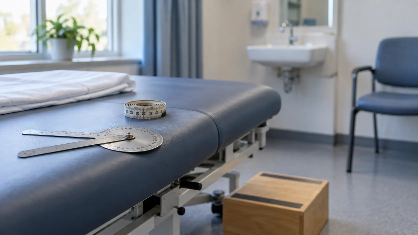
Set-up. Expose both legs to the underwear, with the iliac crests and lower back visible and socks off, and offer a chaperone. You need a goniometer, a tape measure and blocks of known thickness. Examine standing first, then supine on a firm couch, side-lying for the lateral structures and prone for extension and the posterior hip. In a seven-minute station, a workable budget is 60 seconds for gait and standing, 75 for supine inspection and leg length, 45 to feel, 90 to move (including the Thomas test), 100 for special tests and 30 for the neurovascular check, spine and knee. That leaves 20 seconds for the closing sentence.
The other hip is your control. Normal right and left hips have similar ranges of movement, so record every finding as a difference between the two sides rather than as an absolute number.
Look: start with the gait
Watch the patient walk away, turn and come back. Name the pattern and the side:
- Antalgic: stance on the painful side is shortened. This is the commonest finding.
- Trendelenburg: the pelvis drops on the swing side while the weak leg bears weight.
- Compensated Trendelenburg (gluteal lurch): the trunk lurches over the stance leg, which shortens the lever the abductors have to work against. The drop and the lurch are the same deficit, uncompensated and compensated.
- Short-leg: the pelvis and shoulder dip towards the short side as it takes weight.
- Stiff-hip: the limb circumducts because the hip cannot flex during swing.
A stick belongs in the hand opposite the painful hip. With the patient standing, put a thumb on each iliac crest to check for pelvic obliquity. Look for increased lumbar lordosis (the clue to a fixed flexion deformity), gluteal wasting, scars and sinuses.
On the couch, an osteoarthritic hip rests in slight flexion, adduction and external rotation because of anterior capsular and adductor contracture. An acutely effused hip lies in flexion, abduction and external rotation instead, the position of greatest capsular volume. In an older patient who cannot bear weight after a fall, fixed external rotation with shortening is a fractured neck of femur until proven otherwise.
Leg length comes only after the pelvis is square. Check that both anterior superior iliac spines (ASISs) are level before you pick up the tape.
| Measurement pattern | True length (ASIS to medial malleolus) | Apparent length (umbilicus to medial malleolus) | Typical cause |
|---|---|---|---|
| True shortening | Short | Short | Femoral or tibial shortening, arthritic collapse, coxa vara, old fracture |
| Apparent shortening | Equal | Short | Fixed adduction contracture, pelvic tilt from the spine |
| Apparent lengthening | Equal | Long | Fixed abduction contracture |
The Galeazzi test locates the shortening. With the knees flexed to 90° and the heels together, a knee that sits lower and further back points to the femur, while one that is only lower points to the tibia. Blocks under the short leg give the functional answer, but the method is imprecise: the mean error is about half a centimetre. Treat clinical differences under 1 cm as within measurement error.
Feel: temperature, then the three territories
Run the back of your hand over each groin and trochanter. A warm, tender, irritable hip is septic until it has been aspirated. Do not stress-test it.
Feel the femoral pulse at the mid-inguinal point, halfway between the ASIS and the pubic symphysis. Then press the flat of your hand deep, just lateral to the pulse and over the femoral head, while rolling the leg with your other hand. Crepitus, a click or guarding here comes from inside the joint. Palpate the adductor longus origin at the pubic body. With the patient lying on the good side, map the greater trochanter: its tip, the posterior border and the gluteus medius insertion above it. In 65 patients with lateral hip pain, trochanteric palpation was 80% sensitive for MRI-confirmed gluteal tendinopathy, so a trochanter that is not tender makes the diagnosis unlikely.
Ask the patient to show you where it hurts. Pain from inside the joint is deep and hard to point to. The patient often cups the hand over the hip, thumb behind the trochanter and fingers in front reaching towards the groin. This is the C-sign, and it means intra-articular pain even though the hand sits on the side of the hip. One finger on the trochanter suggests the gluteal tendons. Buttock pain points to the posterior capsule, the sacroiliac joint or the lumbar spine.
Move: in degrees, on both sides
Ask for active movement first, then move the hip passively. Watch the patient's face, and use your other hand on the pelvis to catch the point where the movement becomes pelvic rather than hip movement. For flexion, that hand goes flat under the lumbar spine. For abduction and adduction, it rests on the opposite ASIS. Measure rotation in the 90/90 position, remembering that swinging the foot outwards rotates the hip internally. Record each movement as an arc compared with the other side, for example "internal rotation in flexion 10° on the left against 35° on the right".
Osteoarthritis follows the capsular pattern. Internal rotation goes first and is lost most, followed by abduction. Flexion is preserved longest, and loss of extension shows as a fixed flexion deformity. Birrell and colleagues studied new primary-care attenders aged 40 and over with hip pain. Restricted internal rotation was the plane most predictive of radiographic osteoarthritis and flexion the least. Restriction in any one plane was 86% sensitive for moderate disease and 100% for severe disease, but specificity was only 54% and 42%. Restriction in all three planes reversed the trade-off, with specificity of 93% and 88%. Sutlive's preliminary five-item prediction rule includes passive internal rotation of 25° or less. When four or more items were present, the positive likelihood ratio was 24.3 (95% CI 4.4–142.1) in 72 patients, but the rule has not been validated.
FAI loses motion against a hard stop. In FAI, internal rotation in 90° of flexion and flexion itself are reduced, both with a hard, painful end-point, and the abductors are usually normal. In Clohisy's prospective series of 52 symptomatic hips, mean flexion was 97° and mean internal rotation in flexion only 9°. The opposite phenotype has rotation arcs beyond 60°, apprehension and hypermobility. This points to adult hip dysplasia or microinstability, not impingement.
| Hip osteoarthritis | FAI syndrome | Lateral hip pain (gluteal tendinopathy) | |
|---|---|---|---|
| Typical patient | Older adult, start-up pain, a stick | Young active adult, pain on sitting, pivoting and getting out of a car | Middle-aged woman who cannot lie on that side |
| Where it hurts | Groin, often referred to the thigh or knee | Deep groin, with the C-sign | Over the trochanter, can be pointed to with one finger |
| Gait | Antalgic, often with Trendelenburg drop or lurch | Usually near normal | Mild antalgic or Trendelenburg pattern |
| Range of movement | Capsular pattern, internal rotation lost most, fixed flexion deformity | Internal rotation in flexion and flexion lost, with a hard painful end-point | Near full |
| Tests that help | Thomas test, timed Trendelenburg, leg length | FADIR with the patient localising the pain | Trochanteric palpation, 30-second single-leg stance |
| Next step | Weight-bearing AP pelvis and lateral | Imaging to complete the Warwick triad: AP pelvis and Dunn lateral; MR arthrography if the labrum would change management | MRI only if it would change management |
How do you perform the Trendelenburg test?
Stand or kneel behind the patient with the back exposed and a thumb on each posterior superior iliac spine or iliac crest. Ask the patient to stand on the leg being tested and bend the other knee so that the foot clears the floor. Offer your hands for balance. Watch the pelvis, not the shoulders, and hold the position for up to 30 seconds. The test is positive if the pelvis drops on the unsupported side. If the trunk lurches towards the stance leg, that is the compensated sign. A level pelvis that drifts down at 15 or 20 seconds is a positive test of fatigue, so say when the drop happened. Test both sides.
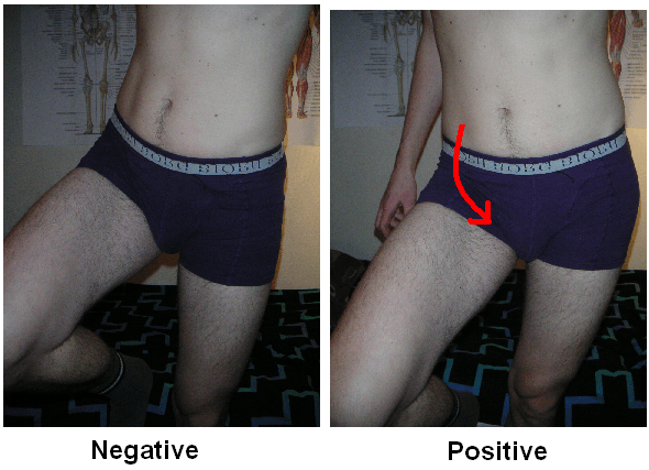
What it means. The abductors cannot produce enough force to keep the pelvis level. Possible causes include pain inhibition from arthritis, a gluteus medius or minimus tendon tear, superior gluteal nerve palsy, coxa vara or a shortened femoral neck lever, and neurological disease. The test shows which side is failing, not why. After a lateral-approach arthroplasty, think of abductor avulsion or nerve injury.
Where the test misleads. Hardcastle and Nade noted that at least four methods of performing the test had been described. They examined 50 normal subjects and 103 patients with hip or spinal disorders and defined a standard method. False positives came from pain, poor cooperation and the rib cage impinging on the iliac crest. False negatives came from the patient using trunk and leg muscles above and below the pelvis, or leaning beyond the standing hip. Do not allow the patient to lean.
How accurate it is. In Bird's study of 24 women with greater trochanteric pain syndrome, assessed with MRI, Trendelenburg's sign was the most accurate of three physical signs for a gluteus medius tear: sensitivity 72.7%, specificity 76.9% and intraobserver kappa 0.676. The same posture can be used with a different endpoint. In Grimaldi's 65 patients, reproduction of lateral hip pain within 30 seconds of single-leg stance had a specificity of 100% and a positive likelihood ratio of about 12 for MRI-confirmed gluteal tendinopathy. That moved a nominal 50% pre-test probability to 98%. The sample was small, so quote the figures with that caveat. In the Trendelenburg test you watch the pelvis; in the single-leg stance test you ask about pain.
The Thomas test: unmasking a fixed flexion deformity
With the patient supine, put your hand flat under the lumbar spine. Flex the opposite hip until the lordosis flattens onto your palm, and ask the patient to hold that knee. If the tested thigh lifts off the couch and cannot be pressed flat, there is a fixed flexion deformity. Measure the angle between the thigh and the couch, then repeat on the other side, because a bilateral deformity looks symmetrical.
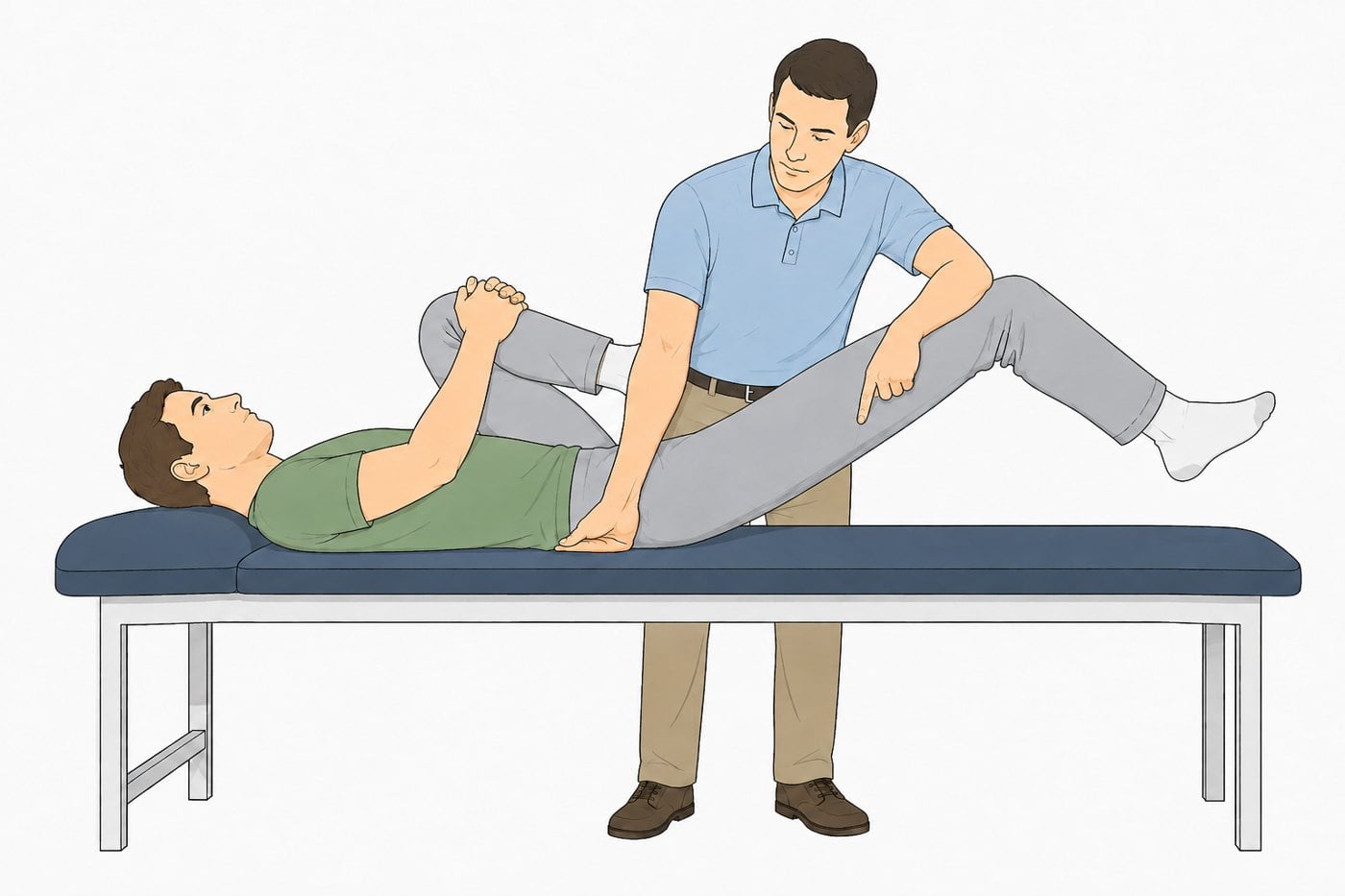
The commonest false negative comes from not flexing the opposite hip far enough, so the lordosis still hides the deformity. A soft couch gives false positives. If the knee of the tested leg hangs flexed, the tight structure may be rectus femoris rather than the capsule. The evidence for the Thomas test covers reliability only. No study has measured its sensitivity or specificity, so report the deformity in degrees and do not attach an accuracy figure to it.
FADIR, FABER and the provocation tests: what does a positive mean?
FADIR. With the patient supine, flex the hip and knee to about 90°. Adduct the hip 15–20° across the midline, then rotate it internally by swinging the ankle outwards. Vary the flexion between 80° and 100° to find the position that hurts. Ask the patient to point: only the familiar deep groin pain counts. Lateral or posterior pain does not.
Reiman's meta-analysis pooled nine studies and found FADIR sensitivity of 0.94 to 0.99, but a diagnostic odds ratio of only 5.71 to 7.82. The authors concluded that the test has screening accuracy only. In Clohisy's series, 88% of symptomatic hips were painful on the test. A negative result still does not clear a convincing history. In Troelsen's 18 hips that had undergone periacetabular osteotomy for dysplasia, checked against MR arthrography, the impingement test had a sensitivity of only 59% and a negative predictive value of 13%. Most painful hips hurt in the FADIR position, including those with arthritis, dysplasia and synovitis.
| Test | Positive finding | What it tells you | Best evidence |
|---|---|---|---|
| Trendelenburg | Pelvis drops on the unsupported side within 30 s | Abductor insufficiency, cause unspecified | Sensitivity 72.7%, specificity 76.9% for a gluteus medius tear (Bird, 24 patients) |
| 30-second single-leg stance | Lateral hip pain within 30 s | Gluteal tendinopathy | Specificity 100%, LR+ about 12 (Grimaldi, 65 patients) |
| Thomas | Thigh lifts off the couch once the lordosis is flat | Fixed flexion deformity | Reliability data only |
| FADIR | Familiar deep groin pain | Intra-articular pain consistent with FAI or labral pathology | Pooled sensitivity 0.94–0.99, DOR 5.71–7.82 (Reiman 2015) |
| FABER | Groin pain in the figure-of-four position | Intra-articular source; posterior pain is not reliably sacroiliac | Sensitivity 0.82, PPV 0.46 against diagnostic injection (Maslowski) |
| Stinchfield | Groin pain on resisted straight-leg raise | Joint loading through iliopsoas | Most specific of four manoeuvres, at only 0.32 (Maslowski) |
| Log roll | Pain or guarding with gentle rotation | An irritable joint: synovitis, effusion, fracture, infection | No accuracy data |
Reiman's earlier meta-analysis of hip tests found only one exception to their weak diagnostic accuracy: the patellar-pubic percussion test for occult fracture, with pooled sensitivity of 95% and specificity of 86%. Every impingement manoeuvre is a screen.
FADIR does not diagnose FAI. The Warwick Agreement defines FAI syndrome as a motion-related clinical disorder of the hip with a triad of symptoms, clinical signs and imaging findings. It was agreed by 22 panel members and one patient from nine countries. A cam on a radiograph without symptoms is not a syndrome, and a positive FADIR without corroborating imaging is not a diagnosis. Our post on hip arthroscopy for FAI and labral repair covers what happens after the triad is complete.
What are the common candidate errors?
- Describing gait without a side. "Antalgic gait" is incomplete; "shortened stance phase on the right" is a finding.
- Measuring leg length before squaring the pelvis. Every number taken after that is wrong, and examiners watch your thumbs on the ASISs.
- Calling movement "restricted" without numbers. The diagnosis lies in the side-to-side difference, especially in internal rotation.
- Missing the fixed flexion deformity because the opposite hip was never flexed enough to flatten the lordosis.
- Calling a wobble a positive Trendelenburg. Give the abductors their 30 seconds, watch the pelvis and say when it dropped.
- Labelling FAI from a positive FADIR, or pushing through guarding and reporting the test as negative.
- Quoting untraceable numbers. Pairings such as "77% sensitive, 41% specific" for FABER do not trace to a verifiable study. Name the study or leave the number out.
- Calling lateral hip pain "bursitis". In Bird's MRI series, trochanteric bursal distension appeared in only two of 24 patients and never without gluteus medius pathology.
- Forgetting the neighbours. The spine refers pain to the groin and the hip refers pain to the knee.
Completing the examination. Say the closing sentence without being prompted: "To complete my examination I would examine the lumbar spine and the knee, perform a neurovascular assessment of both lower limbs, examine the other hip, and request a weight-bearing AP pelvis and a lateral radiograph of the hip." Add MRI or MR arthrography where labral pathology would change management, and ESR, CRP and aspiration where infection is possible. The full station, with every test card and its evidence, is in the comprehensive hip examination guide. For how to structure clinical stations in general, see mastering the clinical examination for orthopaedic exams.
In the exam
Examiners want a synthesis rather than a list of tests: a pattern, the measurements that support it and the investigations you would request.
Frequently asked questions
What is a positive Trendelenburg test?
The patient stands on the tested leg, lifts the other foot and holds that position for up to 30 seconds. The test is positive if the pelvis drops on the unsupported side. If the trunk lurches over the stance leg, that is the compensated sign. Either way, the abductors cannot hold the pelvis level. The cause may be pain inhibition, a gluteal tendon tear, superior gluteal nerve palsy, a short femoral neck lever or neurological disease.
What does a positive FADIR test mean?
A FADIR test is positive when flexion, adduction and internal rotation reproduce the patient's familiar deep groin pain. The pain must be the one the patient came with. It is consistent with FAI syndrome or labral pathology, but arthritis, dysplasia and synovitis also hurt in that position. In a meta-analysis, pooled sensitivity was 0.94–0.99 and the diagnostic odds ratio only 5.71–7.82, so the test screens for the problem but cannot confirm it.
Which hip movement is lost first in osteoarthritis?
Internal rotation. The capsular pattern of the hip restricts internal rotation first and most, then abduction. Flexion is preserved longest, and loss of extension shows up as a fixed flexion deformity. In a UK primary-care study of new attenders aged 40 and over with hip pain, restricted internal rotation was the plane most predictive of radiographic osteoarthritis, and flexion the least predictive.
How do you tell true from apparent leg-length discrepancy?
Square the pelvis first. True length runs from the anterior superior iliac spine to the medial malleolus. Apparent length runs from a midline point, such as the umbilicus, to the medial malleolus. If both are short, the shortening is structural. If true lengths are equal but the apparent length is short, a fixed adduction contracture or pelvic tilt is responsible. A fixed abduction contracture makes the leg look long.
What is the C-sign in hip pain?
The patient shows where the pain is by cupping the hand over the hip, with the thumb behind the greater trochanter and the fingers in front reaching towards the groin, so the hand forms a C. Although the hand sits on the side of the hip, the gesture points to deep, intra-articular pain. A patient who puts one finger on the trochanter is more likely to have gluteal tendinopathy.
References
- Hardcastle P, Nade S. The significance of the Trendelenburg test. J Bone Joint Surg Br 1985;67(5):741-746. DOI (opens in a new tab)
- Bird PA, Oakley SP, Shnier R, Kirkham BW. Prospective evaluation of magnetic resonance imaging and physical examination findings in patients with greater trochanteric pain syndrome. Arthritis Rheum 2001;44(9):2138-2145. PubMed (opens in a new tab)
- Grimaldi A, Mellor R, Nicolson P, et al. Utility of clinical tests to diagnose MRI-confirmed gluteal tendinopathy in patients presenting with lateral hip pain. Br J Sports Med 2017;51(6):519-524. DOI (opens in a new tab)
- Reiman MP, Goode AP, Cook CE, Hölmich P, Thorborg K. Diagnostic accuracy of clinical tests for the diagnosis of hip femoroacetabular impingement/labral tear: a systematic review with meta-analysis. Br J Sports Med 2015;49(12):811. DOI (opens in a new tab)
- Reiman MP, Goode AP, Hegedus EJ, Cook CE, Wright AA. Diagnostic accuracy of clinical tests of the hip: a systematic review with meta-analysis. Br J Sports Med 2013;47(14):893-902. DOI (opens in a new tab)
- Maslowski E, Sullivan W, Forster Harwood J, et al. The diagnostic validity of hip provocation maneuvers to detect intra-articular hip pathology. PM R 2010;2(3):174-181. DOI (opens in a new tab)
- Troelsen A, Mechlenburg I, Gelineck J, et al. What is the role of clinical tests and ultrasound in acetabular labral tear diagnostics? Acta Orthop 2009;80(3):314-318. DOI (opens in a new tab)
- Griffin DR, Dickenson EJ, O'Donnell J, et al. The Warwick Agreement on femoroacetabular impingement syndrome (FAI syndrome): an international consensus statement. Br J Sports Med 2016;50(19):1169-1176. DOI (opens in a new tab)
- Clohisy JC, Knaus ER, Hunt DM, et al. Clinical presentation of patients with symptomatic anterior hip impingement. Clin Orthop Relat Res 2009;467(3):638-644. DOI (opens in a new tab)
- Birrell F, Croft P, Cooper C, et al. Predicting radiographic hip osteoarthritis from range of movement. Rheumatology (Oxford) 2001;40(5):506-512. DOI (opens in a new tab)
- Sutlive TG, Lopez HP, Schnitker DE, et al. Development of a clinical prediction rule for diagnosing hip osteoarthritis in individuals with unilateral hip pain. J Orthop Sports Phys Ther 2008;38(9):542-550. DOI (opens in a new tab)
Study the full topic
Included in Pro
Go deeper with Pro
- Clinical exam guide: comprehensive hip examination
- Clinical exam guide: the young adult hip, FAI and instability
Share this article
Cite this article
OrthoVellum Editorial Team. How to Examine the Hip in the Clinical Exam: OA, FAI and the Trendelenburg Test [Internet]. OrthoVellum; 2026 Oct 12 [cited 2026 Oct 12]. Available from: https://www.orthovellum.com/blog/hip-examination
About this content
Prepared by the OrthoVellum Editorial Team from cited sources, under our editorial policy.
For education and exam preparation; not medical advice or a substitute for clinical judgement and local guidance.
Spotted an error? Tell us; every report is read.
Get new articles by email
New clinical explainers, landmark-trial summaries and exam guidance from the OrthoVellum blog. No account needed; unsubscribe from any email.