Return-to-Play Timing | Type III Controversy | Surgical Decision-Making in the Athlete
- Default to conservative even in elite athletes - the COTS RCT shows no clinical advantage to surgery at 2 years for high-grade injury
- Return-to-play differs sharply by treatment: 6-12 weeks conservative vs 6-9 months after surgery - decisive when counselling mid-season
- Type III is controversial - reserve surgery for failed conservative trial, throwing athletes, and high-demand cosmesis concerns
- Absolute surgical indications remain Type IV-VI (posterior, severe superior, inferior displacement)
- Hook plate requires removal at 3-4 months - a mandatory second surgery to factor into the athlete's season
- “COTS RCT: no clinical advantage to surgery at 2 years for high-grade (III-V) AC injury
- “15-20% re-injury rate in contact sport after conservative treatment; padding reduces but does not eliminate risk
- “Type IV posterior displacement may mimic Type III on AP - axillary view mandatory
- “Throwing mechanics load the AC joint in late cocking - overhead athletes may have persistent horizontal-adduction pain
Overview and Sports Epidemiology
AC joint injuries are among the most common shoulder injuries in contact and collision sports. The injury pattern, the treatment considerations and the return-to-sport timeline all differ significantly from those of the non-athletic population.
Scope. This page covers the athlete: sport-specific epidemiology, biomechanics, in-season management and return to play. For the foundational Rockwood classification, biomechanics and the full general evidence base, see AC joint injuries.
Where the injuries come from. Injury rate, mechanism and grade distribution vary by sport:
- Injury Rate
- 8-12 per 1000 athlete-exposures
- Common Mechanism
- Direct tackle to shoulder
- Type Distribution
- Type II-III most common
- Injury Rate
- 6-10 per 1000 exposures
- Common Mechanism
- Board collision
- Type Distribution
- Type III-V higher rate
- Injury Rate
- 3-5 per 1000 exposures
- Common Mechanism
- Tackling, blocking
- Type Distribution
- Type II-III predominant
- Injury Rate
- 2-4 per 1000 exposures
- Common Mechanism
- Fall onto shoulder
- Type Distribution
- Type III-IV patterns
- Injury Rate
- 2-3 per 1000 exposures
- Common Mechanism
- Direct pressure
- Type Distribution
- Type II-III typical
Mechanism. A direct blow, the fall onto the point of the shoulder with the arm adducted, accounts for 85% of cases. Axial loading, tackling with the shoulder leading, is another; indirect trauma through the outstretched hand is rare (15%). Throwing athletes add a chronic mechanism, repetitive stress, which produces Type I-II sprains.
Athletes have higher rates of Type III-V injury than the general population (60% against 40%), a consequence of the high-energy mechanisms involved.
Career impact. The professional brings contract, insurance and career implications; the college athlete a scholarship and draft prospects; the recreational athlete a question of quality of life and how much activity modification they will tolerate. For all of them the time of season, mid-season or off-season, affects when any surgery can be timed.
Pathophysiology and Mechanisms
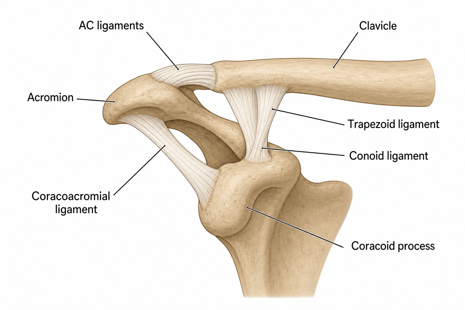
The AC joint is the link between the axial skeleton, through the clavicle, and the appendicular skeleton, through the scapula. In the athlete it is loaded repetitively and at high magnitude.
Horizontal stability comes from the AC ligament complex. The superior AC ligament provides 56% of anteroposterior stability and resists tackle and collision forces; the posterior AC ligament provides 25% and stabilises the throwing follow-through. The capsule contributes proprioception and neuromuscular control, and the deltotrapezial fascia shares the load, which is critical for the overhead athlete.
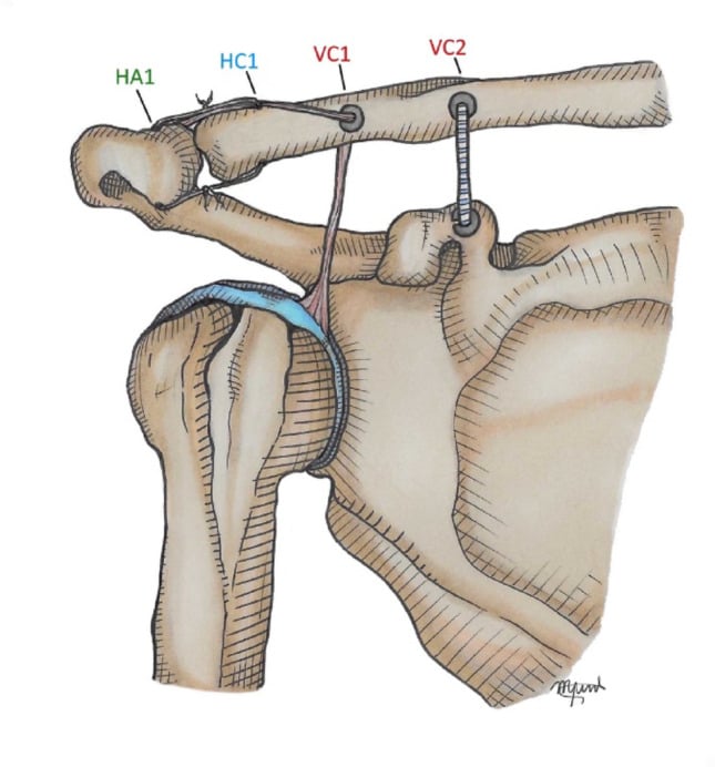
Vertical stability comes from the two coracoclavicular ligaments, and it is vertical stability that fails in high-grade injury. The conoid is medial, 45mm from the AC joint, the stronger of the two and the primary vertical restraint. The trapezoid is lateral, 20mm from the joint, the secondary restraint, and controls rotation. Those two distances are the tunnel positions of an anatomic reconstruction.
How the joint is loaded depends on the sport, and so does the consequence of instability.
Throwing (cricket, baseball, javelin) loads the AC joint differently in each phase:
- Wind-up - minimal AC stress
- Early cocking - scapular positioning, moderate AC load
- Late cocking - peak AC stress: horizontal abduction stretches the posterior AC ligament
- Acceleration - rapid scapular protraction, shear across the AC joint
- Deceleration - high eccentric load on the deltotrapezial complex
- Follow-through - horizontal adduction compresses the joint
Late cocking and deceleration place the maximum stress on the AC joint. Instability alters throwing mechanics and velocity, and the throwing athlete, unlike most contact athletes, may benefit from early surgery.
In throwing athletes, AC instability causes scapular dyskinesis leading to altered glenohumeral mechanics. This can reduce throwing velocity by 10-15% and increase the risk of secondary rotator cuff or labral pathology.
Classification Systems
Rockwood classification. The grade is set by which ligaments have failed and how far the clavicle has displaced, and it drives the entire management decision.
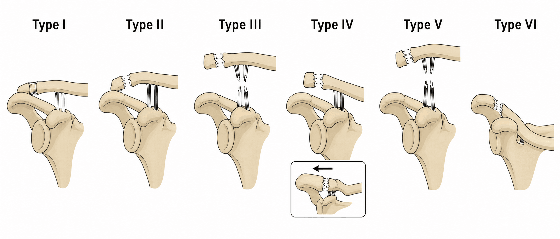
Type III is the controversial grade, and the decision it forces is worked through in Management. Type IV-VI are outside the controversy. They are surgical: none has any role for conservative management, and a high-grade injury can be career-threatening.
Clinical Assessment in Athletes
History. The questions that change management in an athlete:
- Sport and position played
- Level: professional, college or recreational
- Hand dominance, critical in throwing sports
- Time in season: pre-season, mid-season, playoffs or off-season
- Mechanism: direct blow, fall or repetitive
- Previous AC joint injury, which raises the re-injury risk
- Performance impact: pain with specific movements
- Career implications: contract year, scholarship, draft
Examination. The general tests of the AC joint are read here for what they mean to the athlete's sport:
- Technique
- Arm across body to opposite shoulder
- Positive Finding
- AC joint pain
- Athletic Significance
- Predicts pain with tackling, blocking
- Technique
- 90° flexion, 10° adduction, IR, resist downward
- Positive Finding
- AC pain relieved with supination
- Athletic Significance
- Overhead/throwing mechanics affected
- Technique
- Depress lateral clavicle, observe recoil
- Positive Finding
- Clavicle depresses and springs back
- Athletic Significance
- CC ligament integrity - Type III+
- Technique
- Translate clavicle AP with acromion fixed
- Positive Finding
- Increased translation vs contralateral
- Athletic Significance
- AC ligament compromise - instability
- Technique
- Simulated throwing motion (late cocking: horizontal abduction, external rotation)
- Positive Finding
- AC pain during late cocking phase
- Athletic Significance
- Throwing athletes - biomechanics affected
- Technique
- Full push-up position with scapular protraction
- Positive Finding
- AC pain or weakness
- Athletic Significance
- Contact athletes - scapular stability and deltotrapezial function for blocking/tackling
Functional assessment. Compare active range with the other side, test strength in every shoulder movement, look for scapular dyskinesis, and watch the athlete perform the movement patterns of the sport, noting any pain during the simulation. In the overhead athlete, cross-body adduction against resistance predicts pain during sport.
Differential diagnosis. The athlete who has fallen onto the point of the shoulder can have several injuries that mimic or coexist with an AC separation. The cross-body adduction view, the axillary view and a focused examination distinguish them.
- Key distinguishing feature
- Bony tenderness, crepitus; deformity is fracture not joint
- Best test / imaging
- AP/Zanca radiograph shows fracture line, not joint widening
- Why it matters
- Neer IIB pattern is unstable and often needs fixation
- Key distinguishing feature
- Chronic, atraumatic or repetitive load; no acute displacement
- Best test / imaging
- MRI: distal clavicle bone marrow oedema and resorption
- Why it matters
- Managed with load modification, not acute injury pathway
- Key distinguishing feature
- Apprehension, deep joint pain, arm held in fixed position
- Best test / imaging
- Apprehension/relocation test; AP + axillary radiograph
- Why it matters
- Different reduction and rehabilitation pathway
- Key distinguishing feature
- Painful arc, weakness in abduction/ER, night pain
- Best test / imaging
- Jobe/Hawkins tests; ultrasound or MRI
- Why it matters
- Common coexisting pathology in overhead athletes
- Key distinguishing feature
- Pain with O'Brien deep in joint (not localised to AC)
- Best test / imaging
- O'Brien test, MR arthrography
- Why it matters
- O'Brien is positive in both AC and SLAP — localise the pain
- Key distinguishing feature
- Medial-end pain, possible dysphagia/venous congestion
- Best test / imaging
- Serendipity view, CT of SC joint
- Why it matters
- Posterior SC dislocation is a surgical emergency
Investigations in Athletes
Radiographs. An AP and a Zanca view (10-15° cephalic tilt) of both sides. Compare the CC distance with the contralateral side, measure the AC joint width and calculate the displacement percentage.
The axillary lateral is mandatory in every AC joint injury. It is the only view that shows the posterior displacement of a Type IV, which looks identical to a Type III on the AP film.
Stress views, weighted with 5-10 kg, are controversial in athletes. They may help separate a Type II from a Type III, but pain limits them in the acute setting and they rarely change the decision; most centres no longer use them routinely.
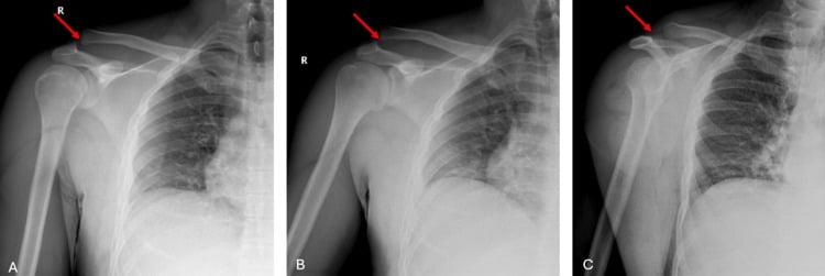
MRI is indicated for chronic AC joint pain (distal clavicle osteolysis), for suspected rotator cuff or labral pathology, for pre-operative planning of a chronic reconstruction, and after failed conservative treatment, when it looks for occult pathology.
Weightlifter's shoulder - chronic repetitive stress causes distal clavicle osteolysis. Seen on MRI as bone marrow oedema and clavicle resorption. Different from acute AC separation. Treatment: activity modification, NSAIDs, consider distal clavicle excision if persistent.
CT is indicated for a suspected fracture of the clavicle, acromion or coracoid, for pre-operative planning of a complex reconstruction, and after failed surgery, to assess the hardware and the bone quality.
Ultrasound can assess AC stability dynamically and measure the CC distance, but it is operator-dependent and not yet standard of care.
The Cross-Body Adduction View and Dynamic Horizontal-Instability Assessment (IIIA vs IIIB)
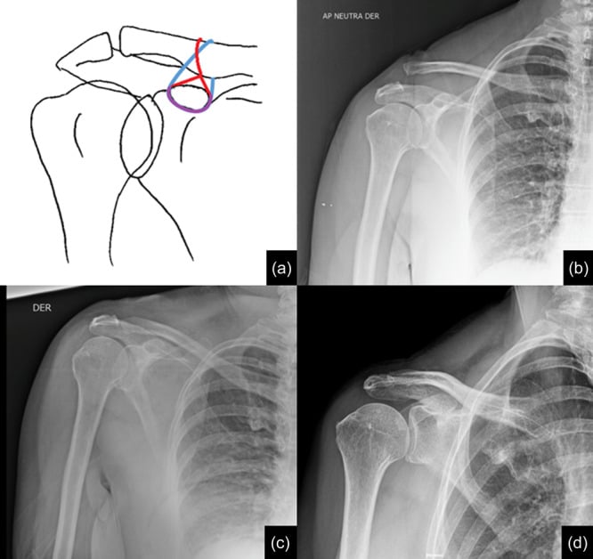
The single most useful refinement in the grade III controversy is to ask whether the joint is horizontally stable. The standard AP and Zanca films show vertical (superior) displacement, but they say nothing about horizontal (antero-posterior) stability, and it is horizontal instability, together with scapular dysfunction, that separates the athlete who may benefit from surgery from the one who will not. Horizontal stability is assessed dynamically, with the cross-body (modified Basamania) adduction view and a focused scapular examination, rather than from the static AP film.
Performing the view. The patient forward-flexes the arm to roughly 90 degrees and adducts it across the body toward the opposite shoulder, and an AP radiograph of the AC joint is taken in that position. In a horizontally stable joint the distal clavicle stays reduced relative to the acromion; in an unstable joint the distal clavicle overrides the acromion, the dynamic shift a static AP film misses. It is the radiographic counterpart of the clinical cross-body adduction test in the examination: pain plus a demonstrable override signals horizontal instability.
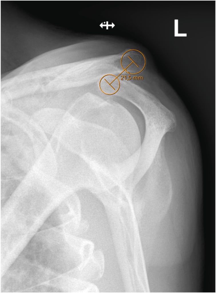
Subdividing grade III (ISAKOS / Beitzel). The view and the scapular examination split the controversial grade into a stable and an unstable subtype:
- Type IIIA (stable)
- No clavicular overriding — joint stays reduced
- Type IIIB (unstable)
- Overriding distal clavicle — dynamic horizontal instability
- Type IIIA (stable)
- No significant scapular dysfunction
- Type IIIB (unstable)
- Therapy-resistant scapular dyskinesis
- Type IIIA (stable)
- Favour non-operative — behaves like a stable injury
- Type IIIB (unstable)
- Consider operative stabilisation, especially in the overhead/throwing athlete
The practical sequence is therefore: confirm the grade on AP/Zanca, exclude a posterior Type IV with the axillary view, then use the cross-body adduction view plus a scapular assessment to decide whether a grade III is a IIIA (trial conservative, as the great majority of athletes are) or a IIIB (the small subgroup in whom early surgery is reasonable). A trial of structured physiotherapy is part of the definition: it is therapy-resistant scapular dysfunction, not dysfunction at presentation, that defines IIIB. Vertical displacement on the AP film alone does not decide the operation.
Management Algorithm
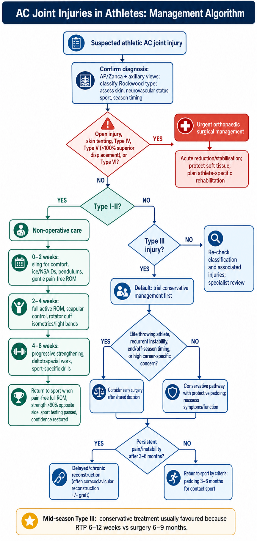
The management decision in the athlete turns on the grade, the demands of the sport, and the time in the season.
The Type III decision. Before 2010 many throwing and contact athletes were operated on, in the belief that anatomic reduction improved outcomes and out of concern for the cosmetic deformity in an elite athlete. The COTS RCT (2015) overturned that default: there is no clinical difference in outcomes at 2 years between hook plate fixation and non-operative treatment, and 80-90% of athletes, contact and throwing athletes included, return to sport conservatively. Surgery carries its own complications (infection, hardware problems, recurrence), and cosmetic deformity does not correlate with performance. Surgery is reserved for the 10-20% who have failed a conservative trial at 3-6 months, and for the specific indications below.
- 1Trial conservativeSling, analgesia, early ROM for 3–6 weeks — the default even in elite athletes.
- 2ReassessPain, strength and sport-specific testing; most athletes are progressing by now.
- 3Escalate selectivelyPersistent symptoms at 3–6 months, or a throwing athlete / off-season timing → reconsider surgery.
The same conservative protocol serves Type I-III athletes.
The aims are pain control and protection of the healing tissues. Training is light cardio only, if tolerated, and nothing for the upper body.
- Ice 20 minutes every 2-3 hours
- NSAIDs (ibuprofen 400mg TDS or naproxen 500mg BD)
- Sling for comfort, weaned by day 3-5
- Avoid provocative movements (cross-body, overhead)
- Pendulum exercises from day 2-3, and pain-free gentle range of motion
The aims are to restore range and begin strengthening: full active range of motion, scapular stabilisation exercises, rotator cuff isometrics and light resistance band work, still avoiding heavy loading and cross-body movement. Training moves to the upper body ergometer and light weights under 5 kg.
A Type I-II athlete who is pain-free through range with strength over 80% progresses to the sport-specific work of Phase 3.
Progressive resistance training, deltotrapezial strengthening (shrugs, rows), sport-specific movement patterns, proprioceptive training, and bench press and push-ups once they are pain-free.
The test is isokinetic strength over 90% of the contralateral side in every movement; a Type II-III athlete who meets it moves to Phase 4.
Full contact practice in protective padding, sport-specific drills at game intensity, an assessment of psychological readiness and a maintenance strengthening programme. The athlete returns to competition only when every return-to-play criterion is met (Postoperative Care and Return to Play).
In-Season Management: Playing Through an AC Injury
The question the athlete and coach ask on the sideline is whether the athlete can keep playing now. That is a separate question from the definitive treatment timeline: it is the short-term, in-season decision about returning to the same match or the same week, and it is answered by grade.
- Same-game / same-week return
- Often possible once pain is controlled and ROM/strength allow safe participation
- How it is managed in-season
- Analgesia, an AC-joint 'donut' pad or strapping, taping; symptom-limited return
- Same-game / same-week return
- Not same-game; default is a short protected period then a graded return within the season
- How it is managed in-season
- Sling for comfort, early ROM, protective padding; return only once pain settles and criteria are met
- Same-game / same-week return
- No — remove from play; these are surgical injuries
- How it is managed in-season
- Protect the limb and arrange surgical management; do not return to contact
Playing through a low-grade injury rests on four principles.
Protect the joint mechanically. A purpose-made AC "donut" pad offloads a direct blow to the point of the shoulder; shoulder padding or strapping is the mainstay for the contact athlete who returns to play.
Control pain without masking a dangerous injury. Simple analgesia and ice allow participation in a low-grade sprain, but pain control must never be used to push a high-grade or undiagnosed (potential Type IV) injury back onto the field.
Re-examine and image before clearing for contact. Exclude a Type IV (axillary view) and a distal clavicle fracture before deciding an athlete can play on; the deformity of a high-grade separation can be mistaken for a "minor" one on the sideline.
Share the decision. The choice to play through, particularly mid-season or in playoffs, is made with the athlete after explaining that continued play carries a re-injury risk and that definitive treatment (almost always conservative) can usually wait for the off-season without compromising the eventual result.
Surgical Techniques for Athletes
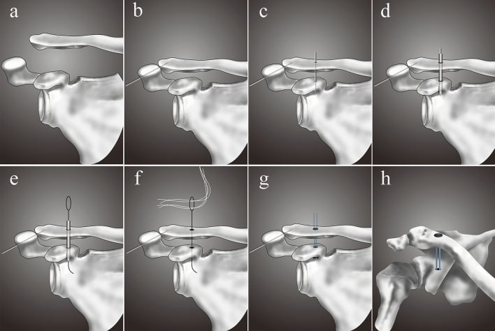
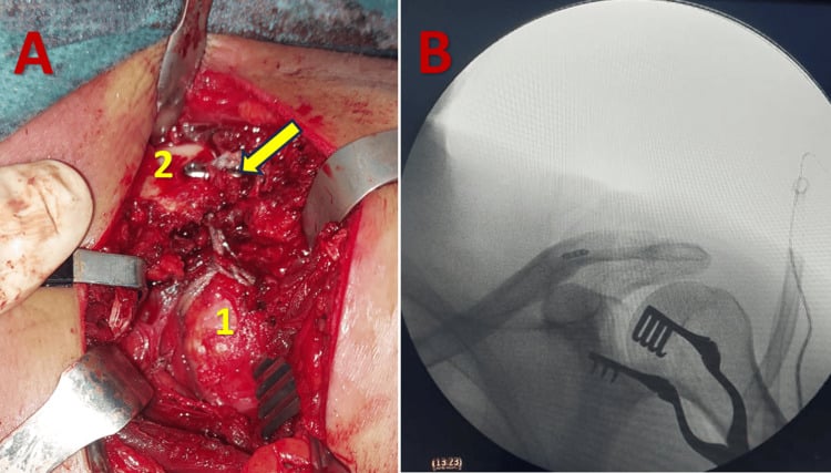
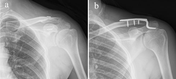
The hook plate is the rigid, technically simple option for the acute injury. In the athlete its indications are an injury under 3 weeks old, a Type III-V in which rapid rigid fixation is wanted, and off-season timing.
Technique. The steps, in order:
- Beach chair position, 30-45° upright
- Superior incision along Langer lines; identify and preserve the lateral cutaneous nerve branches
- Reduce the AC joint with manual pressure
- Hook placed 10-15mm medial to the lateral edge of the clavicle; hook length 15-18mm, to avoid over-stuffing the subacromial space
- 3-4 cortical screws in the clavicle
- Robust deltotrapezial fascia repair with non-absorbable suture (FiberWire, Ethibond)
The deltotrapezial repair is critical for athletes. It provides load sharing and allows an earlier return to play. The rigid fixation of the plate itself allows earlier range of motion than a CC reconstruction.
Complications. Subacromial impingement if the hook is too long or too medial (50% if the plate is not removed), acromion fracture or erosion (5-10%), and loss of reduction after removal (10-15%).
The hook plate is removed at 3-4 months; this is a second operation and it is not negotiable. Plan it for the off-season where possible. After removal allow 4-6 weeks of protection before contact sport. Some loss of reduction after removal is normal and usually asymptomatic.
Complications in Athletes
- Incidence
- 15-20% after conservative treatment (under 10% in the general population)
- Impact on RTP
- Repeat time loss, may require surgery
- Prevention/Management
- Protective padding, strengthening, technique modification
- Incidence
- 10-20% both treatments
- Impact on RTP
- Limits performance, may end career
- Prevention/Management
- Adequate rehab, consider delayed surgery if conservative fails
- Incidence
- 5-10% (usually mild)
- Impact on RTP
- Reduces power in throws, tackles
- Prevention/Management
- Intensive strengthening, scapular stabilisation
- Incidence
- 20-30% chronic instability
- Impact on RTP
- Secondary rotator cuff/labral issues
- Prevention/Management
- Scapular-focused rehab, periscapular strengthening
- Incidence
- 1-2% surgical cases
- Impact on RTP
- Delays RTP 3-6 months, may require hardware removal
- Prevention/Management
- Sterile technique, perioperative antibiotics
- Incidence
- 5-15% depending on technique
- Impact on RTP
- Requires revision surgery, extends RTP
- Prevention/Management
- Appropriate technique, adequate fixation, protect healing
- Incidence
- 2-5% CC reconstruction
- Impact on RTP
- Requires extended immobilisation, delayed RTP
- Prevention/Management
- Avoid excessive tunnel size, adequate bone stock
Career-ending complications are rare, but four may end an athletic career:
- Chronic pain syndrome, after failed conservative and surgical treatment
- Recurrent instability, after multiple surgical failures
- Neurovascular injury, brachial plexus injury during surgery (very rare)
- Chronic shoulder dysfunction from secondary rotator cuff or labral pathology
Cosmetic deformity (a visible bump) occurs in 80% of conservatively treated Type III injuries and 20% of surgical cases. It does NOT correlate with athletic performance or career longevity; multiple studies show elite athletes performing at the same level with the deformity. Counsel athletes accordingly.
Prevention is proper surgical technique, compliance with rehabilitation, a protected return to sport with protective equipment for contact, and monitoring for secondary pathology.
Postoperative Care and Return to Play
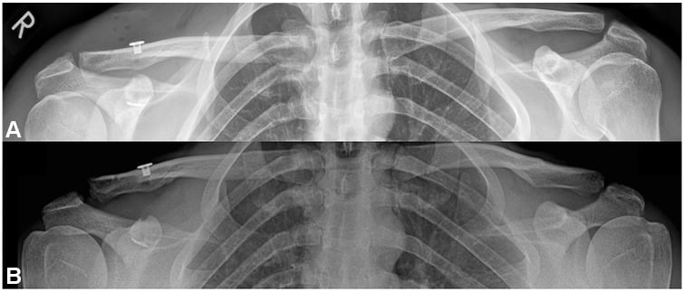
Whether recovery has been conservative or surgical, return to play is gated by the same criteria, not by time alone, and all of them must be met before return to contact or overhead sport:
- Full pain-free range of motion, equal to the contralateral side
- Strength greater than 90% of the contralateral side on dynamometry, in all movements
- Sport-specific testing passed, with drills at game intensity
- Psychological readiness, the athlete's confidence restored
That is the aspiration rather than what the literature actually does, and the numbers are worth knowing. Of 63 studies reporting any explicit return-to-play criterion after AC surgery, time from surgery was the criterion in 95.2%, with 6 months the most frequent timeline (37.8% of studies) and a reported range of 2 to 12 months. Only four studies used a comprehensive functional checklist. So the defensible position is that criteria-based clearance is the principle but is not yet standardised, and the six-month figure quoted so often is a convention derived from what studies report rather than evidence that six months is when tissue is ready.
The practical consequence is that a fixed timeline should never override a failed criterion in either direction: an athlete at six months who cannot pass sport-specific testing is not ready, and one at four months who meets every criterion after a low-demand injury is not obliged to wait out the calendar.
Surgical rehabilitation runs in four phases, each with its own test for progression.
The aims are to protect the repair and prevent stiffness.
Weeks 0-2. Sling full time except for exercises; pendulum exercises only; elbow, wrist and hand range; no active shoulder motion; ice and wound care.
Weeks 2-6. Sling when walking and sleeping; passive range to 90° elevation in neutral rotation; active-assisted range from week 4; pain-free scapular retraction; no lifting and no resisted motion.
Progress when passive range is full, pain is under 3/10 and the wound has healed.
The aims are full active range and the beginning of strengthening: wean the sling by week 6-8, progress to full active range, light resistance (1-2 kg), scapular stabilisation and rotator cuff strengthening, still avoiding heavy lifting and contact.
At 12 weeks range should match the contralateral side, strength should be 50-60% of it, and pain under 2/10 with activities. Progress when active range is full and pain-free and strength is over 50%.
Progressive resistance training, bench press and overhead press (light at first), push-ups and pull-ups, plyometrics late in the phase, sport-specific movement patterns and proprioceptive training.
At 16 weeks strength should be 80-90% of the contralateral side, pain minimal (under 1/10) and functional testing passed. Progress when strength is over 85% and sport simulation is pain-free.
The aim is a safe return to full competition. As a class, non-contact sports return at 4-5 months, contact sports at 6-9 months and throwing sports at 6-12 months with a gradual throwing progression; the sport tabs below give the sport-specific ranges. The return is cleared only when every return-to-play criterion above is met, isokinetic testing is passed, and the surgeon and the athletic trainer have both cleared the athlete.
Return to play by sport. The criteria are common; the timelines and the progressions are not.
In AFL, rugby, ice hockey and American football the conservative return is 2-4 weeks for a Type I, 4-6 weeks for a Type II and 6-12 weeks for a Type III; after a hook plate or a CC reconstruction it is 6-9 months.
The athlete is cleared for contact when contact practice and, for the relevant positions, tackling drills are pain-free, and strength is over 95% of the contralateral side, because the demands of contact are high.
Padding. An AC joint-specific pad of donut design is worn for 3-6 months after a conservative return and 6-12 months after surgery, and may reduce the re-injury risk by 30-40%.
Collision positions (linebacker, prop forward, defenceman) may need a longer RTP timeline and more extensive strengthening than skill positions (quarterback, backs, forwards). Assess demands individually.
Outcomes and Return to Performance
Conservative outcomes fall with the grade, and the cosmetic deformity that follows a Type III is common but functional.
- RTP Rate
- 95-100%
- Time to RTP
- 2-4 weeks
- Same Level Performance
- 95%
- Notes
- Excellent prognosis
- RTP Rate
- 90-95%
- Time to RTP
- 4-6 weeks
- Same Level Performance
- 90%
- Notes
- May have minor cosmetic deformity
- RTP Rate
- 80-90%
- Time to RTP
- 6-12 weeks
- Same Level Performance
- 85%
- Notes
- Cosmetic deformity common but functional
Surgical outcomes by technique:
- Success Rate
- 80-85%
- Same Level Performance
- 80-85%
- Complications
- Removal required, impingement risk
- Success Rate
- 85-90%
- Same Level Performance
- 85-90%
- Complications
- Lower failure vs Weaver-Dunn
- Success Rate
- 80-85% (early data)
- Same Level Performance
- 85%
- Complications
- Limited long-term data
The pooled surgical return-to-sport data are worth quoting exactly, because the two figures usually collapsed into one are different questions. Across 12 studies and 315 patients (mean age 33.8 years), return to any level of sport was 94-100%, but return to pre-injury level was 62-100%, pooling at 89.6% for type III/IV and 86.2% for type V. Nearly everyone gets back on the field; a meaningful minority does not get back to the standard they had. That gap is the honest counselling line for a surgical candidate, and it does not vary much by technique, which is itself the finding, since it removes "this fixation gets you back better" as a reason to choose one construct over another.
Elite athlete performance data show NO significant difference in career longevity, statistics or performance metrics between athletes treated conservatively and surgically for Type III injuries.
Prognosis. The factors that favour a good outcome:
- Young age (under 25 years)
- Type I-II injury
- Compliance with rehabilitation
- Access to specialised sports medicine care
- Off-season injury timing
- Non-throwing sport
The factors that worsen it:
- Type V injury (severe soft tissue disruption)
- Multiple re-injuries
- Associated pathology (rotator cuff, labrum)
- Poor rehabilitation compliance
- In-season injury with a rushed return
- Throwing athlete with a Type III
By sport. In the NFL, 95% return after a Type I-II and 85% after a conservatively treated Type III, with the same performance statistics before and after injury; average time loss is 2-4 weeks for a Type I-II and 6-8 weeks for a Type III. In professional rugby 90% return after a conservatively treated Type III, and re-injury is higher in forwards (20%) than in backs (10%). Among MLB pitchers 80% return after a conservatively treated Type III with a velocity loss under 5%, which is not significant; surgery delays the return but the long-term outcomes are similar, and biomechanics normalise by 6-12 months.
Guidelines, Registries & Global Practice
Global epidemiology
AC joint injury is one of the most common shoulder injuries in young, athletic populations, with a clear male predominance and a peak in the second and third decades of life. The highest rates occur in collision and contact sports (rugby, American football, ice hockey) and in cycling and skiing, typically from a direct fall onto the point of the shoulder. Low-grade injuries (Rockwood I-II) are substantially more frequent than high-grade injuries (III-VI). These patterns are consistent across the major systematic reviews and consensus statements cited below; OrthoVellum reports them as the global picture rather than any single nation's data.
Major guidelines and consensus statements — side by side
- Scope
- Classification of the grade III controversy
- Key position
- Subdivide grade III into stable IIIA (non-operative) and unstable IIIB (consider surgery), using the cross-body adduction view and scapular dysfunction
- Evidence basis
- Level V expert consensus
- Scope
- European diagnosis and treatment
- Key position
- True AP or bilateral Zanca sufficient for diagnosis; cross-body override test for horizontal instability; arthroscopically-assisted suspensory reconstruction without biological augmentation for acute injuries; tendon-graft biological reconstruction for chronic; 3-week acute/chronic cut-off
- Evidence basis
- Modified Delphi (greater than 66.7% agreement)
- Scope
- Grade I-II and most acute grade III
- Key position
- Non-operative first-line for I-III; surgery reserved for IV-VI and selected/failed III
- Evidence basis
- Supported by the COTS Level I RCT
- Scope
- Operative technique
- Key position
- Anatomic CC (and AC) reconstruction preferred over historic non-anatomic transfers; preserve coracoacromial ligament
- Evidence basis
- Expert/biomechanical
Registry note: AC joint injuries are soft-tissue ligamentous injuries treated by repair/reconstruction rather than arthroplasty, so they are not captured by the national joint replacement registries (NJR, AJRR, AOANJRR, SHAR, etc.). The best population-level evidence therefore comes from RCTs (COTS), systematic reviews (Kay, Gawel, Smith) and sports-injury surveillance datasets rather than implant registries.
Practice variation
- Grade III is the genuine area of global disagreement: most North American and UK practice favours an initial non-operative trial (COTS RCT), whereas some European/sports-medicine centres operate earlier on the unstable IIIB subtype, on high-demand throwing athletes, or where horizontal instability is demonstrated.
- Acute vs chronic threshold is set at roughly 3 weeks internationally (ESA-ESSKA): acute injuries are amenable to suspensory/hook fixation, chronic injuries generally require biological graft reconstruction.
- Resource setting: in lower-resource settings, simpler constructs (hook plate, K-wire historically) are used because graft and suspensory implants are costly; in well-resourced centres, arthroscopically-assisted anatomic reconstruction predominates.
- Return-to-play decisions remain mostly time-based (Gawel review) — typically around 6 months after surgery and 6-12 weeks after non-operative care — with a global shift toward criteria-based clearance (full pain-free ROM, strength greater than 90% contralateral, sport-specific testing).
MCQ Practice Points
Q: A 20-year-old college football linebacker sustains an acute Type III AC separation mid-season. What is the most appropriate initial management?
A: Conservative treatment with sling, ice, analgesia, and early ROM. Level I evidence (COTS RCT) shows no clinical difference in outcomes between operative and non-operative high-grade injuries at 2 years. 80-90% of athletes RTP successfully with conservative treatment. Surgery reserved for failed conservative at 3-6 months or specific indications (throwing athletes, off-season timing).
Q: An athlete with Type III AC separation asks about return to contact sport timeline. What is the expected RTP with conservative vs surgical treatment?
A: Conservative: 6-12 weeks. Surgical: 6-9 months. This timeline difference is CRITICAL when counseling athletes, especially mid-season. Surgical treatment commits to season-ending recovery, while conservative allows potential return same season.
Q: What imaging view is ESSENTIAL to differentiate Type III from Type IV AC joint separation?
A: Axillary lateral view. Type IV (posterior displacement) looks identical to Type III on AP view. Axillary view shows clavicle displaced posterior to acromion. Failing to obtain axillary view leads to missed Type IV diagnosis and failed conservative treatment.
Q: Why might throwing athletes have different outcomes with Type III AC separations compared to contact athletes?
A: Throwing biomechanics place high stress on AC joint during late cocking (horizontal abduction) and deceleration phases. AC instability can cause scapular dyskinesis, reduce throwing velocity by 10-15%, and increase risk of secondary rotator cuff or labral pathology. Some throwing athletes may benefit from early surgery, though evidence is mixed.
Q: Why is hook plate NOT suitable for chronic AC joint injuries (over 6 weeks)?
A: Hook plate provides temporary rigid fixation that requires removal at 3-4 months. It works in acute injuries where soft tissue healing is occurring. In chronic injuries, soft tissues have already healed in malaligned position and anatomic CC ligament reconstruction is needed to restore stability. Hook plate would fail in chronic setting.
Q: What Level I evidence exists regarding Type III AC separation management in athletes?
A: The Canadian Orthopaedic Trauma Society (COTS) RCT (2015, J Orthop Trauma) randomised 83 patients with acute complete (grade III-V) dislocations to hook plate vs non-operative treatment. No significant difference in DASH or Constant scores at 6 months, 1 year, or 2 years; the operative group had better radiographic alignment but a higher reoperation rate. Smith et al meta-analysis (2011, J Orthop Traumatol) pooled 6 case series and found no functional difference for grade III (better cosmesis but longer sick leave with surgery). Together these support conservative first-line treatment.
Clinical Decision Scenarios
Practise clinical reasoning and management decisions out loud
“A 22-year-old professional cricket fast bowler sustained a Type III AC joint separation to his bowling arm 2 weeks ago during a match. X-rays show 80% superior displacement with CC distance 18mm (contralateral 12mm). He is currently mid-season with 6 weeks remaining. His team is in playoff contention and wants him to return ASAP. He asks about surgery. How would you manage this case?”
“A 19-year-old university rugby prop forward presents with recurrent right AC joint instability. He had a Type III AC separation 9 months ago treated conservatively. He returned to rugby at 10 weeks wearing protective padding but has had 3 episodes of the shoulder 'giving way' during scrums. X-rays show persistent 60% superior displacement. He is currently off-season with 4 months until pre-season training. What is your management?”
“A 24-year-old professional cyclist presents to you 3 weeks after a high-speed crash. He was diagnosed with a Type III AC separation at another hospital and treated with sling for 2 weeks. He has been trying to return to training but has severe pain and the shoulder 'doesn't feel right.' On examination the lateral clavicle is prominent posteriorly and difficult to palpate anteriorly. You review the images - there is an AP shoulder X-ray showing superior clavicle displacement but no axillary view was done. What is your concern and management?”
TYPE III CONTROVERSY IN ATHLETES
- COTS RCT (Level I): NO clinical difference operative vs conservative at 2 years
- 80-90% of athletes RTP successfully with conservative treatment
- Conservative RTP: 6-12 weeks vs Surgical RTP: 6-9 months
- Default to CONSERVATIVE even in elite athletes
- Consider surgery: throwing athletes, failed conservative 3-6 months, off-season
- Cosmetic deformity does NOT predict athletic performance
RETURN TO SPORT CRITERIA
- Full pain-free ROM (equal to contralateral)
- Strength over 90% contralateral (all movements, isokinetic testing)
- Sport-specific testing passed (throwing velocity, tackling drills, etc)
- Psychological readiness restored
- Protective AC joint padding for contact sports 3-6 months
SPORT-SPECIFIC TIMELINES
- Contact sports (AFL, rugby): 6-12 weeks conservative, 6-9 months surgical
- Throwing sports (cricket, baseball): 8-16 weeks conservative, 6-12 months surgical
- Overhead sports (swimming, tennis): 6-10 weeks conservative, 4-6 months surgical
- Cycling: 4-8 weeks conservative, 4-6 months surgical
- Re-injury rate contact sports: 15-20% (protective padding reduces by 30-40%)
IMAGING ESSENTIALS
- MANDATORY: AP + Zanca + AXILLARY view (all AC injuries)
- Axillary view ONLY way to diagnose Type IV (posterior displacement)
- Type IV looks IDENTICAL to Type III on AP view alone
- Stress views controversial, rarely change management
- MRI for chronic pain (distal clavicle osteolysis, cuff pathology)
SURGICAL DECISION-MAKING
- Absolute indications: Type IV, V, VI
- Relative indications athletes: throwing athletes, failed conservative 3-6 months
- Hook plate: acute only (under 3 weeks), MUST remove 3-4 months
- Anatomic CC reconstruction: chronic injuries, throwing athletes, permanent solution
- TightRope: minimally invasive, no removal, faster RTP, emerging technique
- CRITICAL: robust deltotrapezial fascia repair for all athletes
THROWING ATHLETE SPECIFICS
- Late cocking phase: peak AC joint stress (horizontal abduction)
- AC instability causes scapular dyskinesis, reduces velocity 10-15%
- May benefit from early surgery (controversial, no Level I evidence)
- Interval throwing program: 12 weeks gradual progression
- Monitor velocity, mechanics, pain throughout progression
CRITICAL EXAM TRAPS
- Don't reflexively operate on Type III athletes (80-90% conservative success)
- Don't miss Type IV (no axillary view = missed diagnosis)
- Don't use hook plate for chronic injuries (over 6 weeks)
- Don't forget deltotrapezial repair (critical for athletes)
- Don't rush return (re-injury risk 15-20% if premature)
- Don't ignore cosmetic deformity counseling (expected, doesn't affect function)
Evidence Base
Canadian Orthopaedic Trauma Society (COTS) RCT
- 83 patients with acute complete (Rockwood grade III, IV, V) AC dislocations randomised to hook plate fixation (n=40) vs non-operative treatment (n=43). The non-operative group had significantly better DASH scores at 6 weeks and 3 months, but there was NO significant difference at 6 months, 1 year, or 2 years (mean DASH 5-6, Constant 91-95 in both). Operative group had superior radiographic alignment but a significantly higher reoperation rate.
Smith et al. Meta-analysis of Grade III Management
- Systematic review and meta-analysis of 6 eligible studies (all retrospective case series) on Rockwood grade III dislocation. Operative management gave a better cosmetic outcome (p less than 0.0001) but a longer duration of sick leave (p less than 0.001). There was NO difference in strength, pain, throwing ability, or rate of AC osteoarthritis. Authors concluded there is a lack of well-designed studies to define optimal treatment.
Beitzel et al. ISAKOS Consensus — modified Rockwood (IIIA/IIIB)
- ISAKOS Upper Extremity Committee consensus subdividing the controversial Rockwood grade III. Grade IIIA = stable AC joint with no clavicular overriding on cross-body adduction view and no significant scapular dysfunction (favour non-operative). Grade IIIB = therapy-resistant scapular dysfunction with an overriding clavicle on cross-body adduction view (consider operative). The cross-body (modified Basamania) view and dynamic assessment guide the operative decision.
Kay et al. Return to Sport After Surgery — Systematic Review
- 12 studies (315 patients, mean age 33.8 years) on surgical management of AC dislocation. Return to any level of sport ranged 94-100%; return to pre-injury level ranged 62-100%. Pooled return to pre-injury level was 89.6% for type III/IV injuries and 86.2% for type V. Rates were comparable across injury types and surgical techniques.
Gawel et al. Return-to-Play Criteria — Systematic Review
- 63 studies reporting at least one explicit return-to-play criterion after AC joint surgery. Time from surgery was by far the most common criterion (95.2% of studies), with 6 months the most frequent timeline (37.8%); reported timelines ranged 2-12 months. Only 4 studies (6.3%) used conditional criteria (ROM, strength, clinical/radiographic stability, functional and safety testing, hardware removal).
Shen et al. TightRope vs Hook Plate — Comparative Study
- 35 patients with acute AC dislocation: minimally invasive single-TightRope CC fixation (n=16) vs clavicular hook plate (n=19), mean follow-up 27-30 months. No significant difference in VAS, UCLA, or Constant-Murley scores. Hook plate gave better reduction; TightRope tended toward less pain. Redislocation 1/16 (6.3%) in the TightRope group; acromial osteolysis 1/19 (5.3%) in the hook plate group. The hook plate requires a second operation for removal.