Core Muscle Injury | Rectus–Adductor Aponeurosis | Groin Pain in Athletes
- Athletic pubalgia = core muscle injury at the rectus abdominis–adductor longus aponeurosis of the pubis
- True hernia is absent — the pathology is a tear or attenuation of the aponeurotic plate
- Must differentiate from osteitis pubis (inflammatory), FAI (hip intra-articular), and inguinal hernia
- MRI is the gold standard for imaging — look for rectus tear, adductor tendinopathy, and secondary cleft sign
- Conservative management (6-12 weeks physiotherapy) is first line; surgery reserved for refractory cases
- “Sports hernia = no true hernia on examination — it is a core muscle / aponeurotic injury
- “Pain is groin-based, exertional, improved with rest — differentiates from hip pathology
- “Resisted sit-up or adduction against resistance reproduces pain
- “MRI shows rectus abdominis tear or secondary cleft — do NOT diagnose on ultrasound alone
Athletic pubalgia is NOT a true hernia. It is a core muscle injury involving disruption of the rectus abdominis–adductor longus aponeurotic plate at the pubic symphysis. There is no palpable inguinal hernia on examination.
Three mimics must be excluded: (1) osteitis pubis (pubic symphysis inflammation on imaging), (2) femoroacetabular impingement (hip intra-articular pathology), (3) true inguinal or femoral hernia. Each demands a different treatment pathway.
MRI is the gold standard. Look for rectus abdominis tear at the pubic attachment, adductor longus tendinopathy or partial avulsion, and the "secondary cleft sign" (fluid cleft extending inferolaterally from the symphysis indicating capsular tear). Ultrasound is operator-dependent and less sensitive.
Conservative first, surgery for refractory cases. A minimum 6-12 week structured physiotherapy programme (core stabilisation, adductor strengthening, lumbopelvic control) is first line. Surgery (open or laparoscopic repair, with or without adductor release) is for patients who fail rehabilitation.
- Diagnosis
- MRI: rectus tear or secondary cleft sign
- Treatment
- Conservative rehab 6-12 weeks first
- Key Pearl
- Most resolve with structured physiotherapy
- Diagnosis
- MRI confirmed core muscle injury, no hip OA
- Treatment
- Surgical repair (laparoscopic mesh or open)
- Key Pearl
- Return to sport in 6-8 weeks post-surgery
- Diagnosis
- MRI: pubic bone marrow oedema = osteitis pubis
- Treatment
- Anti-inflammatories, activity modification, rehab
- Key Pearl
- Not a surgical condition — differentiate carefully
GROINAthletic Pubalgia Differential Diagnosis
Hook:Think GROIN for the five key differentials in the athlete with groin pain!
Overview and Epidemiology
Athletic pubalgia is one of the most common causes of chronic groin pain in athletes and is frequently misdiagnosed as a groin strain, osteitis pubis, or hip pathology. The term "sports hernia" is misleading — there is no true hernia. The pathology is a tear or attenuation of the aponeurosis between the rectus abdominis and adductor longus at their pubic attachment. Accurate diagnosis requires MRI and a structured clinical assessment to differentiate from intra-articular hip pathology, which demands a completely different treatment pathway. Up to 30-50% of athletes with chronic groin pain have core muscle injury.
- Incidence: 0.5-6% of athletes per year in kicking and cutting sports
- Sex: Overwhelmingly male (ratio approximately 10:1)
- Age: Peak 20-35 years (competitive sporting years)
- Sports: Soccer, rugby, Australian rules football, ice hockey, tennis
- Bilateral: 20-30% of cases
- Time loss: Average 6-12 weeks from sport
- Recurrence: Up to 20% without structured rehabilitation
- Career threat: Professional athletes may require surgical intervention
- Misdiagnosis: Average delay to correct diagnosis is 4-8 months
- Cost: Significant in professional sport due to time away from play
Pathophysiology
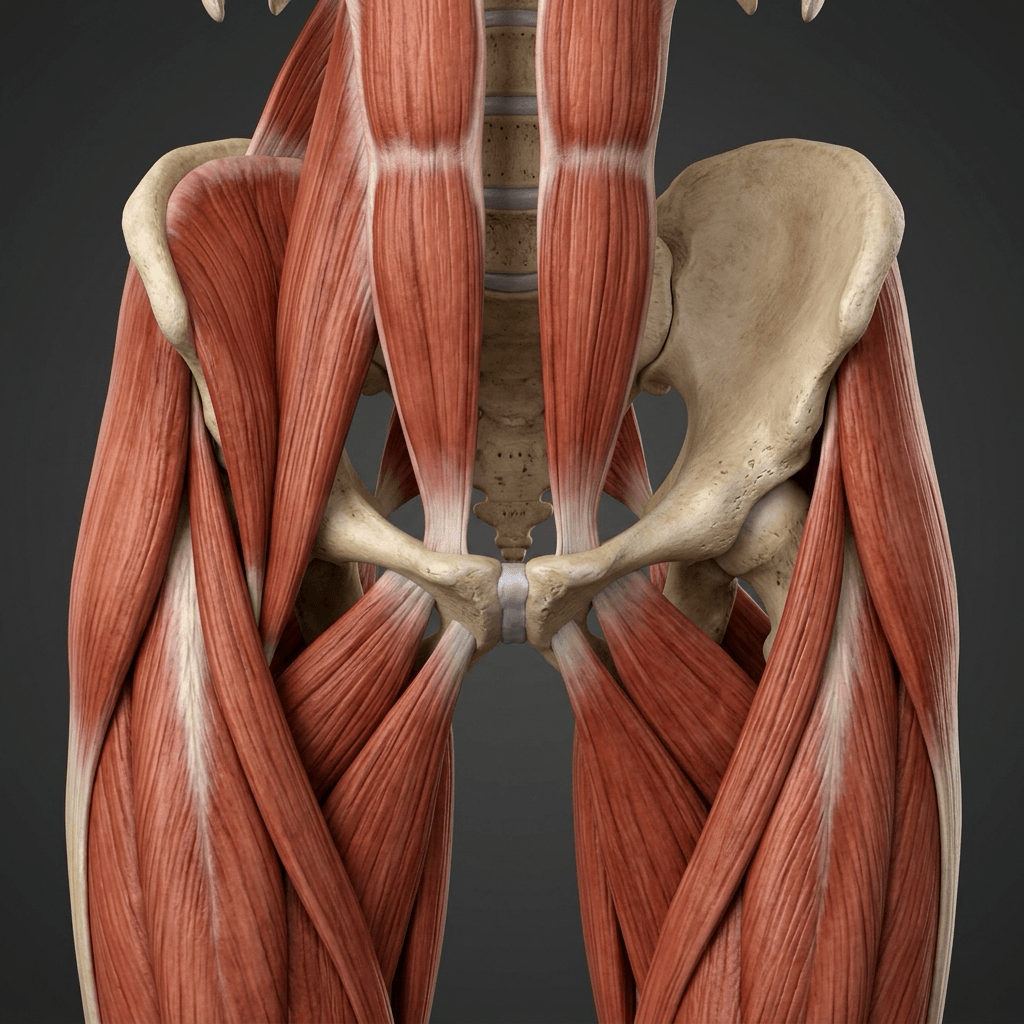
The rectus abdominis and adductor longus share a common aponeurotic attachment on the anterior pubis. The rectus abdominis inserts on the pubic crest and symphysis, while the adductor longus originates from the inferior pubic ramus. These two structures form a functional unit — the pubic aponeurotic plate. During twisting, kicking, and cutting movements, opposing forces (rectus pulling superiorly, adductor pulling inferiorly) create shear stress at this aponeurosis. Repetitive loading causes microtears, attenuation, or frank disruption. There is no true hernia — the posterior inguinal wall may be weakened but no visceral structure protrudes.
- Attachment to Pubis
- Pubic crest and symphysis anteriorly
- Function
- Trunk flexion, intra-abdominal pressure
- When Injured
- Tear or attenuation at pubic attachment
- Attachment to Pubis
- Inferior pubic ramus origin
- Function
- Hip adduction, stabilisation in running
- When Injured
- Tendinopathy or partial avulsion
- Attachment to Pubis
- Pubic tubercle and pectineal line
- Function
- Posterior wall of inguinal canal
- When Injured
- Attenuation contributes to posterior wall weakness
- Attachment to Pubis
- Contributes to inguinal canal anterior wall
- Function
- Trunk rotation, abdominal wall integrity
- When Injured
- Thinning or tear near superficial ring
Normal: Rectus and adductor share balanced load at pubic aponeurosis
Repetitive overload: Kicking, twisting, sprinting creates shear forces
Microtears develop: At rectus insertion, adductor origin, or both
Aponeurotic plate weakens: Progressive loss of integrity
Result: Pain with any activity that loads the pubic aponeurosis (coughing, sit-ups, sprinting)
Overlapping anatomy: Pubic symphysis, hip joint, and inguinal canal are within centimetres
Referred pain: Obturator nerve irritation from adductor pathology refers to medial thigh
Hip pathology coexists: Up to 25% have concurrent FAI or labral pathology
Osteitis pubis overlap: Pubic bone marrow oedema can be present in both conditions
Examination limited: Athletes often have multiplaner groin pain that is hard to localise
Classification and Types
Classification by Anatomical Structure Involved
- Structure Injured
- Inferior rectus at pubic attachment
- MRI Findings
- Rectus oedema or disruption, secondary cleft
- Prevalence
- Most common pattern
- Structure Injured
- Proximal adductor longus tendon
- MRI Findings
- Tendon thickening, peritendinous oedema
- Prevalence
- Very common, often coexists
- Structure Injured
- Both structures at pubic aponeurosis
- MRI Findings
- Bilateral or unilateral multi-structure changes
- Prevalence
- Common in chronic presentations
- Structure Injured
- Conjoint tendon or transversalis fascia
- MRI Findings
- Posterior wall bulge on dynamic ultrasound
- Prevalence
- Less common as isolated finding
The anatomical classification guides surgical planning: isolated adductor pathology may respond to adductor release, while rectus tears may require direct repair or mesh reinforcement.
Clinical Assessment
- Mechanism: Twisting, cutting, sprinting, kicking — repetitive overload
- Pain location: Deep groin, low abdominal, perineal radiation
- Timing: Activity-related, improves with rest, recurs on return
- Provocation: Coughing, sneezing, sit-ups, resisted adduction
- Previous treatment: Failed courses of physiotherapy, NSAIDs, or rest
- Inspect: No visible swelling, no inguinal lump (differentiates from true hernia)
- Palpate: Tenderness over pubic tubercle, rectus insertion, adductor origin
- Resisted sit-up test: Reproduces groin pain (sensitive for rectus involvement)
- Resisted adduction test: Pain with adduction against resistance (adductor pathology)
- Squeeze test: Adductor squeeze at 0, 45, and 90 degrees of hip flexion
Step 1 — Exclude true hernia: Examine standing and coughing for inguinal or femoral hernia. If palpable lump, refer to general surgery — this is NOT athletic pubalgia.
Step 2 — Assess adductors: Resisted adduction in neutral, 45-degree flexion. Pain localised to adductor longus origin suggests adductor tendinopathy.
Step 3 — Assess rectus abdominis: Resisted bilateral sit-up, resisted unilateral hip flexion with trunk rotation. Pain at the pubic insertion suggests rectus involvement.
Step 4 — Assess hip joint: FADIR (flexion, adduction, internal rotation) test for FAI. FABER test for sacroiliac or hip pathology. If positive, order hip-specific imaging.
Step 5 — Assess pubic symphysis: Direct palpation of symphysis. Marked tenderness with bilateral symptoms suggests osteitis pubis.
- Technique
- Supine, resisted trunk flexion from partial sit-up
- Positive Finding
- Pain at pubic insertion of rectus abdominis
- Significance
- Suggests rectus abdominis involvement
- Technique
- Adduction against resistance at 0, 45, and 90 degrees
- Positive Finding
- Pain at adductor longus origin
- Significance
- Suggests adductor pathology
- Technique
- Cough while examiner palpates the inguinal canal
- Positive Finding
- Pain without palpable impulse (no hernia)
- Significance
- Supports posterior wall weakness
- Technique
- Hip flexed 90 degrees, adducted and internally rotated
- Positive Finding
- Deep anterior groin or hip pain
- Significance
- Suggests FAI or labral tear — investigate hip
- Key Features
- Unilateral, exertional groin pain, no hernia
- Discriminating Finding
- Resisted sit-up reproduces pain, no palpable lump
- Imaging
- MRI: rectus tear, secondary cleft sign
- Key Features
- Bilateral pubic pain, insidious onset
- Discriminating Finding
- Direct symphysis tenderness, bilateral symptoms
- Imaging
- MRI: pubic bone marrow oedema, symphyseal sclerosis
- Key Features
- Deep groin pain, prolonged sitting or hip flexion
- Discriminating Finding
- Positive FADIR test, limited internal rotation
- Imaging
- MRI/CT: cam or pincer morphology, labral tear
- Key Features
- Acute onset, localised to adductor longus
- Discriminating Finding
- Sudden injury, tenderness along muscle belly
- Imaging
- MRI: muscle oedema, tear at musculotendinous junction
- Key Features
- Groin lump, cough impulse
- Discriminating Finding
- Palpable reducible mass
- Imaging
- Ultrasound: hernia sac with Valsalva
- Key Features
- Activity-related groin pain, night pain
- Discriminating Finding
- Weight-bearing pain, positive hop test
- Imaging
- MRI: fracture line, periosteal oedema
Up to 25% of athletes with athletic pubalgia have concurrent FAI. If the FADIR test is positive or the patient has limited internal rotation of the hip, hip-specific imaging (MRI with hip protocol) is mandatory before any groin surgery. Operating on the pubic aponeurosis when the primary pain generator is the hip will fail. The two conditions can coexist and may require sequential treatment — address the intra-articular hip pathology first.
The GROIN mnemonic lists "nerve" but the differential otherwise omits it - yet nerve entrapment is a recognised, examinable cause of athletic groin pain that is missed if you only think muscle, bone and joint:
- Obturator nerve entrapment: classically exercise-induced medial-thigh/groin pain with adductor weakness and medial-thigh paraesthesia that builds with exertion and eases with rest - caused by entrapment of the obturator nerve by a thickened fascia over the adductor brevis. It mimics adductor-related pain but has a neuropathic quality and a sensory component, and is confirmed with EMG/nerve studies; treatment is fascial release/neurolysis of the obturator nerve, not aponeurotic repair.
- Ilioinguinal and iliohypogastric nerve entrapment: cause neuropathic lower-abdominal/groin (and scrotal/labial) pain, often after prior hernia repair or lower-abdominal surgery (or from a thickened external oblique aponeurosis), with a positive response to a local-anaesthetic nerve block.
- Genitofemoral nerve: neuropathic groin/scrotal/anterior-thigh pain, again often post-surgical.
Exam point: in the athlete with a neuropathic-quality, dermatomal or sensory groin pain (especially exertional medial-thigh pain with adductor weakness, or pain after previous groin surgery), think nerve entrapment - obturator, ilioinguinal/iliohypogastric or genitofemoral - diagnosed with nerve blocks/EMG and treated by neurolysis, a completely different pathway from core-muscle-injury repair.
PUBICAthletic Pubalgia Clinical Features
Hook:PUBIC — the five hallmarks of athletic pubalgia that separate it from hip pathology!
Investigations
Imaging Protocol
Sequences: Coronal, sagittal, and axial T1 and T2 fat-suppressed sequences through the pubic symphysis and hips
Look for: Rectus abdominis tear or oedema at pubic attachment, adductor longus tendinopathy or partial avulsion, secondary cleft sign (fluid cleft inferolateral to symphysis indicating capsular disruption), pubic bone marrow oedema pattern
Clinical correlation: MRI is the gold standard with sensitivity reported at 85-95% for core muscle injury
Indication: Positive FADIR test, limited hip internal rotation, or deep anterior groin pain
Look for: Cam or pincer morphology, labral tear, chondral damage, ligamentum teres pathology
Important: Hip pathology may coexist with athletic pubalgia — both may require treatment
Indication: Assess for posterior wall deficiency, true inguinal hernia
Technique: Valsalva manoeuvre during real-time scanning of the inguinal canal
Limitation: Operator-dependent, less sensitive for rectus or adductor pathology compared to MRI
Role: Excludes true hernia; does NOT replace MRI for diagnosis
Indication: Patients unable to undergo MRI (pacemaker, metalwork artefact)
Limitation: Poor soft tissue detail compared to MRI — less useful for aponeurotic assessment
Role: Excludes bony pathology (stress fracture, pubic symphysis sclerosis)
The secondary cleft sign on MRI is the most specific finding for athletic pubalgia. It appears as a curvilinear fluid signal extending inferolaterally from the pubic symphysis, representing a tear of the aponeurotic attachment. It is seen in approximately 70-80% of surgical cases. Pubic bone marrow oedema is non-specific and can be present in both athletic pubalgia and osteitis pubis — do not rely on this finding alone to differentiate the two conditions.
The groin packs several potential pain sources within centimetres, and athletic pubalgia, FAI, osteitis pubis and adductor pathology frequently coexist - so when MRI shows multiple abnormalities (or symptoms and imaging do not match), image-guided diagnostic local-anaesthetic injections are a powerful tool to identify the dominant generator before committing to surgery:
- Intra-articular hip injection: significant (greater than half) pain relief implicates the hip joint (FAI/labrum) as the primary source and predicts a good response to hip arthroscopy - operate on the hip first.
- Pubic symphysis / secondary-cleft injection: relief localises pain to the pubic aponeurotic plate / symphysis (athletic pubalgia / osteitis pubis).
- Adductor enthesis injection: relief localises to adductor-related pathology.
Each injection should be ultrasound- or fluoroscopy-guided with documented pre- and post-injection pain scores during a provocative manoeuvre. The principle mirrors the "treat the source, not the scan" rule: a positive hip injection in a patient with both cam morphology and a rectus tear tells you to address the hip first.
Exam point: when groin imaging shows more than one abnormality, use targeted image-guided anaesthetic injections (hip vs symphysis vs adductor) with recorded pain response to identify and treat the dominant pain generator - this is the practical safeguard against the number-one error of operating on the wrong source.
Management Algorithm
Conservative Management (First Line for All Patients)
Goal: Restore lumbopelvic stability, adductor strength, and sport-specific function through structured rehabilitation
Rehabilitation Protocol
Pain control: Relative rest from provocative activities, NSAIDs, cryotherapy
Protected loading: Isometric adductor squeezes (pain-free), transversus abdominis activation
Manual therapy: Hip joint mobilisation, adductor soft tissue work, symphysis mobilisation if indicated
Goal: Pain reduction, baseline activation of core musculature
Progressive loading: Isotonic adductor exercises (Copenhagen adductor programme), single-leg stability work
Core integration: Dead bugs, bird dogs, pallof press with progressive resistance
Running: Straight-line jogging if pain-free
Goal: Pain-free activities of daily living, adductor-to-abductor strength ratio approaching 80-90%
Sport-specific loading: Cutting, twisting, sprinting drills with progressive intensity
Change of direction: Figure-of-eight running, lateral shuffles
Kicking progression: Progressive kicking distance and power for kicking-sport athletes
Goal: Pain-free sport-specific movements at moderate intensity
Full training: Return to full team training, initially non-contact
Match play: Return to competitive sport after pain-free full training
Criteria: Bilateral adductor squeeze test pain-free, adductor strength greater than 90% of contralateral side, sport-specific functional tests passed
Prevention: Ongoing adductor and core maintenance programme
The Copenhagen adductor exercise programme has level 1 evidence for both prevention and treatment of groin problems in athletes. The programme involves isometric and dynamic adductor exercises performed in a sideline position with progressive loading. Studies demonstrate a reduction in groin injuries by approximately 40% when used as a prevention programme. This is the single most evidence-based intervention for athletic groin injuries.
CORESConservative Management Phases
Hook:CORES — the five phases of conservative rehab for athletic pubalgia!
Complications
- Incidence
- 5-15% of surgical cases
- Risk Factors
- Incorrect diagnosis, concurrent FAI, incomplete repair
- Management
- Reassess for missed hip pathology, repeat MRI, revision surgery rarely
- Incidence
- 15-30% without ongoing prevention
- Risk Factors
- Inadequate rehabilitation, premature return to sport
- Management
- Extended rehab programme, adductor prevention exercises
- Incidence
- Variable, often temporary
- Risk Factors
- Complete tenotomy, inadequate rehab
- Management
- Progressive adductor strengthening, most resolve by 6 months
- Incidence
- 1-3% (surgical cases)
- Risk Factors
- Open technique, obesity, diabetes
- Management
- Standard surgical wound management, antibiotics if indicated
- Incidence
- 2-5% of elite athletes
- Risk Factors
- Multiple failed treatments, incorrect diagnosis, psychological factors
- Management
- Multi-disciplinary pain management, psychological support, career counselling
The most common cause of "failed athletic pubalgia surgery" is that the diagnosis was wrong. The true pain generator was FAI, a labral tear, osteitis pubis, or nerve entrapment. Always complete a thorough hip examination and MRI hip protocol before committing to pubic surgery. If any doubt exists about the primary pain source, consider diagnostic injections (hip joint vs pubic symphysis) to differentiate.
Outcomes and Prognosis
- Expected Outcome
- 70-90% resolution of symptoms
- Return to Sport
- 6-12 weeks with criteria-based progression
- Long-term Prognosis
- Excellent if compliance maintained and prevention continued
- Expected Outcome
- 85-95% return to competitive sport
- Return to Sport
- 6-8 weeks post-surgery
- Long-term Prognosis
- Good long-term outcomes with maintained core conditioning
- Expected Outcome
- Variable, depends on hip pathology severity
- Return to Sport
- 3-6 months (longer rehabilitation)
- Long-term Prognosis
- Dependent on hip joint preservation success
- Expected Outcome
- Persistent symptoms, psychological impact
- Return to Sport
- Delayed or absent
- Long-term Prognosis
- Guarded — requires multidisciplinary reassessment
Best prognosis: Acute presentation, accurate early diagnosis, compliant structured rehabilitation, no concurrent hip pathology, male sex
Poor prognosis: Chronic symptoms greater than 6 months, multiple previous failed treatments, concurrent FAI or labral pathology, bilateral symptoms suggesting osteitis pubis
Key threshold: 12 weeks of conservative rehabilitation — if no meaningful improvement by this point, surgical consideration is appropriate. Earlier surgery may be considered in professional athletes with MRI-confirmed pathology.
Guidelines, Registries & Global Practice
- Incidence: 0.5-6% of athletes per year in kicking and cutting sports
- Highest rates: Professional soccer (up to 10-18% career incidence), rugby, ice hockey
- Sex: Overwhelmingly male; female athletic pubalgia is under-recognised and may present differently
- Geographic variation: Higher reported rates in countries with strong football and rugby programmes (UK, Europe, Australia, Scandinavia)
- Economic impact: Significant in professional sport — average 6-12 weeks lost from training and competition
- High-resource: MRI-first diagnostic pathway, structured physiotherapy, surgical options (laparoscopic or open), multidisciplinary team
- Limited-resource: Clinical diagnosis with ultrasound, extended rehabilitation as sole treatment, limited surgical availability
- Universal principle: Structured active rehabilitation is the foundation regardless of resources — the Holmich protocol and Copenhagen programme require no equipment
- Prevention: The Copenhagen adduction programme is free, requires no equipment, and has the strongest evidence for groin injury prevention
- Diagnosis Emphasis
- Clinical examination categorised by anatomical entity; MRI for all persistent cases
- Conservative Treatment
- Minimum 6-12 weeks structured exercise programme before surgical consideration
- Surgical Indication
- Only after failed rehabilitation; multi-disciplinary assessment mandatory
- Diagnosis Emphasis
- MRI pelvis with athletic pubalgia protocol; exclude FAI with hip-specific MRI
- Conservative Treatment
- Physiotherapy-led rehabilitation; Copenhagen programme recommended
- Surgical Indication
- Laparoscopic or open repair in specialist centres
- Diagnosis Emphasis
- MRI pelvis and hip; dynamic ultrasound to exclude true hernia
- Conservative Treatment
- Progressive rehabilitation 6-12 weeks; consider injection therapy
- Surgical Indication
- Open or laparoscopic repair; adductor release if concurrent tendinopathy
- Diagnosis Emphasis
- Assess for bony involvement (pubic ramus stress fracture); MRI essential
- Conservative Treatment
- Rehabilitation first; address biomechanical and training load factors
- Surgical Indication
- Surgical repair principles similar across approaches; expertise matters more than technique
There is no dedicated arthroplasty or implant registry relevant to athletic pubalgia, as the condition involves soft tissue repair without implants. The evidence base is dominated by large case series (Meyers) and a small number of RCTs (Holmich, Haroy). The Doha Agreement (2014) provides the most widely accepted consensus terminology and classification framework, categorising groin pain by anatomical entity: adductor-related, iliopsoas-related, inguinal-related, pubic-related, and hip-related. Athletic pubalgia falls primarily within the inguinal-related and pubic-related categories.
Record in every athlete with groin pain:
- Duration and nature of symptoms (acute vs insidious, unilateral vs bilateral)
- Hernia examination performed (standing, coughing) and result
- Hip examination (FADIR test, internal rotation range) and result
- MRI pelvis findings (rectus tear, secondary cleft, adductor pathology, bone marrow oedema pattern)
- Hip MRI if FADIR positive
- Rehabilitation programme details (duration, compliance, exercises used)
- FADIR test result before any surgical decision
Missed FAI leading to failed athletic pubalgia surgery is a recurring source of poor outcomes and medicolegal claims worldwide.
Controversies & Areas of Uncertainty
No high-quality RCTs directly compare surgical repair with continued conservative management beyond 12 weeks. Surgical evidence comes from large case series with inherent selection bias. The threshold for surgery varies between centres, with some advocating earlier intervention in elite athletes and others insisting on a minimum 6-month rehabilitation trial.
Both techniques report 85-95% return-to-sport rates, but no head-to-head RCTs exist. Laparoscopic repair is preferred by general surgeons (mesh reinforcement of posterior wall), while open repair is preferred by some sports hernia specialists (direct aponeurotic reinforcement). Choice is currently driven by surgeon expertise rather than evidence.
Partial adductor longus release is increasingly favoured over complete tenotomy to minimise post-operative adductor weakness. However, no comparative trials exist, and the long-term functional impact of complete tenotomy in elite athletes remains debated.
The terms "sports hernia," "athletic pubalgia," "core muscle injury," and "inguinal disruption" are used interchangeably, creating confusion. The Doha Agreement (2014) attempted to standardise terminology, but adoption is inconsistent. "Core muscle injury" is increasingly preferred as it accurately describes the pathology without implying a hernia.
MCQ Practice Points
Q: What is athletic pubalgia? A: A core muscle injury involving disruption of the rectus abdominis–adductor longus aponeurotic plate at the pubic symphysis. It is NOT a true hernia — no visceral structure protrudes and no palpable inguinal mass is present. The pathology is a tear or attenuation of the aponeurosis due to repetitive shear forces from twisting and kicking.
Q: What is the gold standard imaging for athletic pubalgia? A: MRI of the pelvis with an athletic pubalgia protocol. Key findings include: rectus abdominis tear or oedema at the pubic attachment, adductor longus tendinopathy or partial avulsion, and the secondary cleft sign (fluid cleft extending inferolaterally from the symphysis). Dynamic ultrasound can exclude true hernia but is less sensitive for core muscle injury.
Q: How do you differentiate athletic pubalgia from osteitis pubis? A: Athletic pubalgia is typically unilateral, with pain reproduced by resisted sit-up or adduction, and MRI shows a rectus tear or secondary cleft sign. Osteitis pubis is typically bilateral, with direct symphysis tenderness, and MRI shows marked pubic bone marrow oedema and symphyseal sclerosis. Osteitis pubis is managed with rest and anti-inflammatories — it is NOT a surgical condition.
Q: What is the first-line treatment for athletic pubalgia? A: Structured physiotherapy for 6-12 weeks comprising: pain control, lumbopelvic stabilisation (transversus abdominis and multifidus), progressive adductor strengthening (Copenhagen adduction programme), and sport-specific functional rehabilitation. The Copenhagen programme has level 1 evidence for treatment and prevention of groin injuries. Surgery is reserved for patients who fail conservative management.
Q: When is surgery indicated for athletic pubalgia? A: Surgery is indicated after failure of a minimum 6-12 weeks of structured, supervised rehabilitation in a compliant athlete with MRI-confirmed core muscle injury and excluded hip pathology. Options include laparoscopic mesh repair, open repair, or combined repair with adductor release. Return-to-sport rates are 85-95% in experienced centres. The number one cause of surgical failure is incorrect diagnosis (missed FAI).
Exam Viva Scenarios
Practise clinical reasoning and management decisions out loud
“A 24-year-old professional soccer player presents with 8 weeks of progressive left-sided groin pain, worse during kicking and sprinting. He has no palpable inguinal hernia. Resisted sit-ups reproduce his pain. X-rays of the pelvis are normal. What is your diagnosis, investigation, and management plan?”
“A 28-year-old elite rugby player has completed 14 weeks of supervised rehabilitation for MRI-confirmed athletic pubalgia (rectus abdominis tear with secondary cleft sign). He continues to have activity-limiting groin pain. His FADIR test is negative and hip MRI shows no FAI. He is desperate to return to competitive rugby. What are your surgical options and how do you counsel him?”
Key Concept
- Athletic pubalgia = core muscle injury (NOT a true hernia)
- Tear or attenuation of rectus abdominis–adductor longus aponeurosis at pubis
- Repetitive shear forces from twisting, kicking, sprinting in athletes
- Male predominance (10:1), peak age 20-35, kicking and cutting sports
Diagnosis
- MRI pelvis is gold standard — look for rectus tear, secondary cleft sign, adductor tendinopathy
- No palpable inguinal hernia on examination
- Resisted sit-up reproduces pain (rectus involvement)
- Must exclude FAI with FADIR test and hip MRI if positive
Differential Diagnosis
- Osteitis pubis: bilateral, symphysis tenderness, bone marrow oedema — NOT surgical
- FAI: positive FADIR, limited internal rotation, hip MRI changes
- True inguinal hernia: palpable lump on examination
- Adductor strain: acute onset, musculotendinous junction tenderness
Management
- First line: structured rehabilitation 6-12 weeks (Copenhagen programme plus core stabilisation)
- Surgery for refractory cases: laparoscopic mesh or open repair, 85-95% return to sport
- Address concurrent hip pathology BEFORE pubic surgery
- Post-surgical rehabilitation: 6-8 weeks progressive return to sport
Key Exam Traps
- Number one cause of failed surgery = missed FAI
- Secondary cleft sign on MRI is the most specific finding
- Osteitis pubis is managed conservatively — never operate for this
- Up to 25% of athletes have both FAI and athletic pubalgia concurrently
Evidence Base and Key Trials
Management of severe lower abdominal or inguinal pain in high-performance athletes. PAIN (Performing Athletes with Abdominal or Inguinal Neuromuscular Pain Study Group)
- Landmark case series establishing the concept of athletic pubalgia as a distinct clinical entity in high-performance athletes
- Identified the rectus abdominis–adductor longus aponeurotic plate as the primary site of injury
- Surgical repair of the identified defect allowed the majority of athletes to return to competitive sport
- Emphasised that the condition is commonly misdiagnosed as a groin strain, hip pathology, or true hernia
Effectiveness of active physical training as treatment for long-standing adductor-related groin pain in athletes: randomised trial
- Randomised controlled trial comparing active training programme (physiotherapy) with passive treatments (massage, stretching, laser)
- Active training group: 79% returned to sport without pain versus 14% in passive treatment group
- The active programme focused on progressive adductor and core strengthening with lumbopelvic stabilisation
- Established structured rehabilitation as first-line treatment for adductor-related groin pain
The Adductor Strengthening Programme prevents groin problems among male football players: a cluster-randomised controlled trial
- Cluster-randomised trial of the Copenhagen adduction exercise programme in professional footballers
- Reduction in groin injury incidence by approximately 40% compared to control
- The programme includes isometric, concentric, and eccentric adductor exercises in a sideline position
- Compliance with greater than 2 sessions per week was associated with the greatest risk reduction
Clinical presentation of femoroacetabular impingement
- Review highlighting that FAI is a frequent cause of groin pain in athletes and often coexists with athletic pubalgia
- Up to 25% of athletes with athletic pubalgia have concurrent FAI
- FADIR test is the most sensitive clinical test for intra-articular hip pathology
- Hip arthroscopy addresses cam and pincer lesions with favourable outcomes in athletes
Athletic groin pain: a systematic review and meta-analysis of surgical versus physical therapy rehabilitation outcomes
- Systematic review and meta-analysis comparing surgical versus physical therapy rehabilitation outcomes for athletic groin pain
- Both surgical and exercise-based rehabilitation showed favourable return-to-sport outcomes
- Physical therapy rehabilitation showed success rates of 70-90% across included studies
- No high-quality RCTs directly comparing surgical versus conservative management exist