Spinous Process Impingement | Interspinous Bursitis | Degenerative Condition
- Kissing spine = close approximation of adjacent spinous processes with pain on extension
- L4-L5 most common level; usually single-level (Filippiadis 2015)
- MRI shows interspinous bursitis as T2 hyperintense signal between spinous processes
- CT shows sclerosis and flattening of articulating spinous process surfaces
- Pain worse with extension, improved with flexion (opposite of disc herniation)
- “First described by Baastrup in 1933
- “Often coexists with facet arthropathy and disc degeneration
- “Interspinous bursa is acquired, not congenital (develops from friction)
- “May cause dorsal epidural cyst from bursal extension
- “Injection is both diagnostic and therapeutic
Overview and Epidemiology
Baastrup disease, the kissing spine syndrome, is a painful degenerative condition caused by close approximation and impingement of adjacent spinous processes, with degeneration of the interspinous ligament between them and development of an adventitious bursa.
History. Christian Ingerslev Baastrup, a Danish radiologist, described it in 1933. He noted the close approximation of the spinous processes on the radiographs of patients with low back pain.
Who. Patients are typically over 60 years, and the finding becomes more common with age. There is no consistent gender predilection: Maes (2008) found no gender association and Filippiadis (2015) states there is none.
How common. In a symptomatic cohort of 539 patients aged 7-89 having lumbar MRI, Maes (2008) found it on 8.2% of scans, rising with age. No figure exists for an asymptomatic population, so the proportion of these findings that are painful is unknown. Only a minority of radiographic findings are held to be symptomatic, but without that comparison the fraction has never been measured.
Where. L4-L5 is the most commonly affected level (Filippiadis 2015). In most cases only one level is affected, though multilevel disease occurs.
What travels with it. Baastrup disease frequently coexists with other degenerative conditions of the spine, reflecting a common degenerative process that affects multiple structures.
- Prevalence
- Very common
- Clinical implication
- May need facet injections also
- Prevalence
- Universal
- Clinical implication
- Part of the same degenerative cascade
- Prevalence
- Common
- Clinical implication
- May have neurogenic claudication
- Prevalence
- Occasional
- Clinical implication
- Contributes to spinous approximation
In the Maes (2008) MRI series, Baastrup changes were significantly associated with older age, central canal stenosis, disc bulging and anterolisthesis, and not with disc degeneration, herniation, Modic changes, scoliosis or gender.
Anatomy of the Interspinous Region
The spinous processes. Posterior projections from the vertebral arch. In the lumbar spine they are thick, broad and quadrilateral, projecting horizontally backward, and their shape changes down the column.
- Spinous process characteristics
- Horizontal orientation
- Spinous process characteristics
- Transitional, slight inferior angulation
- Spinous process characteristics
- Shortest, most horizontal
The interspinous space. Normally it is filled with fat and spanned by two ligaments: the interspinous ligament, which connects adjacent spinous processes, and the supraspinous ligament, which runs along their tips. The space holds only a potential bursa.
The interspinous ligament is composed of three layers:
- Ventral - thin, close to the ligamentum flavum
- Middle - the main bulk, of collagen fibres
- Dorsal - merges with the supraspinous ligament
The bursa is acquired. The interspinous bursa is not congenital. It is an adventitious bursa that develops from repetitive friction between adjacent spinous processes, which distinguishes it from the congenital bursae elsewhere in the body.
Pathophysiology
The driver. Loss of disc height from disc degeneration is the primary driver. As the disc loses height with ageing, the spinous processes approximate and begin to contact during extension, and the sequence below follows.
Loss of disc height with intervertebral space narrowing. This is the initiating event.
Adjacent spinous processes come into closer contact, especially during extension, and the contact pressure between them increases.
The interspinous ligament may undergo myxoid degeneration, developing clefts and cysts.
An adventitious bursa develops from repetitive friction, a protective mechanism that reduces friction.
The bursa may become inflamed, eventually as frank bursitis. The spinous processes develop sclerosis, flattening and enlargement.
The bursa may extend posteriorly, or into the epidural space as a cyst.
The bone. Repeated contact remodels the opposing spinous processes.
- Mechanism
- Reactive bone formation from repetitive contact
- Mechanism
- Remodelling of articulating surfaces
- Mechanism
- Hypertrophy response to stress
- Mechanism
- Degenerative cysts within the spinous process
Why extension hurts. During lumbar extension the spinous processes are brought closer together. In Baastrup disease this causes direct bone-on-bone contact and compresses the inflamed interspinous bursa, generating pain.
Why flexion helps. Lumbar flexion separates the spinous processes and decompresses the interspinous space. This is why patients often prefer sitting, a flexed posture, and have difficulty with prolonged standing or walking, which hold the spine extended.
The dorsal epidural cyst. In some cases the interspinous bursa may extend forward into the spinal canal as a dorsal epidural cyst. It can cause spinal stenosis symptoms, neurogenic claudication, in addition to axial back pain, and it is important to recognise on MRI because it may require surgical excision.
How often is not known. Dorsal epidural cysts are described only in case reports and small series; Chen (2004) reported 10 patients, with no denominator. The "less than 10%" figure in the Filippiadis review refers to how often MRI shows interspinous bursitis in symptomatic patients, not to cysts.
A patient with Baastrup disease who develops leg symptoms consistent with neurogenic claudication needs an MRI to look for a dorsal epidural cyst arising from extension of the interspinous bursa. That is an indication for surgical treatment rather than injection alone.
Classification
No universally accepted classification exists, but the following imaging-based staging is useful.
- Imaging features
- Close approximation of spinous processes; no sclerosis or reactive changes; normal interspinous signal on MRI
- Clinical significance
- Often asymptomatic or minimally symptomatic; may represent early or pre-clinical disease
- Management
- Observation if asymptomatic, conservative measures if symptomatic; generally responds well to non-operative treatment
- Imaging features
- Close approximation with contact; sclerosis of opposing spinous surfaces; flattening and/or enlargement; may have interspinous ligament degeneration
- Clinical significance
- More likely to be symptomatic; established disease with bony remodelling
- Management
- Conservative management first; injection therapy often helpful; surgery for refractory cases
- Imaging features
- All features of Grade 2, with T2 hyperintense signal at the interspinous space (bursitis), bone marrow oedema on STIR (active inflammation) and possibly gadolinium enhancement
- Clinical significance
- Active inflammatory process; MRI-confirmed bursitis indicates the symptom generator
- Management
- Injection into the interspinous bursa, diagnostic and therapeutic; surgery for refractory cases or if an epidural cyst is present
Many patients have radiographic evidence of Baastrup disease without symptoms. Interspinous bursitis on MRI (T2 hyperintense signal) and bone marrow oedema on STIR correlate better with symptomatic disease than kissing spinous processes on a radiograph, and Grade 3 correlates best of all.
Clinical Presentation
The pain. Midline low back pain, aching and sometimes sharp, usually chronic and of insidious onset. Extension makes it worse: standing, walking, walking downhill, arching the back, lying prone. Flexion relieves it: sitting, bending or leaning forward. That is the opposite of the pattern in disc herniation.
What it does not do. It usually does not radiate, though there may be local radiation, and there are no neurological symptoms unless an epidural cyst has formed.
The rest of the history. Ask about occupation, particularly jobs requiring prolonged standing, and about previous spine problems or surgery.
Examination. Inspection may show an exaggerated lumbar lordosis or a flexed antalgic posture. The patient is tender over the spinous processes in the midline, which may feel prominent; paramedian tenderness suggests facet involvement. Extension is limited by pain and flexion is typically full, though associated degeneration may add stiffness.
- Technique
- Passive lumbar extension
- Significance
- Reproduces midline pain
- Technique
- Press on spinous processes
- Significance
- Local tenderness
- Technique
- Extension + rotation
- Significance
- May be positive (also positive in facet)
Neurology. Typically normal in uncomplicated Baastrup disease. Abnormalities suggest associated stenosis, radiculopathy or an epidural cyst.
Rule out serious pathology: night pain (tumour, infection), fever (infection), weight loss (tumour), bladder or bowel dysfunction (cauda equina), progressive neurological deficit. Baastrup disease should be mechanical, extension-aggravated pain without red flags.
The differential. Among the causes of extension-aggravated low back pain, the location of tenderness is what differentiates them. Interspinous ligament sprain also belongs on the list, and Baastrup disease may coexist with other pathology.
- Pain location
- Midline, over spinous processes
- Aggravated by
- Extension
- Key discriminator
- Midline spinous tenderness; T2/STIR interspinous bursitis; relieved by interspinous block
- Pain location
- Paramedian (2-3 cm lateral)
- Aggravated by
- Extension + ipsilateral rotation
- Key discriminator
- Paramedian tenderness; relieved by medial branch block, not interspinous block
- Pain location
- Buttock/legs more than back
- Aggravated by
- Standing/walking (extension)
- Key discriminator
- Neurogenic claudication, relief with flexion/sitting; canal narrowing on MRI
- Pain location
- Midline/deep
- Aggravated by
- Flexion, sitting, loading
- Key discriminator
- Flexion-aggravated (opposite pattern); high-intensity zone/Modic changes
- Pain location
- Midline with instability
- Aggravated by
- Extension, activity
- Key discriminator
- Palpable step-off; slip on lateral/flexion-extension films
- Pain location
- Midline, acute focal
- Aggravated by
- Any loading
- Key discriminator
- Acute onset, often athletic/trauma; lucency on CT, oedema on MRI
BFSSExtension Pain DDx - BFSS
Hook:Extension aggravates all BFSS conditions - location of tenderness differentiates
Investigations
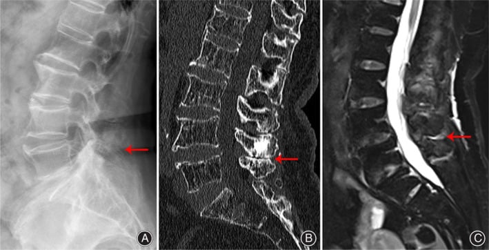
Radiographs are first line, and the lateral view is essential. It shows the kissing spinous processes, sclerosis of their tips and loss of disc height at the affected levels. Its role is screening, showing the bony contact.
CT is best for bony detail: sclerosis, flattening and enlargement of the spinous processes, cystic change within them, and the anatomy needed for surgical planning.
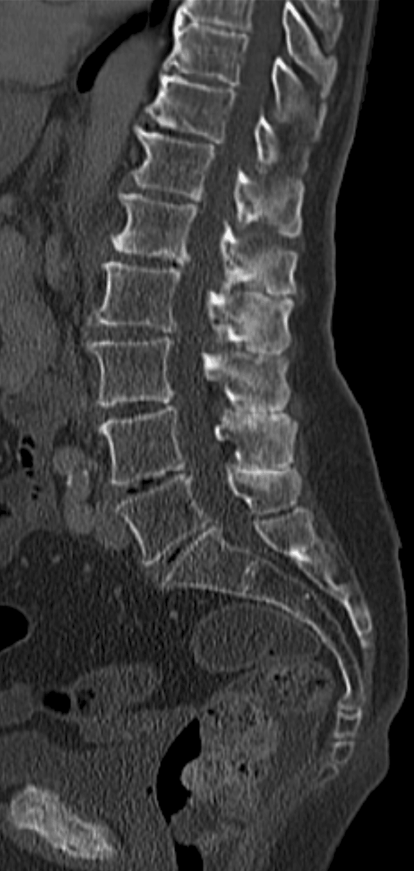
MRI is the most sensitive test for active disease, and the one that shows the bursa.
- Finding
- Spinous process morphology
- Significance
- Baseline anatomy
- Finding
- Interspinous bursa, high-signal fluid
- Significance
- Bursitis, the key finding; confirms symptomatic bursitis
- Finding
- Bone marrow oedema in the spinous processes
- Significance
- Active inflammation
- Finding
- Enhancement of interspinous tissue
- Significance
- Active synovitis or bursitis
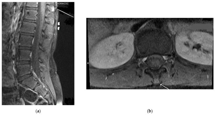
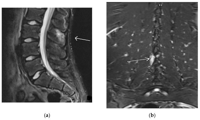
The diagnostic injection. An injection into the interspinous bursa is both diagnostic and therapeutic, and a response confirms Baastrup disease as the pain generator. With the patient prone and under fluoroscopic or ultrasound guidance, a needle is placed into the interspinous space at the affected level or levels, and local anaesthetic and steroid are injected. The response is read in three ways:
- Good relief - Baastrup disease confirmed as the pain source
- Partial relief - there may be coexisting pathology
- No relief - consider another diagnosis
Blood tests are usually not required. If infection or inflammatory arthropathy is a concern, check the ESR and CRP, which are normal in Baastrup disease, and HLA-B27 if spondyloarthropathy is suspected.
Interspinous Bursography and the Bursa-to-Epidural-Cyst Channel
The dorsal epidural cyst raises a question the routine work-up does not answer: how do you prove that a posterocentral epidural fluid collection genuinely arises from the interspinous bursa, rather than being a facet (juxta-articular) synovial cyst or an unrelated lesion? Interspinous bursography is the technique that resolves it.
The technique. Under fluoroscopy a spinal needle is advanced into the interspinous space at the affected level and a small volume of iodinated contrast is injected directly into the adventitious bursa. Opacification confirms that the needle lies within a true fluid-filled bursal cavity rather than intact ligament, and outlines its extent. It is usually combined with the therapeutic injection, so that once the bursa is opacified the local anaesthetic and corticosteroid are delivered into a confirmed target.
What it shows.
- The bursa as a discrete, contrast-filling cavity, the anatomical substrate of the Grade 3 bursitic stage
- In patients with a dorsal epidural cyst, contrast tracking from the bursa anteriorly into the posterocentral epidural space, documenting a direct connection. Chen and colleagues used bursography, alone or combined with CT, to establish this channel in their series of ten patients
- On post-bursography CT, the channel into the canal, which CT displays best when the plain fluoroscopic images are equivocal
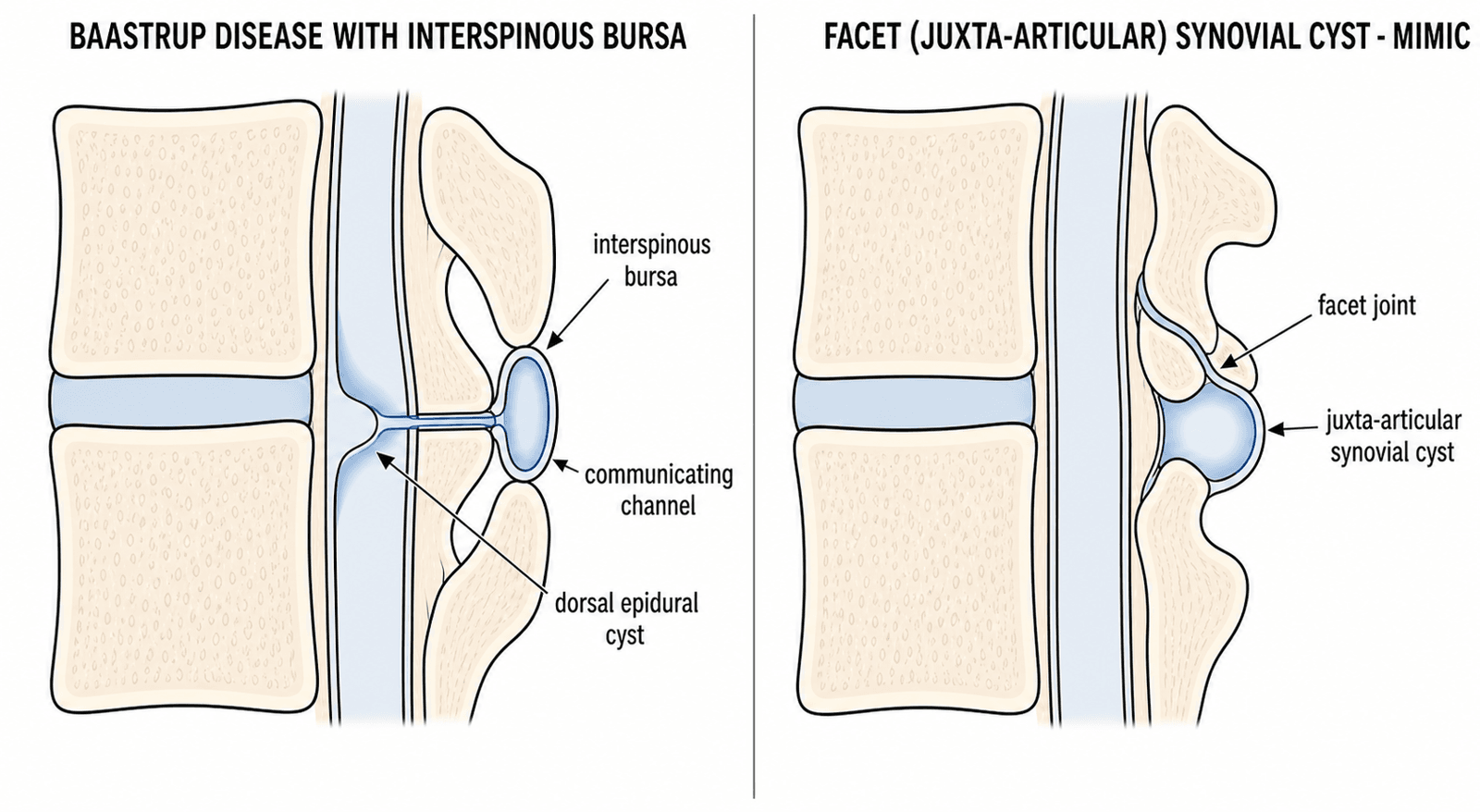
If a Baastrup patient has a posterocentral epidural cyst, do not assume it is a facet (juxta-articular) synovial cyst, which arises laterally from the zygapophyseal joint. Contrast passing from the midline interspinous bursa into the epidural space proves the cyst is an extension of the bursa. Decompressing the cyst alone will then fail, and isolated excision recurs, unless the feeding bursa and the kissing spinous contact are also treated.
Management
The principle. Conservative management is first line, and most patients respond to conservative measures and injection therapy. Surgery is reserved for refractory cases.
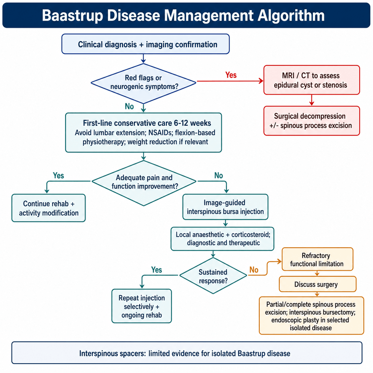
Conservative care is the starting point for every patient.
- Details
- Avoid prolonged extension, use a lumbar support
- Details
- First-line pharmacotherapy
- Details
- Core strengthening, flexion-based exercises
- Details
- Reduces lumbar lordosis and load
Injection. Indicated when conservative management has failed after 6-12 weeks, for diagnostic confirmation, or as a therapeutic trial, using the technique described under Investigations. The only published series (Filippiadis 2015, n=55) reported near-complete relief, mean pain falling from 8.18 to 0.62 on the NVS at 1 year, but it was uncontrolled: treat it as evidence of safety and diagnostic utility rather than a measured effect size. In that series 22% needed a second injection within 7-10 days, and the duration of relief is variable.
When to operate. The choice of surgical technique depends on associated pathology and surgeon preference. When an epidural cyst is present, surgery is often required for decompression.
- Refractory to conservative and injection therapy
- Epidural cyst causing neurogenic symptoms
- Significant functional impairment
Spinous process excision. Either partial excision of just the articulating surfaces or complete resection of the spinous process. Through a midline posterior approach the interspinous bursa is removed and enough of the spinous process excised to eliminate contact, preserving the supraspinous and interspinous ligaments if possible. Morbidity is minimal; the 70-85% good-to-excellent figure for pain relief is a conventional estimate rather than a measured rate (see Outcomes).
Endoscopic interspinous plasty. A full-endoscopic approach removes the interspinous bursa tissue and resects part of the spinous process with minimal soft-tissue disruption. It is minimally invasive, recovery is faster, and it can be an outpatient procedure. It is an emerging technique with promising early results, to be considered for isolated Baastrup disease, though the evidence is a three-patient series (Lin 2021).
Interspinous spacers are the third option, discussed in the section that follows.
Complications
Of the disease. Beyond the dorsal epidural cyst, ongoing degeneration may affect other levels, and Baastrup disease may develop at them.
Of injection. Infection, which is rare, bleeding, temporary numbness, and steroid side effects if injections are repeated.
Of surgery. Each complication and how it is prevented or managed:
- Prevention/Management
- Sterile technique, prophylactic antibiotics
- Prevention/Management
- Careful dissection
- Prevention/Management
- Preserve ligaments, avoid excessive resection
- Prevention/Management
- Adequate excision of bursa and bone
- Prevention/Management
- May need to address multiple levels
Natural history. Without treatment Baastrup disease typically follows a chronic, waxing and waning course, and progressive degeneration may lead to increasing symptoms over time. With appropriate treatment, whether conservative, injection or surgery, the prognosis is generally good, and most patients achieve adequate pain control with conservative measures.
Postoperative Care
After spinous process excision:
Mobilise the same day or the next morning. A drain is rarely required, oral analgesia is usually sufficient, and sitting and standing are tolerated.
Walking as tolerated, with a wound check and suture removal at 10-14 days. Avoid heavy lifting, extension and bending; return to sedentary work is often possible by 1-2 weeks.
A gradual return to normal activities, with physiotherapy for core strengthening and flexibility, avoiding repetitive extension movements. Return to manual work typically takes 4-6 weeks.
Follow-up at 6 weeks, 3 months, then as needed, with an ongoing core-strengthening programme and avoidance of prolonged extension.
Outcomes
- Success rate
- 50-70%
- Duration of benefit
- Variable
- Notes
- First-line for all patients
- Success rate
- 60-70%
- Duration of benefit
- 3-12 months
- Notes
- Diagnostic and therapeutic
- Success rate
- 70-85%
- Duration of benefit
- Long-term
- Notes
- Definitive treatment
- Success rate
- 60-75%
- Duration of benefit
- Variable
- Notes
- Device complications possible
Read these as teaching figures. Every success-rate range in this section is a conventional teaching figure, not a measured rate from a defined cohort. No comparative or controlled outcome data exist for Baastrup disease: the surgical evidence is case reports and small series (Lin 2021 n=3, Corr 2023 n=1), and the one injection series (Filippiadis 2015 n=55) was uncontrolled.
Satisfaction is estimated the same conventional way: 50-60% satisfied after conservative management, 60-70% good or excellent after injection, and 70-85% good or excellent pain relief after surgery.
Prognostic factors. Isolated disease without stenosis, single-level involvement, a good response to the diagnostic injection and no epidural cyst are favourable. Multilevel involvement, associated spinal stenosis or facet arthropathy, workers' compensation claims and a poor response to injection are unfavourable.
Guidelines, Registries & Global Practice
Baastrup disease has no dedicated society guideline; it is managed under the broader frameworks for non-specific and degenerative low back pain. There is no condition-specific implant registry. The themes below apply worldwide.
Global Epidemiology
- Figure
- 8.2% of lumbar MRIs
- Source / context
- Maes 2008, N=539, mixed-age symptomatic cohort
- Figure
- L4-L5, then L5-S1
- Source / context
- Greatest lumbar extension range
- Figure
- Over 70 years
- Source / context
- Degenerative, rises with age
- Figure
- No consistent predilection
- Source / context
- Maes 2008 found no association; older series suggested mild male excess
- Figure
- Minority of radiographic cases
- Source / context
- Most "kissing spines" are incidental
Society Guidance: How Baastrup Fits Low Back Pain Pathways
- Relevant recommendation
- No routine imaging in primary care; risk-stratified care; exercise and self-management first
- Implication for Baastrup
- Reserve MRI for failed conservative care or red flags; do not over-investigate incidental kissing spines
- Relevant recommendation
- Multimodal conservative care; image-guided injection as targeted diagnostic-therapeutic tool
- Implication for Baastrup
- Supports interspinous block to confirm the pain generator before surgery
- Relevant recommendation
- Confirm symptomatic level before any intervention; surgery only for refractory, anatomically concordant pain
- Implication for Baastrup
- Mandates a positive diagnostic block prior to spinous process resection
- Relevant recommendation
- Image guidance (fluoroscopy or ultrasound) for all interspinous infiltration
- Implication for Baastrup
- Defines standard of care for the injection step
The common thread across societies: conservative first, confirm the level with an image-guided block, operate only on concordant refractory disease.
High- vs Limited-Resource Practice
- Well-resourced settings: MRI (sagittal T2/STIR) to confirm bursitis and exclude mimics, fluoroscopic/ultrasound-guided infiltration, and endoscopic or open spinous process surgery available.
- Limited-resource settings: Diagnosis often rests on lateral radiograph plus the clinical extension-pain pattern and a landmark-guided (non-image) infiltration as both test and treatment. Open partial spinous process excision is the realistic surgical option where endoscopy is unavailable.
Areas of Uncertainty / Controversy
- Symptomatic attribution: Because kissing spines are frequently incidental, the chief controversy is whether Baastrup disease is a true pain generator or a bystander. A positive diagnostic interspinous block is the only reliable arbiter.
- No accepted classification: The Grade 1-3 imaging staging used here is descriptive, not validated against outcomes.
- Surgical evidence is weak: All operative data are small case series or reports (Corr 2023, Lin 2021); no comparative trials exist, so surgery remains a last resort for image-confirmed, refractory, isolated disease.
- Interspinous spacers: Devices such as X-STOP have been withdrawn in several markets; evidence for spacers in isolated Baastrup disease (as opposed to stenosis) is minimal.
MCQ Practice Points
Q: What is the pathognomonic MRI finding in symptomatic Baastrup disease?
A: T2 hyperintense signal within the interspinous space representing interspinous bursitis. This is an adventitious bursa that develops from repetitive friction between kissing spinous processes. Bone marrow edema on STIR sequences in the spinous processes indicates active inflammation.
Q: How do you differentiate Baastrup disease from facet arthropathy clinically?
A: Both cause extension-aggravated low back pain, but:
- Baastrup disease: MIDLINE tenderness over spinous processes
- Facet arthropathy: PARAMEDIAN tenderness over facet joints (2-3cm lateral to midline)
Both improve with flexion and worsen with extension, standing, and walking downhill.
Q: At which level does Baastrup disease most commonly occur?
A: L4-L5 - the most commonly affected level (Filippiadis 2015). This level has the greatest range of motion in the lumbar spine and is subjected to the highest mechanical stress during extension. In most cases only one level is affected; multilevel involvement occurs in advanced cases. (Beware unverifiable precise percentages for level distribution - the cited reviews give no figure.)
Q: What is the first-line treatment for symptomatic Baastrup disease?
A: Conservative management: Activity modification (avoid extension), NSAIDs, and physical therapy focusing on core strengthening and flexion-based exercises. Interspinous bursa injection (corticosteroid + local anesthetic) is both diagnostic and therapeutic. Surgery (spinous process excision) is reserved for refractory cases.
Clinical Decision Scenarios
Practise clinical reasoning and management decisions out loud
“A 68-year-old retired teacher presents with 2-year history of midline low back pain. The pain is worse when standing and walking, and improved when sitting. Examination shows tenderness over L4 and L5 spinous processes. Lateral X-ray shows close approximation of L4-L5 spinous processes with sclerosis.”
“The same patient returns 6 months later with new symptoms of bilateral leg heaviness and numbness after walking 200 meters, relieved by sitting. MRI now shows a fluid-filled cyst extending from the L4-L5 interspinous space into the dorsal epidural space, causing thecal sac compression.”
“A 72-year-old man presents with chronic midline low back pain. MRI shows interspinous bursitis at L3-L4, L4-L5, and L5-S1 with bone marrow edema at all levels. He has failed conservative management and wants to discuss injection options.”
Definition
- Close approximation of adjacent spinous processes
- Interspinous bursitis develops from friction
- First described by Baastrup 1933
- L4-L5 most common level
Clinical Features
- Midline low back pain
- Worse with EXTENSION (standing, walking)
- Improved with FLEXION (sitting)
- Tender over spinous processes
- Usually no neurological symptoms
Imaging Findings
- X-ray: Kissing spinous processes, sclerosis
- CT: Sclerosis, flattening, enlargement
- MRI T2: Interspinous bursitis (hyperintense)
- MRI STIR: Bone marrow edema
- MRI most sensitive for active disease
Differential Diagnosis
- Facet arthropathy (paramedian tenderness)
- Discogenic pain (flexion-aggravated)
- Spinal stenosis (leg symptoms)
- Often coexists with other degeneration
Management
- Conservative: Activity modification, NSAIDs, PT
- Injection: Interspinous bursa (diagnostic/therapeutic)
- Surgery: Spinous process excision if refractory
- Epidural cyst requires surgical excision
Key Exam Points
- Extension pain = BFSS (Baastrup, Facet, Stenosis, Spondy)
- Location of tenderness differentiates
- MRI confirms active bursitis (T2 hyperintense)
- Interspinous bursa is ADVENTITIOUS (not congenital)
- Epidural cyst = complication needing surgery
Interspinous Spacer Devices: Rationale and Why the Evidence Is Weak
The rationale. Baastrup pain is generated by direct spinous-on-spinous contact and compression of the inflamed bursa during extension. An interspinous process device (IPD) is placed between the two spinous processes to prevent that contact and maintain the interspinous space:
- Distracting the interspinous space, physically separating the kissing surfaces so they no longer impinge
- Limiting segmental extension at that level, blocking the movement that provokes pain while preserving flexion
In principle this directly reverses the mechanism, which is why spacers were trialled and why they can also help coexisting dynamic (extension-dependent) stenosis. They are minimally invasive and reversible. Examples are X-STOP, Coflex, Wallis and DIAM. Several early devices such as X-STOP have been withdrawn in multiple markets, X-STOP itself in many countries.
Why they often fail in Baastrup disease.
- Spinous process fracture is the dominant failure mode. The device transmits load through the spinous process, which in Baastrup disease is already remodelled, sclerotic and frequently osteoporotic in the elderly typical patient. A poor bony anchor fractures under the distraction load, and the fracture reproduces or worsens midline pain.
- Migration, loosening and heterotopic ossification, with loss of the initial distraction over time, among other device-related complications.
- The bursa is not removed. A spacer that separates the tips can still leave the symptomatic adventitious bursa in situ, so relief may be incomplete compared with excising the bursa and the articulating bone.
The evidence. Outcomes are variable, and the evidence for spacers in isolated Baastrup disease, as opposed to neurogenic claudication from stenosis, is minimal. The sclerotic Baastrup spinous process is a particularly unfavourable host for a load-bearing implant, and most surgeons therefore favour partial spinous process excision with bursectomy for refractory isolated disease.
The examiner's trap: "Would you use an interspinous spacer for this patient's Baastrup disease?" Name the mechanism (distraction plus an extension block offloads the kissing contact), then the catch: the diseased spinous process is sclerotic and often osteoporotic, so spinous process fracture is the classic failure. Targeted bursectomy with partial spinous process excision is the more durable option.
Evidence Base
Original Description by Baastrup (1933)
- First description of close approximation of adjacent lumbar spinous processes
- Associated with low back pain in a subset of patients
- Radiographic finding best seen on lateral lumbar X-ray
- Gave rise to the descriptive term 'kissing spines'
Prevalence and Associations on MRI
- Cross-sectional review of 539 symptomatic patients on lumbar MRI
- Interspinous bursitis (Baastrup disease) present in 8.2% (44 of 539)
- Significantly associated with older age, central canal stenosis, disc bulging and anterolisthesis
- NO significant association with disc degeneration, herniation, Modic changes, scoliosis or gender
Imaging Spectrum: Pictorial Review
- L4-L5 is the most commonly affected level; higher occurrence over age 70 with no gender predilection
- Hallmark is close approximation/contact of adjacent spinous processes
- Associated findings: oedema, cystic lesions, sclerosis, flattening, enlargement, bursitis, occasional epidural cysts/fibrotic masses
- Therapy spectrum: conservative care, percutaneous infiltration, surgical bursa excision or osteotomy
Posterior Epidural Cysts in Baastrup Disease
- Report of 10 patients with posterocentral epidural cysts arising from interspinous bursal fluid
- Interspinous bursal fluid can extend into the posterocentral epidural space and cause central canal stenosis
- Interspinous bursography (alone or with CT) documents the communicating channel between bursa and cyst
- Explains the mechanism linking axial Baastrup disease to neurogenic symptoms
Fluoroscopy-Guided Interspinous Infiltration
- Consecutive series of 55 patients, 67 fluoroscopy-guided interspinous infiltrations (corticosteroid plus local anaesthetic)
- Mean pain score fell from 8.18 to 0.62 on the NVS scale (mean reduction 7.56, p less than 0.001) at up to 1 year
- A second infiltration was needed in 22% (12 of 55) within 7-10 days
- No clinically significant complications
Full-Endoscopic Interspinous Plasty
- First report of full-endoscopic interspinous plasty for Baastrup disease (3 patients, local anaesthesia)
- Back-pain VAS improved from 7-8 preoperatively to 1-2 at 12 months
- Oswestry Disability Index improved markedly in all three cases
- CT confirmed an enlarged interspinous gap (1-4 mm) with little recurrent osteoproliferation
Partial Spinous Process Decompression
- Case report and literature review of surgical decompression for isolated symptomatic Baastrup disease
- Diagnosis confirmed with CT, MRI, SPECT and a positive local anaesthetic infiltration test
- Partial spinous process resection performed only after conservative therapy failed
- Highlights the lack of consensus on a consistent treatment strategy in the literature
References
- Baastrup CI. On the spinous processes of the lumbar vertebrae and the soft tissues between them, and on pathological changes in that region. Acta Radiologica. 1933;14(1):52-55. (Historical primary source; predates PubMed indexing.)
- Maes R, Morrison WB, Parker L, Schweitzer ME, Carrino JA. Lumbar interspinous bursitis (Baastrup disease) in a symptomatic population: prevalence on magnetic resonance imaging. Spine (Phila Pa 1976). 2008;33(7):E211-5. PMID: 18379391. doi:10.1097/BRS.0b013e318169614a
- Filippiadis DK, Mazioti A, Argentos S, et al. Baastrup's disease (kissing spines syndrome): a pictorial review. Insights Imaging. 2015;6(1):123-128. PMID: 25582088. doi:10.1007/s13244-014-0376-7
- Chen CKH, Yeh L, Resnick D, et al. Intraspinal posterior epidural cysts associated with Baastrup's disease: report of 10 patients. AJR Am J Roentgenol. 2004;182(1):191-194. PMID: 14684538. doi:10.2214/ajr.182.1.1820191
- Filippiadis DK, Velonakis G, Malagari A, et al. Fluoroscopy-guided infiltration for pain reduction in patients with Baastrup's disease: clinical experience and results. Skeletal Radiol. 2015;44(9):1327-31. PMID: 25930945. doi:10.1007/s00256-015-2154-0
- Lin WT, Xie FQ, Lin SH, et al. Full-endoscopic approach for chronic low back pain from Baastrup's disease: interspinous plasty. Orthop Surg. 2021;13(3):1102-1110. PMID: 33783125. doi:10.1111/os.12988
- Corr F, Grimm D, Rothoerl RD. Partial spinous process decompression in Baastrup's disease: a case report and literature review. Cureus. 2023;15(1):e34070. PMID: 36843812. doi:10.7759/cureus.34070