Rim Fracture-Dislocation | Volar vs Dorsal | Carpal Subluxation
- Fracture-subluxation - the carpus subluxates with the rim fragment (key distinguishing feature)
- Volar Barton = Thomas Type II Smith fracture - overlapping classification
- Shear mechanism - axial load with wrist in flexion (volar) or extension (dorsal)
- Always unstable - casting fails, surgery required for all displaced fractures
- Buttress plating - plate prevents fragment and carpus from displacing
- “Barton = rim fracture WITH carpal subluxation (vs die-punch = articular depression)
- “Volar Barton is more common than dorsal Barton
- “Match plate to fracture side - volar plate for volar Barton, dorsal for dorsal
- “Anatomic reduction of articular surface is critical - greater than 2mm step predicts arthritis
Overview and Epidemiology
Barton's is one of several eponyms for the same bone end, and they are not interchangeable: the parent account of the injury, its classification and the non-eponymous patterns is distal radius fractures. Of the eponyms, Smith's is the extra-articular volar-angulated pattern most often confused with a volar Barton's, chauffeur's is the radial styloid shear, and die-punch is the impacted lunate facet — which matters here, because a depressed lunate facet and a volar lunate facet shear fragment are different problems needing different implants.
The injury. A Barton's fracture is a fracture-dislocation of the distal radius: a fracture of the volar or dorsal articular rim with subluxation of the carpus. The carpus subluxates with the fractured rim fragment, and that single feature separates Barton's from every other distal radius fracture. A Barton's is intra-articular by definition.
Terminology. The names overlap:
- Volar Barton: volar rim fracture with volar carpal subluxation, the more common type
- Dorsal Barton: dorsal rim fracture with dorsal carpal subluxation, the less common type, and the pattern Barton originally described
- Reverse Barton: the volar Barton, the "reverse" of the dorsal pattern Barton described, and not the other way round
- Thomas Type II Smith fracture: the intra-articular variant in the Thomas classification of Smith fractures, which is identical to a volar Barton
Who. Barton's fractures account for 5-10% of distal radius fractures, and the volar type is about three times as common as the dorsal. Peak incidence is in young adults aged 20-40 after high-energy trauma (falls from height, motor vehicle accidents, sports injuries); the pattern is also seen in the elderly with osteoporosis after a simple low-energy fall. There is no significant gender predominance.
John Rhea Barton (1794-1871), an American surgeon at Pennsylvania Hospital, described this fracture pattern in 1838. He also invented the Barton bandage and performed the first hip arthroplasty, an interposition arthroplasty for hip ankylosis, in 1826.
Anatomy and Pathophysiology
The two rims. The distal radius has two distinct articular rims. The volar rim is the stronger and carries the attachment of the volar radiocarpal ligaments; the dorsal rim is thinner and carries the dorsal radiocarpal ligaments.
The ligaments. The volar radiocarpal ligaments are the stronger set:
- Radioscaphocapitate ligament
- Long radiolunate ligament
- Short radiolunate ligament
- Radioscapholunate ligament
The dorsal set is weaker:
- Dorsal radiocarpal ligament
- Dorsal intercarpal ligament
When the rim fractures, the radiocarpal ligaments stay attached to the fragment, and those ligaments connect to the proximal carpal row, so when the rim displaces the entire carpus subluxates with it. The intact portion of the radius offers no bony block: the fractured rim acts as a "ramp" down which the carpus slides. The carpus and the rim fragment therefore move as a single unit, and no cast can prevent that displacement.
Mechanism. The load is axial, and the position of the wrist decides which rim breaks. With the wrist in flexion the shear force concentrates at the volar rim, which fractures along an oblique line, and the volar radiocarpal ligaments pull the carpus volarly with the fragment. With the wrist in extension the shear concentrates at the dorsal rim, and the dorsal ligaments carry the carpus dorsally with the fragment.
Classification Systems
The classification is straightforward: identify which rim is fractured and confirm carpal subluxation in the same direction.
- Rim
- Volar
- Subluxation
- Volar
- Mechanism
- Flexion + axial load
- Frequency
- More common (75%)
- Rim
- Dorsal
- Subluxation
- Dorsal
- Mechanism
- Extension + axial load
- Frequency
- Less common (25%)
Clinical Assessment
History. Ask about the mechanism and the position of the wrist at impact, the energy involved, hand dominance, occupation and functional demands, previous wrist injuries and medical comorbidities.
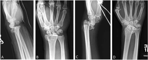
Examination. The deformity, swelling and tenderness sit on the side of the fractured rim, and the carpus is displaced in the same direction as the rim fracture. That carpal displacement is the defining finding, and it must be confirmed on the lateral radiograph.
- Volar Barton
- Volar prominence, wrist appears flexed
- Dorsal Barton
- Dorsal prominence, wrist appears extended
- Volar Barton
- Predominantly volar
- Dorsal Barton
- Predominantly dorsal
- Volar Barton
- Volar distal radius
- Dorsal Barton
- Dorsal distal radius
- Volar Barton
- Median nerve (volar)
- Dorsal Barton
- PIN/radial sensory (dorsal)
- Volar Barton
- Volarly subluxated
- Dorsal Barton
- Dorsally subluxated
Neurovascular assessment. The median nerve is the one most at risk in a volar Barton's: test sensation over the palmar surface of the thumb, index and middle fingers and the power of abductor pollicis brevis (opposition weakness), and remember that an acute carpal tunnel syndrome may develop. Assess the ulnar nerve if there is medial displacement, palpate the radial artery at the wrist and check capillary refill in every digit.
Associated injuries. Assess for:
- DRUJ instability (piano key sign after reduction)
- TFCC injury
- Scapholunate ligament injury
- Ulnar styloid fracture
- Carpal fractures
- Discriminating feature
- Volar rim fragment WITH volar carpal subluxation
- Articular
- Yes (rim)
- Carpal relationship
- Subluxated volarly with fragment
- Key pitfall
- Treating as simple Smith and casting
- Discriminating feature
- Dorsal rim fragment WITH dorsal carpal subluxation
- Articular
- Yes (rim)
- Carpal relationship
- Subluxated dorsally with fragment
- Key pitfall
- Using a volar plate (cannot buttress dorsal rim)
- Discriminating feature
- Frank dislocation, radiocarpal ligament rupture, both styloids fractured
- Articular
- Variable
- Carpal relationship
- Dislocated (not just subluxed)
- Key pitfall
- Mistaking high-energy dislocation for a benign rim fracture
- Discriminating feature
- Volar angulation, extra-articular
- Articular
- No
- Carpal relationship
- Normal (carpus follows shaft)
- Key pitfall
- Calling any volar injury a Barton's
- Discriminating feature
- Dorsal angulation, dinner-fork deformity
- Articular
- Usually no
- Carpal relationship
- Normal
- Key pitfall
- Overlooking volar lip on lateral view
- Discriminating feature
- Central lunate-fossa articular depression
- Articular
- Yes (central)
- Carpal relationship
- Normal
- Key pitfall
- Confusing impaction with rim shear
- Discriminating feature
- Lateral styloid fragment
- Articular
- Yes (lateral)
- Carpal relationship
- Normal (or scapholunate injury)
- Key pitfall
- Missing associated SL ligament tear
Investigations
Radiographs. Three views, and the lateral is the critical one:
- PA view: the rim fragment, radial shortening and articular involvement
- Lateral view: the direction of subluxation and which rim is involved
- Oblique views: additional fragment detail
What to look for. In a volar Barton's the volar rim fragment is visible on the lateral, the carpus sits anterior to the radius shaft, the lunate may appear tilted volarly, and the PA shows disruption of the volar cortex. A dorsal Barton's is the mirror image: a dorsal rim fragment on the lateral, the carpus posterior to the shaft, the lunate tilted dorsally, and dorsal cortical disruption on the PA.
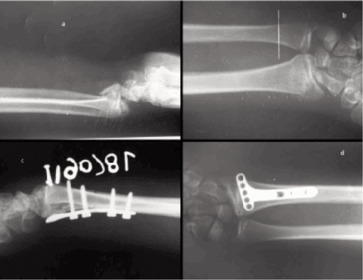
What to measure. These guide surgical planning and implant selection:
- Size of the rim fragment as a percentage of the articular surface
- Degree of subluxation, in mm of carpal translation
- Articular step-off, in mm
- Comminution of the fragment
- Radial height loss and radial inclination
- Associated injuries (ulnar styloid, TFCC)
CT is recommended for every Barton's fracture being treated surgically. It gives the exact fragment size as a percentage of the joint surface and the orientation of the fracture in the coronal plane, detects additional fracture lines and comminution, assesses articular congruity and associated carpal injuries, and plans the plate and screw placement.
MRI. MRI is not routine for an acute Barton's fracture. Consider it for persistent pain after healing, when it evaluates the TFCC and ligamentous injuries.
Management Algorithm
Non-operative treatment is rarely appropriate for a Barton's fracture, because the carpal subluxation cannot be maintained with casting alone. Most surgeons treat every displaced Barton's fracture as an operative injury and proceed directly to fixation.
The indications are very limited:
- A truly non-displaced fracture without subluxation, which is rare and may not be a true Barton's
- A patient unfit for surgery with acceptable alignment
- A palliative situation
If it is attempted. Reduce closed under anaesthesia and hold in a sugar-tong splint, converting to a long arm cast when the swelling subsides. Position the wrist in neutral rotation with slight extension for a volar pattern and slight flexion for a dorsal one, and take weekly radiographs for the first 3 weeks, because the failure rate is high.
Surgical Technique
Setup. Supine on an arm table with a tourniquet, image intensifier available, under regional or general anaesthesia.
The FCR approach.
- Longitudinal incision over the FCR tendon (6-8cm)
- Incise the FCR sheath and retract FCR ulnarly, which protects the median nerve
- Retract FPL radially
- Incise pronator quadratus in an L-shape and elevate it from the radial border
- Expose the fracture site directly
Reduction.
- Clear haematoma and debris from the fracture
- Identify the volar rim fragment
- Reduce the carpus by traction and direct manipulation
- Reduce the rim fragment to the articular surface
- Provisional K-wire fixation
- Confirm the reduction with fluoroscopy
Fixation.
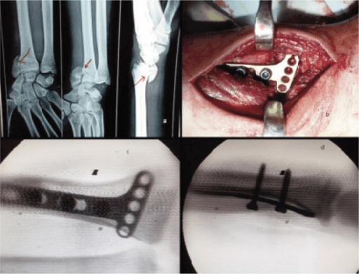
- Select an appropriate volar locking plate
- Position the plate to support the volar rim (the buttress effect)
- Fix the shaft first, which allows the plate to be adjusted
- Insert the distal locking screws into the rim fragment
- Confirm the screws capture the fragment and do not penetrate the joint
- Final fluoroscopy: PA, lateral and tilted lateral
Plate position. The plate must extend far enough distally to buttress the rim, balanced against the watershed line, since a plate placed too distal causes tendon problems. Small fragments may need specialised low-profile implants. Careful attention to plate position optimises the fixation and minimises complications.
The commonest reason a volar Barton's (or any volar marginal fracture) fails despite a volar plate is an unrecognised, separate volar lunate facet fragment, the "critical corner":
- It is a small fragment of the volar-ulnar corner of the distal radius, carrying the short radiolunate ligament, the key restraint preventing volar carpal subluxation.
- It lies distal to the watershed line, so the distal screws of a standard volar locking plate cannot reach or capture it. The plate buttresses the main rim but the corner, and with it the carpus, subluxes volarly, and the construct fails early.
- Recognise it: scrutinise the lateral and a CT for a small, separate volar-ulnar fragment; persistent volar carpal translation after plating is the warning sign.
- Fix it specifically: a more distal plate is not the answer, because it crosses the watershed line (see the Soong grading under Complications). Use a fragment-specific implant: a volar rim or hook ("tine") plate, a spring or wire-form fixation, or a separate distal screw construct that captures the corner without sitting prominently on the volar surface.
Exam point: a volar Barton's that re-subluxes after a textbook-looking volar plate = a missed volar lunate facet (critical corner) fragment needing fragment-specific fixation.
Complications
- Incidence
- 10-20%
- Prevention/Management
- Anatomic reduction (less than 2mm step), stable fixation
- Incidence
- Variable
- Prevention/Management
- Adequate fixation, early motion
- Incidence
- 5-15%
- Prevention/Management
- Appropriate plate position, low-profile dorsal implants
- Incidence
- 5-10%
- Prevention/Management
- Address associated ligament injuries
- Incidence
- 5%
- Prevention/Management
- Careful approach, release if symptomatic
- Incidence
- 10-20%
- Prevention/Management
- Early motion protocol, hand therapy
- Incidence
- 10-15%
- Prevention/Management
- Proper plate sizing, removal if symptomatic
- Incidence
- 5-10%
- Prevention/Management
- Address ulnar styloid, TFCC repair if needed
Post-traumatic arthritis is the most significant long-term complication of a Barton's fracture. Studies show that an articular step-off greater than 2mm significantly increases the risk of symptomatic arthritis, which makes anatomic reduction of the articular surface the most important factor in preventing it.
Tendon complications. On the dorsal side EPL rupture is the most common, with EDC irritation; the prevention is appropriate plate position, a low-profile implant and EPL transposition. On the volar side the tendon at risk is FPL, and the reason is where the plate sits relative to the watershed line, set out in the pearl below.
Carpal instability. Instability follows either an associated ligament injury that was not addressed or an inadequate reduction that allowed chronic subluxation. If symptomatic it requires ligament reconstruction.
Stiffness. Stiffness is more common after prolonged immobilisation, an early motion protocol reduces the risk, and hand therapy is essential for the best outcome.
The flip side of "get the plate distal enough" is "do not cross the watershed line", the named concept behind flexor tendon rupture after volar plating:
- The watershed line is the most volar (palmar) bony ridge of the distal radius, just proximal to the joint; the flexor tendons, especially FPL, glide directly over it. A plate placed distal to this line sits proud beneath the tendons and abrades them.
- The Soong grading of volar plate prominence (Soong et al., the topic's evidence card) stratifies risk: Grade 0 = plate proximal to the watershed line and not volar to the volar rim; Grade 1 = plate at or over the volar rim but proximal to the critical line; Grade 2 = plate volar to the volar rim, the most prominent. Grade 2 carries the highest FPL rupture risk.
- The tension to resolve: the critical-corner fragment tempts you to place the plate too distal (raising the Soong grade and FPL risk), while tendon safety pushes the plate proximal (risking loss of the corner). The solution is a low-profile or fragment-specific implant that captures the distal fragment without sitting volar to the watershed line.
Exam point: late spontaneous FPL (then index FDP) rupture after volar plating = a prominent (Soong Grade 2) plate distal to the watershed line; manage with early plate removal once united.
Postoperative Care and Rehabilitation
The principle. Stable internal fixation is what permits early wrist motion, within the first 1-2 weeks, and that early motion reduces stiffness, improves the outcome, and is the key advantage of operative management over casting. The protocol balances protection against mobilisation, hand therapy optimises the result, and every review looks for pain, weakness or numbness as the signs of a complication.
- Volar splint in neutral position
- Elevation and ice
- Active finger ROM immediately
- Wound check at 48-72 hours
- Oedema control measures
- Remove splint, convert to removable wrist orthosis
- Begin active wrist ROM (flexion, extension, radial/ulnar deviation)
- Suture removal at 10-14 days
- Hand therapy referral
- Continue finger ROM
- Progress active ROM
- Begin forearm rotation
- Gentle passive ROM after week 4
- Oedema control, scar management
- X-ray at 6 weeks to confirm healing
- Discontinue orthosis if healed
- Progressive strengthening
- Grip strengthening exercises
- Return to light activities
- Full ROM goal by 10-12 weeks
- Return to full activities
- Sport-specific rehabilitation if needed
- Consider hardware removal if symptomatic
- Final outcome assessment
Outcomes and Prognosis
What to expect. Surgical treatment of a Barton's fracture produces good to excellent results in the majority of patients. The determinants of that outcome are these.
Favourable factors
- Anatomic articular reduction (less than 2mm step)
- Stable fixation achieved
- Early motion protocol
- Young age
- Single fragment pattern
- No associated carpal injury
Unfavourable factors
- Articular incongruity (greater than 2mm step)
- Comminuted fragment
- Associated carpal ligament injury
- Elderly with poor bone quality
- Delayed treatment
Function. Most patients achieve a functional range of motion, within 20-30 degrees of normal, and grip strength typically recovers to 70-80% of the contralateral side. Return to work takes 4-8 weeks for desk work and 8-12 weeks for manual work; return to sport takes 3-4 months.
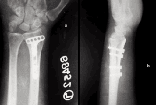
In the long term. The risk of post-traumatic arthritis correlates with the quality of the reduction, and symptomatic arthritis may require a salvage procedure, fusion or arthroplasty. Hardware removal is needed in 15-20% for irritation.
Guidelines, Registries & Global Practice
Global epidemiology
Barton's fracture is an uncommon pattern, accounting for a small proportion of the very large global burden of distal radius fractures (one of the most common fractures worldwide, with a characteristic bimodal distribution: younger patients sustaining high-energy injuries and older patients with osteoporotic fragility falls). Volar Barton's is encountered more frequently than the dorsal (reverse) variant, which is genuinely rare, with the largest published surgical series numbering only ~20 cases (Lozano-Calderón et al., PMID 16818974). The defining articular-rim-plus-subluxation pattern is consistent across populations; resource setting affects access to CT and to low-profile fragment-specific implants rather than the injury itself.
Major guidelines, side by side
The dedicated evidence base for Barton's fractures is limited to retrospective series, so guidance is largely extrapolated from the wider distal radius and partial-articular fracture literature. The strongest unifying recommendation across bodies is that displaced partial articular (rim) fractures with carpal subluxation are surgical and that fixation should be driven by fracture pattern.
- Position relevant to Barton's
- Operative fixation favoured where it restores articular congruity; for unstable patterns, technique should be driven by fracture pattern (no single construct superior beyond 3 months). Age ~65 used as a proxy for activity in deciding surgery for the broader fracture group.
- Evidence level
- Moderate–Strong (CPG)
- Position relevant to Barton's
- Classifies as partial articular 23-B3 (coronal/frontal-plane rim); recommends buttress (anti-glide) plating on the side of the fragment as the governing biomechanical principle.
- Evidence level
- Expert consensus / principle-based
- Position relevant to Barton's
- Displaced intra-articular fractures should be managed at units able to deliver definitive fixation with early senior review; emphasis on early definitive surgery and rehabilitation.
- Evidence level
- Consensus standard
- Position relevant to Barton's
- Aligns with AO buttress principle; advocates anatomic articular reduction and stable fixation permitting early motion.
- Evidence level
- Expert consensus
Registry evidence
Major arthroplasty registries (NJR, AJRR, AOANJRR, SHAR, Norwegian, NZJR) do not track fracture-fixation implants such as distal radius plates, so there is no registry-level survivorship data specific to Barton's fixation; the evidence base is therefore confined to single-centre series and comparative cohorts (e.g. Tang et al., PMID 22868605).
Global practice variation
- Implant choice: volar locking plates are the default for volar Barton's worldwide; in limited-resource settings, non-locking buttress plates or K-wire/external-fixation constructs remain in use and still achieve union, though with modestly lower functional scores (Tang et al., PMID 22868605).
- Dorsal Barton's: managed by dorsal buttress plating where low-profile implants are available; fragment-specific or wire fixation is substituted where they are not.
- Imaging: CT for pre-operative planning is routine in high-resource centres but selectively used elsewhere; the lateral radiograph remains the universal diagnostic view.
Across every major board, Barton's fractures test the same four points: (1) differentiating the rim-with-subluxation pattern from Smith and Colles, (2) explaining why the radiocarpal ligaments make it inherently unstable, (3) the buttress (anti-glide) plating principle, and (4) selecting the surgical approach to match the fractured rim. Expect a lateral radiograph and a request to compare the three patterns.
MCQ Practice Points
Q: What distinguishes a Barton's fracture from other distal radius fractures? A: Barton's fracture is a rim fracture with carpal subluxation. The carpus subluxates with the rim fragment because the radiocarpal ligaments remain attached to the fragment.
Q: A volar Barton's fracture is synonymous with which Thomas classification? A: Thomas Type II Smith fracture. Both describe a volar rim fracture-subluxation of the distal radius.
Q: What plate position is required for a dorsal Barton's fracture? A: Dorsal buttress plate. The plate must be on the same side as the fracture to provide buttress effect. A volar plate cannot buttress a dorsal rim fragment.
Q: What mechanism typically causes a volar Barton's fracture? A: Axial load with wrist in flexion. This creates a shear force that fractures the volar rim. Extension mechanism causes dorsal Barton's.
Q: What articular step-off threshold is associated with increased post-traumatic arthritis? A: Greater than 2mm. Studies (Knirk and Jupiter) show significant increase in radiographic arthritis with greater than 2mm articular incongruity.
Clinical Decision Scenarios
Practise clinical reasoning and management decisions out loud
“A 35-year-old man falls from a ladder onto his outstretched left hand with the wrist flexed. X-rays show a fracture of the volar rim of the distal radius with volar subluxation of the carpus. How would you manage this injury?”
“A 28-year-old woman falls during gymnastics, landing on her outstretched hand with the wrist extended. X-rays show a fracture of the dorsal rim of the distal radius with dorsal subluxation of the carpus. How does your management differ from a volar Barton's?”
“An examiner shows you lateral radiographs of three different distal radius fractures and asks you to differentiate them. One shows volar angulation with no articular involvement, one shows a volar rim fracture with volar carpal subluxation, and one shows dorsal angulation. Identify each and explain the key distinguishing features.”
DEFINING FEATURES
- Rim fracture (volar or dorsal) WITH carpal subluxation
- Carpus subluxates with fragment (ligaments attached)
- Volar Barton = Thomas Type II Smith fracture
- Always intra-articular (partial articular AO 23-B3)
VOLAR VS DORSAL
- Volar Barton: Volar rim + volar subluxation (3x more common)
- Dorsal Barton: Dorsal rim + dorsal subluxation
- Mechanism: Volar = flexed wrist, Dorsal = extended wrist
- Plate MUST match fracture side (buttress principle)
WHY ALWAYS UNSTABLE
- Radiocarpal ligaments remain attached to rim fragment
- When rim fractures, ligaments pull carpus with it
- Rim fragment acts as ramp allowing subluxation
- No cast can maintain reduction
SURGICAL TREATMENT
- Volar Barton: FCR approach, volar buttress plate
- Dorsal Barton: Dorsal approach, low-profile dorsal plate
- Goal: Anatomic reduction (less than 2mm step)
- Early motion within 1-2 weeks
COMPLICATIONS
- Post-traumatic arthritis (greater than 2mm step = high risk)
- Tendon complications (FPL volar, EPL dorsal)
- Carpal instability if reduction not maintained
- Stiffness if prolonged immobilization
EXAM DIFFERENTIALS
- Smith: Volar angulation, NO subluxation, extra-articular (Type I/III)
- Colles: Dorsal angulation, NO subluxation
- Barton: Rim fracture WITH subluxation (key feature)
- Die-punch: Articular depression, no subluxation
Evidence Base
Knirk & Jupiter. Intra-articular fractures of the distal end of the radius in young adults
- Landmark study of 43 intra-articular distal radius fractures in young adults (mean age 27.6). Radiographic post-traumatic arthritis developed in 91% of joints healing with residual radiocarpal incongruity versus only 11% when the joint healed congruously. Accurate articular restoration was the single most critical determinant of outcome.
Soong, Earp, Bishop, Leung & Blazar. Volar locking plate implant prominence and flexor tendon rupture
- Two cohorts (168 plated radii) graded by plate prominence relative to the volar 'watershed' line. Three flexor tendon ruptures occurred (4% prevalence) in the cohort with prominent (Grade 2) plates; none occurred in the lower-profile cohort. Introduced the widely used Soong grading of volar plate prominence.
Lozano-Calderón, Doornberg & Ring. Fractures of the dorsal articular margin of the distal radius with dorsal radiocarpal subluxation
- Series of 20 dorsal Barton's fractures (the pattern Barton originally described), 18 treated with dorsal buttress plating. At a mean of 30 months, 18/20 achieved excellent/good Gartland-Werley scores with grip strength 85% of the contralateral side. Most were accompanied by a spectrum of volar injuries (ligament tears, volar lip fractures, articular impaction).
Tang, Yang, Chen et al. Volar anatomical plates versus locking plates for volar Barton's fractures
- Comparative study of 33 volar Barton's fractures. All united; radiographic parameters (radial inclination, volar tilt, ulnar variance) were equivalent between anatomical and locking plates. Good/excellent functional results were higher with locking plates (94.1%) than anatomical plates (75%).
Harness, Ring & Jupiter. Volar Barton's fractures with concomitant dorsal fracture in older patients
- Identified a variant of volar Barton's fracture with a subtle additional fracture of the dorsal metaphyseal cortex. Where this dorsal line was unrecognised and an undercontoured volar buttress plate was used, all 5 patients lost palmar tilt with dorsal translation of the articular fragment.
Ilyas & Mudgal. Radiocarpal fracture-dislocations
- Review distinguishing high-energy radiocarpal fracture-dislocations (with disruption of the radiocarpal ligaments and usually both styloids) from rim-fragment Barton's fractures. Closed reduction is usually achievable but open reduction and internal fixation is typically required for anatomic restoration.